23.1 Introduction
Nevus of Ota (nevus fuscoceruleus ophthalmomaxillaris) and Ito (nevus fuscoceruleus acromiodeltoideus) are lesions that are examples of dermal melanocytosis. Dermal melanocytosis results from abnormal migration of neural crest cells with subsequent pigmentary changes from the ectopic melanocytes in the dermis.
23.2 Epidemiology
Nevus of Ota and Ito are more common in patients with African or Asian ancestry although they can occur in all races. Females are affected more often than males [1]. There are two peaks of presentation, one at birth, with a second peak near puberty. A few cases of later onset have been reported in the literature [2].
23.3 Clinical Findings
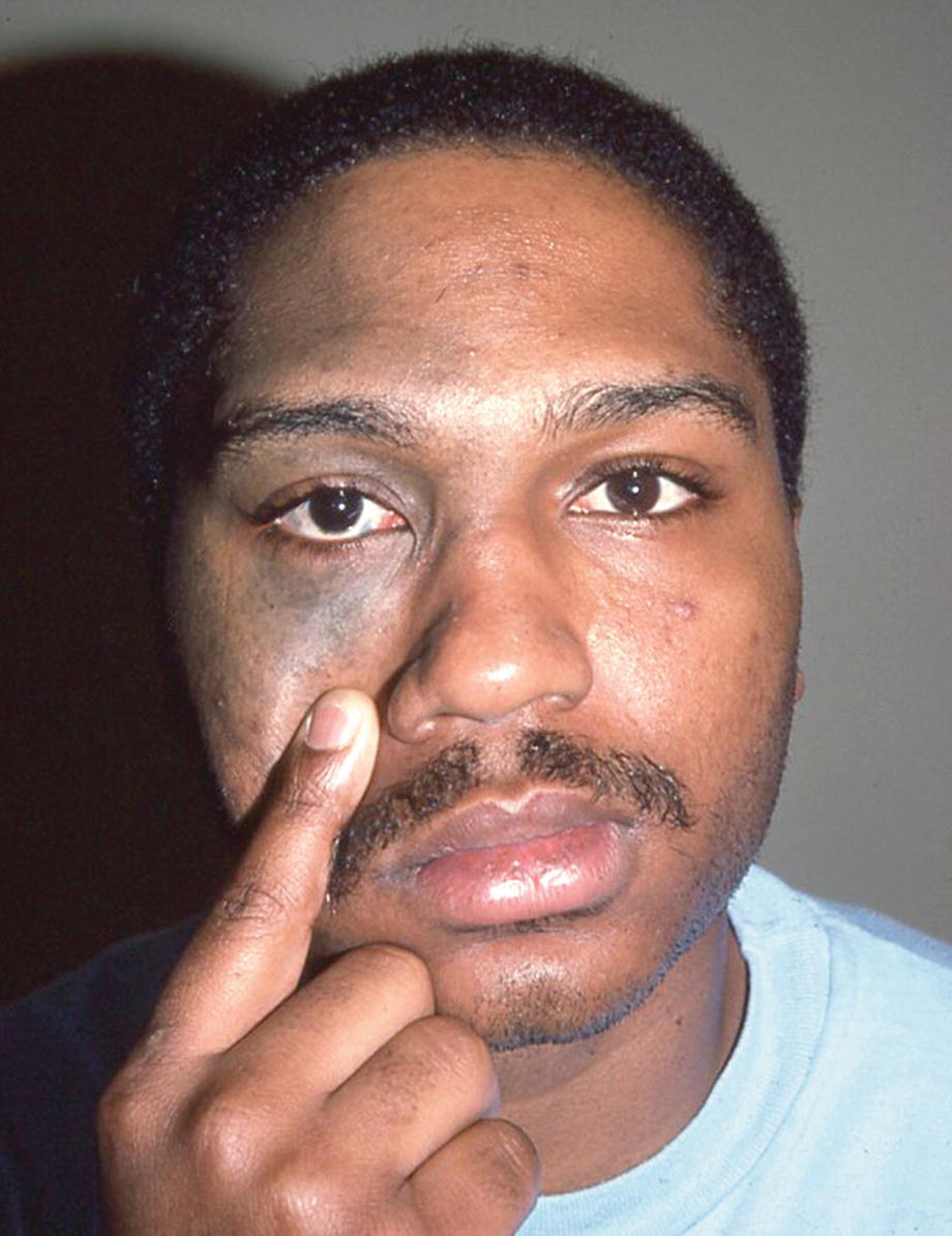
Patient with nevus of Ota. Also has ocular pigmentation
23.4 Laboratory
Histopathology shows pigmented dendritic melanocytes dissecting dermal collagen bundles in the dermis.
23.5 Treatment
A variety of lasers have been used to treat nevus of Ota and Ito [3]. Because of the increased risk of glaucoma in eyes with oculodermal melanocytosis, routine ophthalmologic exams are recommended [3].
23.6 Prognosis
Adolescents without complications such as glaucoma have a good prognosis. Malignant melanoma occurring in the skin, meninges, and/or eye associated with these lesions is quite rare.