26.1 Introduction
The term “pyogenic granuloma” is unfortunately somewhat of a misnomer. The lesion is not typically pyogenic nor is it a granuloma. The lesion has also been labeled a lobular capillary hemangioma – an acquired vascular lesion.
26.2 Epidemiology
Pyogenic granulomas are most often noted in childhood and adolescence. The etiology is theorized to be neovascularization. This can occur following trauma or can also be seen arising within a nevus flammeus [1].
26.3 Clinical Findings
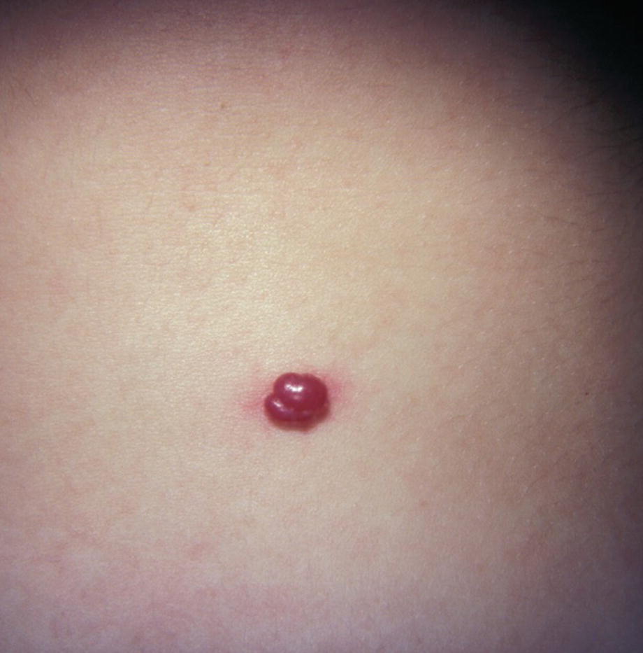
Lobulated pyogenic granuloma

Pedunculated pyogenic granuloma
26.4 Laboratory
Histopathology shows a proliferation of capillaries and fibroplasia.
26.5 Treatment
Surgical excision with curettage and/or cautery was previously considered the best option; however, recently, topical beta blockers have been noted to be useful [2].
26.6 Prognosis
Incomplete excision may result in recurrence.