12
The air scribes with their tiny jackhammer tips were silent one morning in late April 2009 as I worked my way from prep station to prep station, looking at the previous evening’s results. I was particularly eager to see what Pepson Makanela, one of our top preparators, had done. He had been working on the new block I recovered from the site in January.
The small cross section of bone I had spotted was near the end of a humerus, and it did mirror the break in the part of the child’s arm we already had. Pepson had been carefully working on this arm bone, exposing more and more of the shaft. If it was complete, it would give us a direct comparison of the juvenile’s arm with the adult’s, telling us a lot about how the two skeletons might differ, and maybe even about the growth and development of these hominins.
Sitting down in the empty chair at Pepson’s station to get a closer look at how his work had progressed, I saw something that I didn’t expect. There, just off to the side of the humerus shaft, about three-quarters of the way up the arm, a new patch of fossil bone shone light against the darker rock that surrounded it. I recognized it immediately—it was a tiny window onto a fossil maxilla—the part of the face just above the canine tooth.
A face would be a tremendously important find for us. It might be just what we needed to give us a broader comparison with skulls of Homo. As it lay there in the rock, I was fairly certain it would only be a fragment, though. The block was so small. Still, a fragment was better than nothing at all.
When Pepson arrived for work, I asked him to concentrate on this small area, to expose a bit more, and to be particularly careful about the delicate areas of the face that he was approaching. I showed him a cast of the Taung Child’s face so he would know what he might expect. I checked back on his progress throughout the day. As the careful preparation progressed, it became clear that quite a lot of the face was there. The excitement grew in the prep lab. A stream of scientists and students stopped by, watching as first the base of the eye socket was revealed and then, by the end of the day, a tooth row began to emerge. Gleaming white teeth in exceptional condition—I was thrilled!
I brought Jackie and the kids to the lab on Saturday to show them our newest discovery. I took a few family photos of the three of them posing with the newest addition to our family, and then I showed them what I thought was preserved in the block. I was pretty confident it would be half of a face, and I used the cast of the Taung Child’s skull to demonstrate how the Malapa child’s face must be sitting within the rock.
“I can tell you how much is in there,” Jackie said confidently.
I turned to her with curiosity. My wife, Jackie, is a radiologist with access to a CT scanner, which uses x-rays to look inside things, taking snapshots from many angles that are then reconstructed by the computer into either black and gray slices of sections through a body or, with the right software, a ghostly three-dimensional image. But medical CT scanners use relatively weak beams, protecting human patients from possible harm. Sticking rocks into these scanners had never been very effective because they were so much denser than living bone, with a bit of metal content, leaving a useless image with no real detail. I had tried it numerous times over the years and always failed. For that reason, I had never even considered trying CT on the dense Malapa material.
“We have a new scanner that I think can penetrate that rock and give good results,” she continued.
“Can we try Monday?” I asked, eagerly. If we could see into the rock, it would certainly help with decisions about how to prepare the precious fossil.
Monday morning, Jackie and I sat in a darkened room before a computer screen. Calling up the black-and-white image of the block, she picked a random slice somewhere near the center and adjusted the contrast for the best focus. As the image began to resolve itself at her skilled hands, my mouth literally dropped open. Jackie had picked a slice that directly passed through a point almost where the midline of the face would be.
There, as clear as a photograph, was the cross section of a complete skull!
My mind raced through the fossil record, thinking about the hominin fossil skulls I knew, most reconstructed from hundreds of tiny fragments. Such fossils were often highly distorted, buried under tons of rock, often crushed or trampled even before the sediment had enclosed them. As Jackie and I scrolled through the remarkably clear images, we realized that we were not looking at a fragmented or distorted skull. The image before us appeared to be an almost perfectly complete, undistorted juvenile skull with all its teeth intact. It seemed almost miraculous that something like this could have been preserved. Here we had a skull together with much of its skeleton, and the skeleton of a second individual besides.

THIS SKULL WOULD PROVE to be the key to understanding what the Malapa hominins were and what they were not. Using the new CT images as a guide, the preparators slowly revealed the face. Fossil after fossil followed as we worked our way through the Malapa material. First, another mandible, which must belong to the adult skeleton. Then more vertebrae and pelvic bones from both individuals. As we added one piece after another to their anatomies, the portraits were becoming clearer.


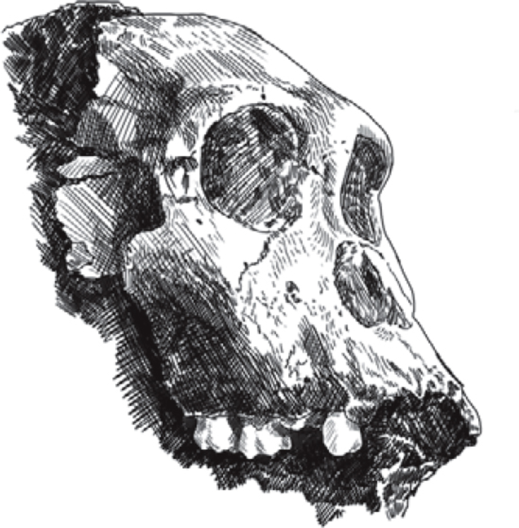
The Malapa skull only partially prepared from the surrounding matrix
By midyear, we knew that the child was probably a male. His still growing body was almost the size of the adult skeleton, which, by the same logic, was probably a female. The small canine tooth of the adult suggested a female, too. If this child had been a human, the development of bones and teeth would make us think he was between around 9 and 13 years old when he died. We could not, at this stage, know exactly how fast these early hominin individuals would have grown up, but comparisons from other immature fossil specimens gave some reason to think that they grew up faster than children today. As we built the two skeletons, piece by piece, we decided to give them each a unique designation, to assist us in talking about them. We called the male child “MH1,” short for “Malapa Hominid 1.” The second skeleton naturally became “MH2.”
The complete skull was what we had wanted to help figure out what these skeletons were. But even with all that new information, we still weren’t sure whether this child was Homo, Australopithecus, or something else. The skull was small. With a brain around 420 cubic centimeters in size, it was similar to skulls of australopith species, including Australopithecus africanus. It was smaller than any specimen of Homo except for that of the Flores skeleton, and controversial as that was, it couldn’t help much with our decision. Brain size had always been seen as a key distinguishing feature of our genus, but even after decades of searching across the whole of Africa, few of the earliest representatives of Homo had any evidence of brain size at all. Homo habilis and Homo rudolfensis merited their place with brains between around 600 and 800 cubic centimeters in size, but no fossil from earlier than around two million years ago preserved such evidence. Some earlier finds had been named Homo over the years, but always on the basis of teeth and jaws. And the teeth and jaws of this little skull were indeed the most Homo-like of its features. As the skull emerged from the rock, its face showed us something different from africanus faces, something a bit more human. Our basic problem was that the fossil record of early Homo was much less complete than the Malapa skeletons.
I arranged a trip for the entire team to travel up to East Africa in September, thinking that the direct comparison with the East African fossils assigned to both genera would help us identify the Malapa species once and for all. Meanwhile, the geologists and geochronologists had been busy. They had thrown every possible method at the problem of getting a date for the fossils. There were, by that time, two geological teams working in parallel. One team was using geophysical methods to date the sediments and flowstones that surrounded the bones. The other team was examining the fossil fauna, to try to identify extinct species found nearby that might help estimate the age of the deposits. These two independent streams of research would eventually join to create what we hoped would be an accurate age estimate for the Malapa fossils.
Paul Dirks, Jan Kramers, and Robyn Pickering focused their energies on trying to date the white flowstones, using a method that Jan and Robyn had worked on for years—uranium-lead dating. The uranium-lead dating technique assesses two separate radioactive decay chains: uranium-238 decaying to lead-206, termed the “uranium series,” and uranium-235 decaying to lead-207, the “actinium series.” Minute quantities of the two types of uranium became part of the calcite crystals within flowstones when they were originally laid down. The chain of decay of these uranium isotopes into the different isotopes of lead provides a way to estimate the age of the flowstone formation. We were in luck at Malapa—the geological team found that there was a flowstone that could be studied with this technique near the fossil hominins. Combining these findings with studies of the fauna found near the hominin skeletons, we could estimate the age of the skeletons: two million years old.
So now we had an approximate age for the fossil skeletons, but we still needed to decide what they were.