INTRODUCTION
Severe acute respiratory syndrome-coronavirus-2 (SARS-CoV-2) is the cause of an outbreak at the world level with the expression of uncommon viral pneumonia
that firstly occurred in Wuhan, China. The virus belongs to genera Betacoronavirus, keeping in view genomic structures and phylogeny interactions with other viruses responsible for severe acute respiratory syndrome (SARS) and the Middle East respiratory syndrome (MERS). They all have notable differences in their genomic and phenotypic structures [1]. All Coronaviridae family viruses are pleomorphic, having RNA as an essential genetic material. The striking feature is the possession of crown-shaped (thus named corona) peplomers or spikes that surround the genetic material. These spike proteins have facilitated the pathways leading to membrane fusion of the virus with the host cells [2]. These peplomers have a special affinity for binding angiotensin-converting enzyme 2 (ACE2), receptors located abundantly on lungs and the heart tissues. S proteins possess highly variant amino acid sequences than those of Open reading frames (ORFs) [3].
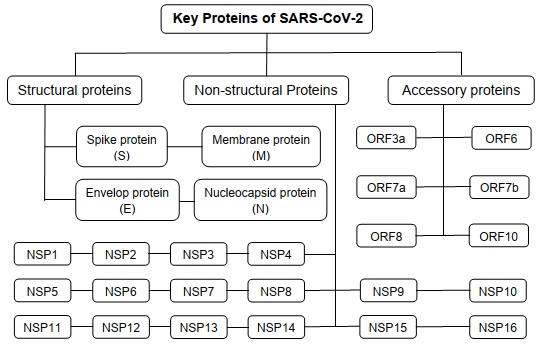
SARS-CoV-2 has been identified as a single standard +RNA comprising 2.9kb nucleotides in length encoding 26 proteins and one RNA-dependent RNA polymerase (RdRp). The proteins of this virus contain large polyproteins, i.e., ORF1a and ORF1ab, which proteolytically slash to form 16 non-structural proteins. Besides these, there are 04 structural proteins, sixteen proteins categorized as non-structural, while 06 of those proteins called accessory proteins (Fig. 1). Though these proteins are not necessary for in vitro replicating viruses, it is important for in vivo virus-host interactions [4].
Fig. (1))Structural summary of the proteins expressed by SARS-CoV-2.
Hemagglutinin esterase lacks gene in SARS-CoV-2. Although it is present in a lineage A of Betacoronavirus named human coronavirus (hCoV) HKU1 [5]. Spike protein plays a role in binding with receptors and attachment to the membrane, which indicates host tropism and disease spread [6]. In SARS-CoV-2, the S gene is different, having <75% nucleotide sequence resemblance compared to all formerly reported SARS-related coronaviruses [4]. However, the other 03 proteins of the structural category are fairly maintained compared to spike protein and are essential for overall coronavirus function.
The glycosylated spike protein is considered responsible for the induction of an immune response in the host. This spike (S) protein facilitates attack on the host cell by attaching to ACE2 protein receptors found on the host cell membrane. Envelope (E) protein is responsible for performing many functions in the viral replication cycle involving viral assembly, the release of the virion, and viral pathogenesis [7]. Membrane (M) protein is an essential membrane protein of the coronavirus that plays a significant role in viral assembly and induction of apoptosis [5]. The nucleocapsid (N) protein of coronaviruses binds directly to viral RNA, making stability. So it is found to provoke antiviral RNAi in SARS-CoV-2 [8]. These proteins provide a coating to the RNA in protein assemblage (Fig. 2) responsible for budding, forming an envelope, and pathogenicity of the COVID-19 virus [9].
Fig. (2))Structural diagram of the SARS-CoV-2.
Besides these, some non-structural proteins (NSP1-NSP16) in SARS-CoV-2 play an essential role in COVID-19 pathogenesis. Receptor recognition is one of the major differential factors among coronaviruses as a basic primary step of pathogenesis. RdRp acts in combination with non-structural proteins to keep up genome reliability [4]. It is reported that the single guide RNAs of CoVs help in the translation of all structural and accessory proteins. Hence, the genetic and phylogeny of the COVID-19 virus is important to understand its role in the pathogenesis of the disease. The current chapter will significantly focus on the structure of these proteins and their role in pathogenesis, starting from viral attachment leading to the infection in the host compared to other Betacoronaviruses.
Classification Track of the SARS-CoV-2
SARS-CoV-2 finds its place in “order” Nidovirales, “family” Coronaviridae, and “subfamily” Orthocoronaviridae, and “genus” Coronavirus. They are 4 genera in their classification, i.e., Alphacoronaviruses, Betacoronaviruses, Gammacorona viruses, and Deltacoronaviruses. Former 02 coronaviruses serve as the etiology of infection in mammals. Gammacoronaviruses and Deltacoronavirus infect avian species and both avian and mammalian species, respectively. Alphacoronaviruses own human coronavirus NL63, porcine transmissible gastroenteritis coronavirus, and porcine respiratory coronavirus. On the other hand, Betacoronaviruses have a virus SARS-CoV-2 that causes COVID-19. SARS-CoV and MERS-CoV caused SARS, and MERS, bat coronavirus HKU4, bovine coronavirus (BCoV), and mouse hepatitis coronavirus (MHV), respectively. Gamma and Deltacoronaviruses are reported to have avian infectious bronchitis coronavirus and porcine Deltacoronavirus, respectively. All these viruses are enveloped, large, and + sense RNA viruses [4].
Structural Biology of COVID-19 Proteins
SARS-CoV-2 (NC_045512.2) contains 11 genes containing 11 ORFs, including ORF1ab, ORF2, ORF3a, ORF4, ORF5 ORF6, ORF7a, ORF7b, ORF8, ORF9, and ORF10. The proteins of SARS-CoV-2 contain 02 large polyproteins, e.g., ORF 1a and ORF1ab, which proteolytically slash to form 16 non-structural proteins. Besides these, ORF2, ORF4, ORF5, and ORF9 encode 4 proteins of the structural category of proteins of the SARS-CoV-2 (Table 1) abbreviated S, E, M, and N proteins, respectively. Additionally, SARS-CoV-2 also possesses 06 proteins a category of “accessory” including ORF3a; ORF6; ORF7a; ORF7b; ORF8; and ORF10 [10] (Table 2). These proteins are derived from certain genes and contain a specific number of amino acids, as shown in Table 1.
| S. No. | Gene | Gene ID | No. of Amino Acids | Protein | References |
|---|---|---|---|---|---|
| 1. | ORF1ab | 43,740,578 | 7,096 | ORF1ab poly protein* | [3, 4] |
| 1. | ORF1a | 43,740,578 | 4,405 | ORF1a poly protein* | [10] |
| 2. | ORF2 (S) | 43,740,568 | 1,273 | Spike protein (S) | [11] |
| 3. | ORF3a | 43,740,569 | 275 | ORF3a protein | [10, 12] |
| 4. | ORF4 (E) | 43,740,570 | 75 | Envelope protein (E) | [7] |
| 5. | ORF5 (M) | 43,740,571 | 222 | Membrane protein (M) | [5] |
| 6. | ORF6 | 43,740,572 | 61 | ORF6 protein | [13] |
| 7. | ORF7a | 43,740,573 | 121 | ORF7a protein | [10, 14] |
| 8. | ORF7b | 43,740,574 | 43 | ORF7b protein | [10, 14] |
| 9. | ORF8 | 43,740,577 | 121 | ORF8 protein | [10] [15] |
| 10. | ORF9 (N) | 43,740,575 | 419 | Nucleocapsid protein (N) | [16, 17] |
| 11. | ORF10 | 43,740,576 | 38 | ORF10 protein | [10] |
The Structural Proteins
This protein category comprises 4 candidates named S, E, M, and N proteins.
Spike Protein
S protein (surface glycoprotein) is a transmembrane protein having a molecular weight of about 150 kDa, containing 1273 amino acids. It is responsible for the adherence of the pathogen to the host cell. S protein makes large bulges from the virus surface out of all structural proteins to give the crown-shaped appearance to coronaviruses. Besides facilitating entry to the virus, the spike is a determining severe factor for the host range of virus and tissue tropism and chiefly responsible for activating host immune responses. Spike consists of 3 segments: a) large ectodomain, b) single-pass transmembrane anchor, and c) short intracellular tail. Former consists of S1 as receptor-binding subunit while S2 serves as membrane-fusion subunit. Spike has been found as a clove-shaped trimmer with 3 S1 heads while one trimeric S2 stalk as identified by electron microscopy [10]. S protein of SARS-CoV-2 owns two subunits S1 and S2. The former subunit is accountable for virus-host range and tropism; however, the latter subunit is helping the fusion of membranes of host and the viral permitting entrance of the genome to the host cells. These proteins identify the human ACE2 receptor to predilected cell surfaces of humans [18]. S protein-derived entry into cells is strongly prevented by SARS-CoV spike mouse polyclonal antibodies. Remarkably, the furin cleavage site presents the S protein of SARS-CoV-2 (Fig. 3) which is not found in the S protein of pathogen SARS-CoV. This modification here can probably describe the variance in the pathogenicity of these two viruses [10]. While the TMPRSS2 gene (Transmembrane protease, serine 2), a protease is present on the host cell and helps binding S protein after priming with it. Both furin and TMPRSS2 are necessary for activation of SARS-CoV-2 pathogen in human airway cells on one end to serve as promising drug targets for the treatment of COVID-19 cases. Linkage of S protein with ACE2 is playing important concern because it is responsible for starting the infection development. Cryo-electron microscopy structure analysis has discovered that S protein of SARS-CoV-2 has almost 10−20 times greater binding ability for ACE2 compared to SARS-CoV [6]. Due to this reason, transmissibility and contagiousness of the SARS-CoV-2 are higher than SARS-CoV [19].
Fig. (3))Diagram of Spike (S) protein of SARS-CoV-2 showing trimeric segments.
Envelope Protein
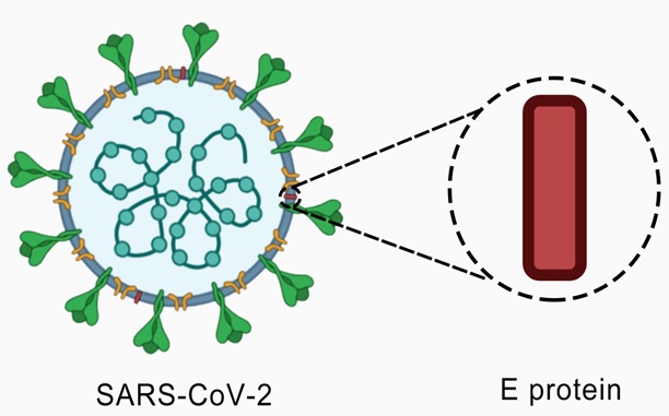
This protein is a small central membrane protein in coronaviruses, containing 75 amino acids. It can oligomerize to develop an ion channel. The E protein of SARS-CoV-2 (Fig. 4) is responsible for doing many functions in the viral replication cycle involving viral assembly, the release of the virion, and viral pathogenesis [7].
Fig. (4))Diagram of Envelope (E) protein of the SARS-CoV-2.
Interestingly, when sequence alignment of the E proteins from both types of SARS viruses was studied. It was observed that the SARS-CoV pathogen has a glutamate residue (E69) with a negative charge which resembles a positively charged arginine in SARS-CoV-2 (R69) [7].
Membrane Protein
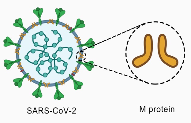
The SARS coronavirus M protein is essential that plays a significant role in viral assembly. It contains 222 amino acids. M protein of SARS-CoV-2 is the most fundamental candidate of structured protein that presents a vital role in confirming the virus envelope (Fig. 5). It extends the membrane bilayer three times leaving behind a short NH2-terminal domain external to the virus while keeping a lengthy COOH-terminus (cytoplasmic domain) inside it. This protein can bind to all other proteins of its category. N proteins stabilize by binding with M protein and promote the accomplishment of viral assembly by making the N protein-RNA complex inside the internal virion. Additionally, the SARS coronavirus M protein can also induce apoptosis. The M protein interrelates with the N protein to encapsulate the RNA genome [5].
Fig. (5))Diagram of Membrane (M) protein of SARS-CoV-2.
Nucleocapsid Protein
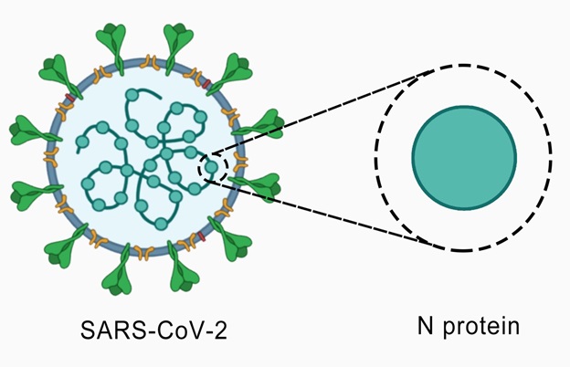
N protein of coronaviruses comprised of 419 amino acids. The protein is an essential part of SARS-CoV-2 confining in the endoplasmic reticulum-Golgi region that is structurally adhered to the nucleic acid material of the virus. This protein adheres to RNA to perform functions relevant to the viral genome, viral replication cycle, and cellular reaction of host cells against infections inflicted by the virus [16]. N protein is also densely phosphorylated and predicted to result in structural changes increasing affinity for viral RNA [17]. It is very much immunogenic and expressed in large quantities during infection, binding directly to viral RNA and making it stable (Fig. 6).
N protein enters the host cell with viral RNA to help the replication process, proceeded assembly, and release of virus particles. This protein contains 2 different RNA-binding domains (N-terminal domain NTD and C-terminal domain CTD) associated with an impoverished structure linkage region comprising a serine/arginine-rich domain [20]. The SARS-CoV N-NTD and the N-CTD can bind with the viral RNA genome because of positive amino acids [21]. It is expected to possess 2 well-folded domains in which both the terminals of N protein are fairly saturated in b-strands. However, CTD is comprised of some of the short helices [16].
Fig. (6))Diagram of Nucleocapsid (N) protein bound to RNA of SARS-CoV-2.
Sequence analysis of N protein exhibited nearly 90% similarity with SARS-CoV because of most well-kept regions in two core domains and linker. Phylogenetic analysis of N proteins exhibited SARS-CoV-2 belonging to lineage B of Betacoronavirus, same as SARS-CoV and bat coronaviruses. Remarkably, the fusion of the SARS-CoV N protein is highly muddled. There are many disordered regions without bounding to nucleic acid in recombinant full-length N protein of the SARS-CoV-2 [22]. This particular disordered region can help the protein quickly bind to different allies and sustain an accurate structure of N protein [23]. Moreover, N protein has been found to provoke antiviral RNAi. In a new study, the N protein was found to prevent cyclin-CDK (cyclin-cyclin dependent kinase) complex that will ultimately cause hypophosphorylation of retinoblastoma protein resulting in inhibition of S phase (genome replication) development in the cell cycle.
Non-Structural Proteins (NSPs)
The ORF1ab of the COVID-19 virus expresses polyproteins which are made up of 16 NSPs. Their role in pathogenesis is being explained in the following subheadings.
NSP1 (Leader Protein)
NSP1 protein of SARS-CoV-2, also known as leader protein made up of 180 amino acids. This protein strongly inhibits host gene expression by binding to the 40S ribosome of the host cell to deactivate translation and stimulates selective degradation of host mRNA. In contrast, viral mRNA remains undamaged [24].
NSP2
NSP2 is the second protein of the polyprotein of SARS-CoV-2, containing 638 amino acids. This protein is well-maintained in SARS-CoV. It can bind to 02 host proteins named prohibitin 1 and prohibitin 2 (PHB1 and PHB2). Then it is responsible for cell cycle development, cell relocation, cellular differentiation, apoptosis, and mitochondrial biogenesis. In short, NSP2 acts to disturb the host cell environment [25].
NSP3 (Papain-like Protease)
NSP3 is the papain-like protease protein consisting of 1945 amino acids, the most significant encoded protein of SARS-CoV-2 having about 200 kDa. Due to its long sequence, it has numerous conserved domains, including ssRNA binding, ADP-ribosyl (ADPr) binding, G-quadruplex binding, ssRNA binding, protease (papain-like protease) and NSP4 binding, and transmembrane domain [25]. PL1 protease (papain-like protease 1) can cut the site between NSP2 and NSP3. Additionally, the PL1 domain can provoke the release of the first 03 non-structural proteins (NSP1, NSP2, and NSP3) from the N-terminal region of polyprotein1a and polyproteins 1ab of coronaviruses [26].
NSP4 (Contains Transmembrane Domain 2) and NSP5 (3C-like Proteinase)
NSP4 is made up of 500 amino acids and interrelates with NSP3. It probably has host proteins to reorganize the membrane in SARS-CoV-2, so it is necessary to replicate the virus [25]. NSP5 consists of 306 amino acids. It splits at 11 distinct sites to produce mature and intermediate non-structural proteins (NSPs) [25].
NSP6 (Putative Transmembrane Domain)
NSP6 contains 290 amino acids. NSP6 protein can produce autophagosomes from the endoplasmic reticulum to assist in the association of replicase proteins. Moreover, NSP6 restricts autophagosome/lysosome development, resulting in the inhibition of autophagosomes to distribute the viral constituents of disruption in lysosomes [10]. In the case of SARS-CoV, it has been reported to produce membrane vesicles [10].
NSP7, NSP8, and NSP9
NSP7 is made up of 83 amino acids. It forms a complex with NSP8 and NSP12 to produce the RNA polymerase activity of NSP8 [27]. NSP8 comprises 198 amino acids. It is a peptide cofactor that makes a heterodimer with NSP7. In addition to the NSP7-NSP8 heterodimer, an NSP8 monomer unit also make complexes with NSP12 to produce RNA polymerase complexes necessary for viral pathogenicity [27]. NSP9 contains 113 amino acids. It interrelates with the DEAD-box RNA helicase 5 (DDX5) cellular protein to make a complex essential for viral replication [13].
NSP10, NSP11, and NSP12 (RNA-dependent RNA polymerase)
NSP10 is made up of 139 amino acids. It interacts with NSP14 to enhance the activity of NSP14 as an S-adenosylmethionine (SAM)-dependent (guanine-N7) methyltransferase (N7-MTase) in SARS-CoV-2 and NSP16 (2′-O-methyltransferase) [3]. NSP11 contains 13 amino acids. The function of NSP11 is unclear until now. NSP12 makes replicas of viral RNA. It comprises 932 amino acids. NSP12 makes a complex with an NSP7-NSP8 heterodimer and shows reduced processing in RNA synthesis because NSP7 and NSP8 reduce the detachment rate of NSP12 from RNA [28].
NSP13 (Helicase)
NSP13 is made up of 601 amino acids that enhance the helicase enzyme activity of unwinding the RNA duplex in SARS-CoV-2. Furthermore, NSP13 of SARS-CoV-2 exhibits 5′-triphosphatase activity, which can introduce the 5′-terminal cap of the viral mRNA. This 5′-terminal cap is the site to recognize translation and necessary for splicing, nuclear export, translation, and help to stabilize mRNA [29].
NSP14 (3′ to 5′ Endonuclease, N7-Methyltransferase)
NSP14 is made up of 527 amino acids. NSP14 of coronaviruses can exhibit 3′-5′ exoribonuclease and N7-methyltransferase activity. The guanine-N7-methyltransferase activity is part of introducing the 5′-cap of the SARS CoV-2, which defends mRNA degradation by using the host guanylyl transferase enzyme [30].
NSP15 (endoRNAse)
NSP15 (endoribonuclease), consisting of 346 amino acids, cuts RNA at uridylates at the 3′-position to form a 2′-3′ cyclic phosphodiester product in SARS CoV-2. This protein especially targets and damages the viral polyuridine structures to inhibit the immune sensing system of the host from identifying the virus. NSP15 uses manganese as a cofactor to stimulate endoribonuclease activity. It has been reported that NSP15 destroys viral dsRNA to avoid its recognition by the host [10].
NSP16 (2′-O-Ribose-Methyltransferase)
NSP16 contains 298 amino acids and forms a complex with NSP10 in SARS-CoV. There is 5′-cap in viral RNA, which defends it from mRNA degradation by 5′-exoribonucleases, stimulates mRNA translation, and inhibits the identification of viral RNA by the innate immune system [12].
Accessory Proteins
Apart from the above-mentioned proteins, there are 06 accessory proteins found in SARS-CoV-2, which are helpful to perform specific functions.
ORF3a Protein
The ORF3a protein in SARS-CoV-2 is an ion channel protein that causes activation of NLRP3 inflammasome (NOD, LRR- and pyrin domain-containing protein 3). It is made up of 1381 amino acids. ORF3a interrelates with TNF receptor associated-factor 3 (TRAF3), resulting in activation of apoptosis-associated speck-like protein (ASC) ubiquitination. It can finally cause activation of caspase 1 and IL-1β maturation [12].
ORF6 Protein
The ORF6 protein is an accessory protein having 61 amino acids and exhibits a significant role in the pathogenesis of SARS-CoV-2. ORF6 can interact with the NSP8 to stimulate the activity of RNA polymerase [13].
ORF7a and ORF7b Protein
ORF7a is an accessory type I transmembrane protein of SARS coronavirus comprising 122 amino acids [14]. The ORF7b accessory protein of SARS-CoV is present in the Golgi section, containing 41 amino acids [14].
ORF8 Protein
SARS-CoV-2 has a single ORF8 protein, while SARS-CoV has 2 ORF8 proteins: ORF8a and ORF8b. ORF8 protein contains 41 amino acids. In SARS-CoV, the ORF8b protein attaches to the associated domain of interferon regulatory factor 3 (IRF3) to deactivate interferon signaling [15].
ORF10 Protein
ORF10 protein is made up of 38 amino acids, and its function is unidentified. Remarkably, SARS-CoV contains an ORF9b protein (NP_828859.1) absent in SARS-CoV-2 [10].
Pathogenesis in Real-Time Replication of COVID-19
There are 26 proteins of different categories, i.e., structural, non-structural, and accessory, with their specific role in pathogenesis (Table 2). Pathogenesis has been detailed into various subsections with underlying mechanisms where these proteins play their role.
| S. No. | Protein | Full Name | Number of Amino Acids | Role in Pathogenesis | References |
|---|---|---|---|---|---|
| 1. | S protein | Spike protein | 1273 | Binding of virus to host cell receptor ACE2 | [10] |
| 2. | E protein | Envelope protein | 75 | Virus assembly, release, and pathogenesis | [7] |
| 3. | M protein | Membrane protein | 222 | Responsible for virus assembly | [5] |
| 4. | N protein | Nucleocapsid protein | 419 | Binds directly to viral RNA and provide stability | [10, 16, 17] |
| 5. | NSP1 | Leader Protein | 180 | Responsible for inhibiting signaling of IFN and causes blockage of host innate immune response by promoting cellular deprivation and ultimately causes blockage of host RNA translation | [10, 24] |
| 6. | NSP2 | - | 638 | Binds to PHBs 1, 2 | [25] |
| 7. | NSP3 | Papain like Protease | 1945 | Stimulating expression of cytokines and cause breakage of the viral polyprotein | [26] |
| 8. | NSP4 | Contains Transmembrane Domain 2 | 500 | Membrane rearrangement | [10, 25] |
| 9. | NSP5 | 3C-like proteinase | 306 | Cleaves at 11 sites of (3C-like proteinase) NSP polyprotein | [10, 25] |
| 10. | NSP6 | Putative Transmembrane Domain | 290 | Contributes to the formation of double-membrane vesicles by trans membrane scaffold protein and generate autophagosomes | [10] |
| 11. | NSP7 | - | 83 | Processivity lock RNA polymerase by making a hexadecameric complex with NSP8 | [10, 27] |
| 12. | NSP8 | - | 198 | Processivity lock for RNA polymerase by making the complex of hexadecameric shape | [10, 27] |
| 13. | NSP9 | RNA binding protein phosphatase | 113 | Binds to helicase | [13] |
| 14. | NSP10 | - | 139 | Stimulation of ExoN and 2-O-MT activity | [3, 10]. |
| 15. | NSP11 | - | 13 | Not identified yet | [10, 28] |
| 16. | NSP12 | RNA Dependent RNA Polymerase | 932 | Copies viral RNA (RNA polymerase) methylation (guanine) | [10, 28] |
| 17. | NSP13 | Helicase | 601 | Unwinds duplex RNA (Helicase) | [29] |
| 18. | NSP14 | 3′ to 5′ Endonuclease, N7-Methyltransferase |
527 | Stimulation of activity of ExoN and 2-O-MT | [10, 30] |
| 19. | NSP15 | endoRNAse | 346 | Degrades RNA to (endoRNAse/endoribonuclease) evade host defense | [10] |
| 20. | NSP16 | 2′-O-Ribose-Methyltransferase | 298 | Avoids MDA5 recognition and inhibits innate immunity regulation | [12]. |
| 21. | ORF3a | - | 1381 | Activates NLRP3 | [10, 12] |
| 22. | ORF6 | - | 61 | Interacts with NSP8 to activate RNA polymerase | [13] |
| 23. | ORF7a | Type I transmembrane protein | 122 | Inhibits transmembrane protein BST-2 glycosylation | [10, 14] |
| 24. | ORF7b | - | 41 | Present in the Golgi compartment and combined with viral particles | [10, 14] |
| 25. | ORF8 | - | 41 | Helps in the deactivation of interferon signaling | [10] [15] |
| 26. | ORF10 | - | 38 | Unidentified | [10] |
Receptor Recognition by Spike Protein
Coronaviruses exhibit a problematic configuration to identify receptors [31]. Alphacoronavirus HCoV-NL63 and SARS-CoV both identify Zn peptidase ACE2 [32]. All coronavirus receptors show specific physiological functions in addition to their role in viral attachment. There are 2 major regions in coronavirus a) S1 N-terminal domain (S1-NTD) and S1 C-terminal domain (S1-CTD). They act as the receptor-binding domain (RBD) by binding alone or in combination with host receptors. S1-NTDs bind sugar in all coronaviruses except MHV S1-NTD of Betacoronavirus, which identifies as a protein receptor CEACAM1. S1-CTDs can identify protein receptors ACE2, APN, and dipeptidyl peptidase 4 (DPP4) [33].
Mechanism of Membrane Fusion by Spike Protein
Spike protein is included in class I viral membrane fusion proteins. In vitro activators like trypsin cleavage and urea incubation cause pre-fusion SARS-CoV spike protein to change to the post-fusion status where S1 separates while S2 converts into dumbbell-shaped having a rod-like appearance in the middle but globular form at every end. These trimeric S2 molecules further combine on one side to give rise to rosettes form in the solution. Compared to influenza virus HA, the six-helix bundle made by coronavirus is remarkably long, which is an indicator for the profuse collection of energy that can be distributed during the structural change of S2, and it helps in the membrane fusion process. Generally, it is reported that coronavirus spikes fusion membranes like those of other class I membrane fusion proteins. It has exceptional features including their bigger size, double cleavage sites, internal fusion peptide, and long six-helix bundle [34].
Activators for Membrane Fusion by Spike Protein
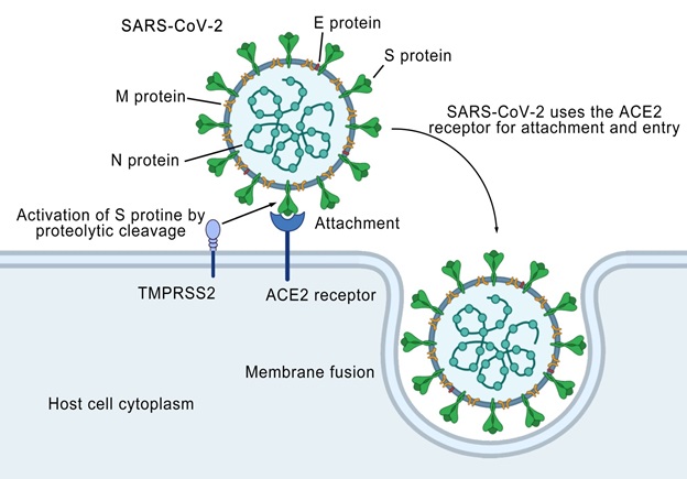
Spike protein comprises 2 functional units viz. S1 and S2. S1 unit contains N-terminal and a C-terminal RBD that directly interact with the host cells [32]. S2, on the other hand, facilitates the virus fusion (by the formation of homo-trimers) to host cellular membranes. S2 sub-unit, containing fusion machinery, converts to a stabilized perfusion state aided by RBD from S1 subunits [10]. The cleavage of both subunits occurs, leading to permanent conformational transformations with the help of host proteases. It has been reported that apart from spike glycoproteins, the clathrin-dependent and clathrin-independent endocytic pathways mediate SARS-CoV entry to host cells [35]. These pathways are yet to be investigated to play a key role in SARS-CoV-2 pathogenesis. The spike glycoproteins present at the outer membranes of the virus bring conformational changes leading to successful internalization in host cells after attaching with ACE2 host receptors facilitated by TMPRSS2 (Fig. 7). Following this discovery, it has been recently established that the utilization of serine protease inhibitors can block the function of ACE2 and TMPRSS2 [36]. Using S proteins for immunogenetics is based on the ability to initiate T cell-mediated immune protection [37]. Therefore, there is an urgency to optimize receptor targets for antiviral and anti-COVID 19 vaccine strategies for animal models before use in clinical settings.
Fig. (7))Structural mechanism showing S protein attacks on the host cell and gets entry after activation and attachment. The virus is attaching it to the ACE2 receptor protein present on the superficial membrane of host cells. A protease, TMPRSS2, facilitates this attachment. The linkage of viral S protein with ACE2 on the host cell surface results in the entry of the virus into the host cell leading to the development of infection.
The gene enrichment analysis of ACE2 has revealed the mechanisms responsible for the successful viral attack and replication within host cells. It has been shown that alveolar type 2 (AT2) cells were highly expressed in COVID-19 patients, depicting the success of novel coronavirus in jeopardizing host cells for transmission and reproduction of virus [18]. The modeling of S proteins for risk factor analysis and target drug designing against SARS-CoV-2 has opened arenas for putative drug targets based on in silico analysis. The study reported that despite genetic diversity, the RBD domain of the S protein of COVID-19 supports strong electrostatic interactions with human ACE2. This finding highlighted the potential of SARS-CoV-2 to infect and transmit disease in humans [3]. Sufficiently low metastability of S2 sub-unit, supported by a low S1 affinity for target receptors, both depending on proteolytic ability, could halt the successful receptor binding and cell transfection. The vital role of S proteins in pathogenesis and infectivity within host cells has been sought for vaccine and treatment candidates against coronaviruses.
Evolution of Spike Proteins
S proteins are generally different from other coronaviruses generally, and Betacoronaviruses (including SARS-CoV) earns them a genetically distinguished nomenclature [38]. Residues from SARS-CoV-2 RBD, crucial for ACE2 binding, are highly conserved or share very similar properties as SARS-CoV. This homology highlights the convergent evolution between both SARS and SARS-CoV-2 coronaviruses [18]. The S protein mutations have been historically giving rise to mutant genetic variations. The typical examples of this phenomenon are feline infectious peritonitis virus (FIPV) serotype II and porcine epidemic diarrhea virus (PEDV). This recombination may not only alter the genetic makeover but modify the tropism for host species [39].
Different coronaviruses possess various binding sites within S1 subunits that are recognized for binding and host cell internalization. For instance, human coronavirus HKU1 and OC43 attach via S domain-A (SA) to acetyl sialoside present on the host cell surface made of glycolipid and glycoproteins. In contrast, the MERS coronavirus utilizes the SA domain to bind non-acetylated surfaces of receptors which probably promote SB domain binding to receptors and host peptidase. However, SARS-CoV and SARS-r CoV undergo direct interaction with ACE2, mediated by SB, to attack host cells. It is well noted that SARS-CoV-2 is as well adapted as SARS-CoV to selectively bind human ACE2 receptors for viral entry [10]. The S proteins of SARS-CoV-2 are trimers that are evolutionarily conserved and highly glycosylated in nature. Therefore, it is required to probe into the evolutionary patterns of S1 and S2 subunits of S proteins making binding to ACE2 receptors very distinct. This binding is highly significant as its detailed understanding could contribute to bringing forward some alterations in the 3D structure leading to attenuation of the virus particles.
Invasion and Replication of SARS-CoV-2
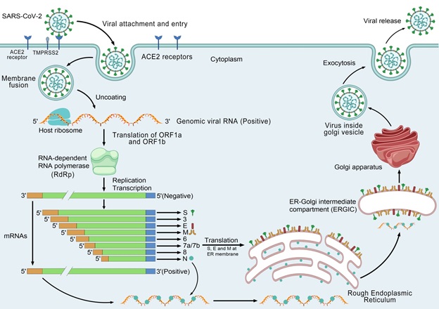
S protein belonging to coronavirus is an important contributing factor for invading viruses into host cells [40]. The SARS-CoV-2 may enter the host via the respiratory tract or mucosal surfaces such as the conjunctiva. SARS-CoV-2 binds to epithelial cells of the nasal cavity, whereas the upper respiratory tract is an efficient replication site of this novel coronavirus. SARS-CoV-2 causes a less abrupt onset of symptoms like other conventional human coronaviruses that are the extensive cause of common seasonal colds [2]. During the prodrome period, the infected patients produce a copious virus in the upper respiratory tract. SARS-CoV-2 can also replicate in the lower respiratory tract and causes radiological evidence of lower respiratory tract involvement in patients having no clinical pneumonia [41]. S protein attaches to cellular receptor ACE2 for SARS-CoV [42] and SARS-CoV-2 [43]. This is further evident by a fluorescent study that SARS-CoV-2 applies the ACE2 receptor the same way as the SARS-CoV [44]. Bronchoalveolar lavage fluid from a positive COVID-19 patient also confirmed the use of ACE2 as an entry receptor by SARS-CoV-2 [36]. ACE2 protein remarkably presents alveolar epithelial cells and gives rise to the regulation of transmission within humans and cross-species [43]. The S-glycoprotein on the SARS-CoV-2 surface attaches to the ACE2 cellular receptor of human cells and holds two subunits (S1 and S2). Subunit S1 is responsible for virus-host range and cellular tropism by RNA binding domain aiding the attachment, whereas S2 causes the virus-cell membrane fusion through heptad repeats 1 (HR1) and HR2, the tandem domains [3]. Lysine 31 residue present on the human ACE2 receptor identifies 394-glutamine residue located in the RBD region of the virus. Single N501T mutation in the virion S protein of SARS-CoV-2 may have shown a significant increase in binding affinity for ACE2. A study suggested that the binding efficiency of ACE2 and SARS-CoV-2 S protein is found 10-20 folds higher as compared with SARS-CoV. The attachment of SARS-CoV-2 into cells is being determined by fusions of membranes between virus and plasma [43]. A recent study on SARS-CoV-2 has reported that S protein priming is required to facilitate the process of entry, which is provided by the TMPRSS211 produced in the host cell (Fig. 7) [43]. In addition to the mechanism of membrane fusion, entry of the virus is facilitated by clathrin-dependent endocytosis and independent endocytosis [45]. After membrane fusion, the virus eliminates the RNA genome into the cytoplasm as an ss+ RNA followed by the translation of two viral replicase polyproteins, pp-1a and pp-1ab, which encode the NSPs, finally giving rise to RTC (replication-transcription complex) in double-membrane vesicle [36]. Continuous replication of RTC gives rise to nests of sub-genomic RNAs that finally encode proteins from the structural category and those of the proteins' accessory category [43]. Additionally, the viral genome encodes some non-structural proteins, including RdRp, 3CLpro, and PLpro, which are slashed into effector proteins by using viral proteinases [46]. PLpro acts as a deubiquitinase that can cause immune suppression by deubiquitinating specific host cell proteins like interferon factor 3 (IF-3) and NF-κB [47]. 3CLpro causes proteolysis of viral polyprotein into functional units while RdRp makes a complete negative-stranded RNA template which can be used by RdRp to further produce RNA of viral genome. Newly developed envelope proteins are introduced into membrane of endoplasmic reticulum, and hence genomic RNA and nucleocapsid protein combine to form nucleocapsid that subsequently assemble into virions. The virus then grows in endoplasmic reticulum-Golgi intermediate compartment that finally releases from host cells via vesicles having enclosed with virus particles [43] as shown in Fig. (8).
Fig. (8))The entire mechanism of action of proteins from attachment to viral release. Firstly, the S protein attaches to ACE2, followed by conformational alteration in S protein facilitating the union of viral envelop with cell membranes via the endosomal pathway. The virus eliminates RNA into the host cell to translate it into viral replicase polyproteins pp1a and pp1ab. These polyproteins are slashed by viral proteinase enzymes giving rise to small products. Polymerases also produce subgenomic mRNA sequences using intermittent transcription, translated into viral proteins of significant importance. Proteins further combine with the RNA genome to construct virions in the endoplasmic reticulum and Golgi bodies transported through vesicles. These vesicles finally attach to the host cell membrane to release the virus.
Antigen Appearance in COVID-19 Infection
Upon entering the virus into cells, the antigen-presenting cells (APC) take up the viral antigen necessary for the body's immunity against viruses. After this, peptides of antigenic nature are taken up by MHC or human leukocyte antigen and, after that, further reach virus-specific Tc lymphocytes [43].
Activation of Humoral and Cellular Immunity against Covid-19 Infection
The appearance of an antigen consequently activates all kinds of the body's immune responses with the help of virus-specific immune cells. Just like acute viral infections, antibodies including IgM and IgG produce against the SARS-CoV-2 virus. Shortly before the termination of the 12th week, SARS-specific IgM antibodies disappear, whereas IgG3 antibodies persist for a prolonged duration which is an indication of defensive character [48]. In contrast, S and N specific antibodies are considered SARS-related IgG antibodies [40]. Recent research has reported the reduced amount of cluster of differentiating immune cells (CD4+ and CD8+) T cells in the blood circulation of the patients. However, their amount are increased like human leukocyte antigen–DR isotype [3]. SARS-CoV patients exhibit a severe reduction of CD4+ and CD8+ T cells in acute kinds of disorders. Even in the absence of antigen, CD4+ and CD8+ memory T cells are observed for 4 years in some SARS recovered patients while playing viral share in the proliferation of T cell, delayed-type hypersensitivity response, and produce interferon-gamma (IFN-γ) [49].
Cytokine Storm in COVID-19
Acute respiratory distress syndrome (ARDS) is predicted to be the main reason for deaths in COVID-19 patients reported by Lancet [50]. ARDS is a general immunopathological event in SARS, COVID-19, and MERS infections [3]. The dominant mechanism for ARDS is cytokine storm which occurs due to excessive release of chemokine 2, (CCL2); chemokine 3, CCL3; chemokine 5, CCL5; chemokine 8, CXCL8; chemokine 9, CXCL9; chemokine 10, CXCL10), and the pro-inflammatory cytokines (IFNα; IFNγ; TGFβ; IL-1β; IL-6; IL-12; IL-18; IL-33;) by immune effector cells in SARS infections [51]. MERS-CoV infected patients also exhibit increased levels of some cytokines like IL-6, IFN-a, and chemokines CCL5, CXCL8, and CXCL-10 in serum similar to SARS-CoV infection [52]. However, SARS-CoV-2 infected patients with many T-lymphocytes, macrophages, and dendritic cells cause excessive release of pro-inflammatory cytokines, which can eventually lead to ARDS [53]. This cytokine storm can cause hyperactivation of the immune system in the body resulting in ARDS and failure of organs that finally result in the expiry of the COVID-19 of critical situations comparable to MERS and SARS infection [3].
Coronavirus Immune Evasion
The causative agents of SARS disease and MERS disease use various approaches to bypass immunity for better survival in host cells. Pathogen-associated molecular patterns are well-maintained microbial structures that pattern recognition receptors can identify. But, both pathogens, i.e., SARS-CoV and MERS-CoV, can stimulate the formation of double-membrane vesicles devoid of PRRs (pathogen recognition receptors), so they can multiply in these vesicles and avoid the host recognition of their ds RNA [54]. IFN-I (IFNα- and IFN-b) expresses a defensive response in the case of SARS-CoV and MERS-CoV infection [55]. Similarly, SARS-CoV-2 also uses passive and active mechanisms to avoid the IFN-1 response in which passive mechanisms involve forming double-membrane vesicles, as mentioned above. At the same time, active mechanism occurs due to the direct effect of proteins of the pathogen on transcription factors and intracellular signaling molecules that control the IFN cascade. Particularly the ORF6 proteins of SARS-CoV-2 play their role to prevent the effect of INF regulatory transcription [53].
Factors Affecting SARS-CoV-2 Pathogenesis
It is necessary to rule out the origin of pathogen and reservoir to develop preventive strategies to combat this disease. This formidable outbreak appeared in the Huanan Seafood Wholesale Market of Wuhan, Hubei, China. The market provided palm civets, bats, raccoon dogs, frogs, snakes, marmots, and other wild animals that are frequently sold [56]. Initially, it appeared as a perception that patients with COVID-19 may have visited the Huanan Seafood Market or may have eaten the infected birds or animals [57]. Primary studies also reported some COVID-19 positive environmental specimens in Wuhan, and the snake was considered a possible key reservoir, but some researchers disagreed. Afterward, the role of bats and some other wild animals, including pangolin, was speculated to support the rapid spillover events of COVID-19 [58]. Recent research expressed that the SARS-CoV-2 is related to subgroup Betacoronavirus having mostly identical genomic sequences to a SARS-like bat coronavirus (bat-CoV), indicating bat natural host of origin. Genomic analysis showed that the genome of SARS-CoV-2 shares nearly 96% identity to the bat-CoV RaTG13, while it is almost 80% identical to SARS-CoV [38]. These speculations were somehow supported by evidence of genetic homology in ACE2 receptor binding mechanisms. Also, the people that had traveled to or had any interaction with the workers/routine visitors of the market were reportedly contracting the disease. Still, later on, some people contracted the disease even without visiting the seafood market [59]. The exact origin, location, and potential natural reservoir of pathogen SARS-CoV-2 are still unclear, and they might come from some wild animal in the market. But it is thought that the virus has zoonotic nature, and bats may be suspected as key reservoirs because of the identical genome to the bat-CoV, and they might share the same ancestors [56]. It was previously presumed that COVID-19 might have transplacental or vertical transmission. This notion supported by various research cohorts-based results has been nullified [60]. Apart from these, some bad health habits, i.e., tobacco smoking and alcohol consumption, may also be important risk factors. The disease has been confirmed in passive smokers; however, active smokers may be adversely affected, leading to inadequate immune response and viral containment. It could be concluded that smoking causes degeneration of protective cilia (alveolar injury) and suppresses the local immunity, eventually leading to more chances of viral internalization and subsequent disease [61].
Host Factors on the Genetic Basis
It was speculated that the COVID-19 infection severity and its outcomes in Asian, Caucasian, European populations might differ [56]. Similarly, it was presumed that there might be some difference in the susceptibility of males and females to contract coronavirus disease. Both notions have been supported and rejected by different research groups making these factors highly controversial [62]. Co-morbidity and immunosuppression in any individual can make the viral pathogenesis shorter and more severe [63]. The elderly or chronic disease (hepatitis, cancer, other renal or hepatic disorders) patients with low profile immune system functionality is also at higher risk of contracting the disease. This could be attributed to compromised immunity and weakened local responses to the virus [64]. Some preliminary examinations also suggested severe obesity and smoking as associated risk factors [65]. However, some researchers reported more risk in males than females in Italy, which may be due to a high incidence of smoking habits [9]. Studies found that SARS-CoV-2 infections lead to fatal pneumonia in old-aged patients, and bacterial infection may also cause secondary bacterial pneumonia [66].
Organ failure due to COVID-19 depends upon the percentage of expression of ACE2 receptors by different cells. These receptors make way for viruses to the organ. The proportion of ACE2 <1% is associated with a low risk of organ failure, while an increase in proportion is directly linked with a higher risk of getting the disease. Nasal and bronchi do not express ACE2; hence have low or no risk of getting organ failure. The highest proportion of ACE2 expression is noted in the ileum giving rise to nearly 30% of the expression of ACE2 receptors hence categorized as high-risk organ failure. The respiratory tract, urinary bladder, esophagus, kidney, and heart are categorized under high-risk organs because the expression of ACE2 appears to be 2%, 2.4%, >1%, 4%, and 7.5%, respectively. Cardiac injury is the most important disease complication in the wake of COVID-19, where 13% of the increase in creatinine kinase [4]. Such patients are observed to have higher levels of mRNA and ACE2 expression that take the patient to organ failure. Kidney failure was found among 14.4% of COVID-19 patients presenting increased levels of serum creatine [61]. Hospital-based mortality becomes 16.1%, while patients showing higher baseline serum creatinine show double mortality than those who did not have a higher baseline creatinine issue. Effective cell adhering and uninterrupted virus entry, low chance of virus elimination, compromised T cell function, susceptibility against cytokine storms, and excessive inflammation are key steps of enhanced progression and pathogenesis of SARS-CoV-2. However, ACE2 expression decreases with insulin administration [67]. In contrast, ACE2 function is reduced by hypoglycemic candidates such as thiazolidinedione, glucagon-like peptide-1, ACE inhibitors, angiotensin receptor blockers, antihypertensive drugs, and statins up-regulated. Compared with the normal control group, the rodent diabetes model exhibited a strong expression of ACE2 in vital organs of the body in the study [68]. According to their Mendelian randomized study, the lungs of patients with diabetes mellitus (DM) determined in COVID-19 disease and characteristics showed increased expression of ACE2. It has also been found that protein concentrations of overexpressed protease enzymes (such as furin) are too high in diabetic patients, promoting the cleavage of spike proteins, hence permitting entry of the virus.
TMPRSS2 is a protease enzyme that is produced by the host and resultantly potentiates priming of S protein of SARS-CoV-2. This process facilitates binding with ACE2, and ultimately entry of virus in the tissue. Some TMPRSS2 inhibitors are approved for clinical application to block entry thus may be helpful in the treatment protocol. Camostat mesylate is a drug that inhibits the expression of this gene [36].
Viral Factors
SARS-CoV-2 is an air-borne virus capable of sustaining on inanimate objects for hours. The virus possesses great genetic diversity, having reported at least 7 distinct types to date. Important virulence factors like an invasion of immune checks from the host under double vesicle formation, inactivation of translational machinery by 8 different proteins of the virus, host cap machinery mimicry, blocking various STAT signaling pathways, and downregulation of gene expression for rendering antigen presentation unsuccessful. These pathways are mediated by both non-structural and structural proteins [69]. It is necessary to take care of and devise strategies for these targeted pathways of viral infection.
RNA-dependent RNA-polymerase (RdRp)
RdRp is responsible for the replication of the genome of COVID-19. Antiviral drugs like Ribavirin & Remdesivir are used for Ebola virus infection, Hepatitis C infection, and RSV infection with the hypothesis of the drug serving as nucleotides analog that restricts the process of replication by a virus that blocks nucleotide formation of SARS-CoV-2 virus. There are some reactions in terms of increased transaminases and renal injuries [5].
Papain-like Protease (PLpro)
These proteins result in the proteolysis of viral polyprotein into active viral protein. Lopinavir has been tried against HIV in combination with ritonavir that works to inhibit protease. Adverse reactions were nausea, vomiting, GIT disturbances, pancreatitis, and cardiac abnormalities [70].
Coronavirus Protease (3CLpro)
This protein causes proteolysis of polyprotein into an active protein. Lopinavir antiviral drugs previously have been used in combination with ritonavir to inhibit protease. Nausea, GIT issues, pancreatitis, vomiting, and cardiac abnormalities were limiting factors [70].
Spike Protein (S Protein)
These proteins help adhere the virus to the host receptor, which facilitates the entrance of the virus. Otherwise, ACE2 catalyzes angiotensin II to angiotensin1–7and angiotensin1–9, which serve as anti-inflammatory and antioxidants, to play a vital role in treating acute respiratory distress. Arbidol antiviral drug is supposed to prevent binding of S protein to the host cell, thus blocking the entry of bacteria into host tissues. The drug has been used against Influenza while elevated transaminase, allergic response, and GIT disorder are observable side effects [58].
Comparison of Different Coronaviruses
Novel virus belongs to the same Betacoronavirus that owns bat SARS-like coronaviruses, and MERS-CoV. However, SARS-CoV differs in some respects from other coronaviruses [23]. It causes milder infection and is transmitted much more in the community, whereas MERS-CoV and SARS-CoV spread is nosocomial [71].
Virology and Genome
Phylogenetic analysis reveals SARS-CoV-2 (belonging to Clad I and Clad IIa; bats are animal reservoirs; the intermediate host is unknown) to be more related to SARS-CoV (belonging to Clad I and Clad IIb; bats are animal reservoirs; the intermediate host is palm civets) and bat SARS-like coronavirus [69]. Phylogenetic association between pangolin CoVs and SARS-CoV-2, although they share a common animal origin. Phylogenetic studies are still scarce to correctly state pangolin as an intermediate host of SARS-COVs. There are amino acid differences between SARS-CoV and SARS-CoV-2, while their mode of binding to ACE2 nearly remains similar. The SARS-CoV-2 shares 96.2% similarity to the bat-CoV RaTG13. This similarity reaches 79.5% with SARS-CoV and 82% identical to MERS-CoV pathogen (belonging to Clad II; bats are animal reservoirs; intermediate host camels) in genomic sequence [60]. MERS virus shows up stable genome since the time of origin in 2012 while other viruses of this genus keep on evolving with new combinations.
S1/S2 of S protein has been proved to be a controlling loop to stabilize the virus when structural cleavage is intact, reaching the downstream S2′ site, which monitors exposure to peptide fusion and their functionality. SARS-CoV-2 S protein contains clear insertion in a genome that maps the S1/S2 priming loop, and this distinction is not found in any other coronaviruses in Betacoronavirus lineage B. This distinct insert in the genome of SARS-CoV-2 gives rise to potential changes in the virus every time. Moreover, SARS-CoV-2 S1/S2 site is considered an exposed flexible loop that makes an easily approachable site for protease cleavage, which presents a bigger share in the S protein function of the SARS-CoV-2 pathogen. The priming loop is predicted to alter the pathway of entry of the pathogen predominantly. There is no attribute found in any other virus belonging to Betacoronavirus lineage B. Cleavage of S protein by furin (Golgi-resident protein) while virus assembly and delivery to cell surfaces. Another source of change in structure is FCoV, a loop that is a proteolytically sensitive structural loop present in the hypervariable region of the spike. This makes the spike region a significant driver of virus evolution [16]. Cryo-electron microscopy found SARS-CoV receptor-binding motif (RBM) down confirmation to be packed closer to the NTD of the S protein. At the same time, that of SARS-CoV-2 RBD angled to the center of the trimmer in down conformation. While similar binding modes for both SARS-CoV and SARS-CoV-2 RBDs compensate for the difference of amino acids in the proteins of both CoVs [18].
The SARS-CoV-2 amino acid sequence of S protein resembles 76.47% of SARS-CoV, showing the same structural configuration and electrostatic properties in the interaction interface [3]. Substitution of only 380 amino acids between SARS-CoV and SARS-CoV-2, which are commonly found in genes of non-structural proteins, has been found by genomic comparison between SARS-CoV-2 and SARS. In contrast, 27 mutations have been found in S protein are liable for receptor adherence and entry into the host cell [43]. This might explain the low pathogenic nature of SARS-CoV-2 pathogen than SARS-CoV but needs more investigations [72]. SARS-CoV-2 has emerged as a human pathogen by a mutation in S protein at RBD, resulting in increased pathogenicity by increased affinity to the receptor and determining the tropism. Therefore, it is assumed that SARS-CoV-2 behaves similarly [73]. Genomic studies revealed that the same human cell receptor, ACE2, is being shared by SARS-CoV-2 and SARS-CoV to enter the cell. However, the primary receptor that MERS-CoV uses is DPP4 for cell entry. The ACE2 receptors are ectoenzymes that are found anchored to various tissues' cell membranes, especially in the lower respiratory tract, gastrointestinal tract, kidney, and heart [66]. SARS-CoV-2 holds 6 major amino acid changes in the RBD region of S glycoprotein at position 344, position 360, position 472, position 479, position 480, and position 487 compared to SARS-CoV where no substitution of amino acids was present in RBD that interacts directly with ACE2 human receptor [74].
Epidemiology
COVID-19 transmission among humans is the greatest challenge to control this disease. Studies have shown that asymptomatic carriers have a high virus load in the upper respiratory tract which participates in a high prevalence of COVID-19. The transmission of a virus is indicated by R0 (reproductive number), indicating the average no. of new infections produced by an infected person from a mass that has never seen the disease before. R0>1 indicates that the infection is likely to enhance whereas, R0<1 indicates that the outbreak will decline. The World Health Organization has estimated higher R0 of SARS-CoV-2 (2-2.5) compared to MERS (<1) and SARS (1.7-1.9), suggesting the higher pandemic potential of novel infection [69]. Serial interval (time of onset of disease and time of onset of disease in secondary case) in SARS-CoV-2 is less than 4 days that suggest transmission of disease before the clinical case. Lower serial interval and higher reproductive number lead to a higher spread of COVID-19. Whereas the death rate of SARS-CoV-2 has been recorded to be 2.3%, SARS-CoV gives rise to 9.5%, while the MERS virus results in 34.4% deaths of patients as reported by the Chinese Centre for Disease Control and Prevention in China [65]. The binding attraction of the SARS-CoV-2 S-protein & ACE2 receptor is higher than the SARS disease virus (2003 strain). This might explain the rapid development and transmission among humans by the SARS-CoV-2 pathogen. It is also predicted that if one nucleotide mutation in RBD of SARS-CoV-2 happens, it would further enhance pathogenesis [73]. The in vitro inoculation of SARS-CoV-2 on the epithelial cell surface of human airways causes cessation of cilia movement and cytopathic effect [60]. In type I and type II pneumocytes as well as enterocytes, there is higher replication of SARS-CoV-2. In the lungs epithelium, downregulation of ACE2 receptors due to SARS-CoV results in the pathogenesis of lung injury and finally expresses as ARDS clinical presentation [66]. However, MERS-CoV recognizes DPP4 as a receptor [75].
Key Protein Differences in Clinical Findings
There are similarities in coronaviruses' clinical manifestation, which included fever, dyspnea, dry cough, and CT scans of different anomalies. Nevertheless, there exist some differences too. Earlier studies revealed increased serum pro-inflammatory cytokines; IP10, MCPI, IFNγ, IL1B, IL6, and IL12 in SARS-CoV patients leading to extensive lung damage via pulmonary inflammation [76]. The increased serum concentration of pro-inflammatory cytokines; TNFα, IFNγ, IL15, and IL17 are associated with MERS-CoV infection. In contrast, COVID-19 patients revealed a higher amount of IFNγ, MCP1, IL1B, and IP10 that might lead to activated T-helper-1 (Th1) cell responses. Patients admitted to ICU show higher TNFα, IP10, MCPI, GCSF, and MIP1A concentrations than those who do not need intensive care, which shows an association of cytokine storm with the lethality of disease [24]. Furthermore, in COVID-19 patients, initiation of increased Th2 cytokine (IL4 and IL10) secretion has been found that suppress inflammation different from SARS [76]. Increased incidence of liver injury in patients that are suffering from SARS and MERS has been reported. Whereas liver anomalies with mild to moderately increased transaminase, prolongation of prothrombin time, and hypoalbuminemia have also been noted in SARS-CoV-2 infection patients [77]. Other clinical findings include increase lactic dehydrogenase, creatine kinase, aminotransferase, C-reactive protein level, and WBCs count (mainly neutrophils), along with the decreased number of lymphocytes. However, RBCs and platelets were similar to that of SARS and MERS infections [78].
CONCLUSION
SARS-CoV-2 that resulted in the pandemic COVID-19, possesses 26 key proteins of three categories named structural proteins (4 members), non-structural proteins (16 members), and accessory category of proteins (6 members). The SARS-CoV-2 and SARS-CoV share similar host ACE2 receptors, while MERS coronavirus uses DPP4 for cell entry. The genomic structural comparison shows the substitution of 380 amino acid mutations in SARS-CoV-2 compared to other SARS-like coronaviruses that play a key role in infectious ability and potential differentiation mechanisms of SARS-CoV-2 to cause formidable infection. Besides the role of these key proteins, some viral and host factors are also important for developing COVID-19 infection. These proteins and protein subunits necessarily are targeted to develop treatment or control measures against COVID-19.
