Self-assessment questions
These questions test your ability to present an abdominal X-ray and recognise pathology. They are presented in the same format as an objective structured clinical examination (OSCE) or Viva station, so in order to make it as real as possible there are no multiple choice questions (MCQs).
There are 16 questions, each based on one abdominal X-ray. Remember to use the ABCDE approach when presenting and remember there may be more than one pathology on a single radiograph.
Part (a) of each question tests your ability to correctly present the X-ray using the ABCDE method and at the same time recognise pathology.
Parts (b) and (c) are typical questions you may get asked in an OSCE and do not necessarily test facts learnt from this book. They are designed to test/teach you more general knowledge relating to the patient’s pathology.
The answers can be found on pages 99–106.
Example question
- Present this radiograph.
“This is a supine AP abdominal radiograph of Mrs VB. The radiograph is anonymised and therefore the date of the examination is unknown.”
“The pubic symphysis is included on this radiograph, however the hemi-diaphragms are not visualised. Ideally I would like to see both hemi-diaphragms”
- “There is no evidence of free gas.”
- “The bowel gas pattern is within normal limits.”
- “There is no abnormal calcification.”
- “There is no fracture or bony abnormality.”
- “There is no evidence of previous surgery, medical devices or any foreign body.”
“In summary, this is a normal abdominal radiograph.”
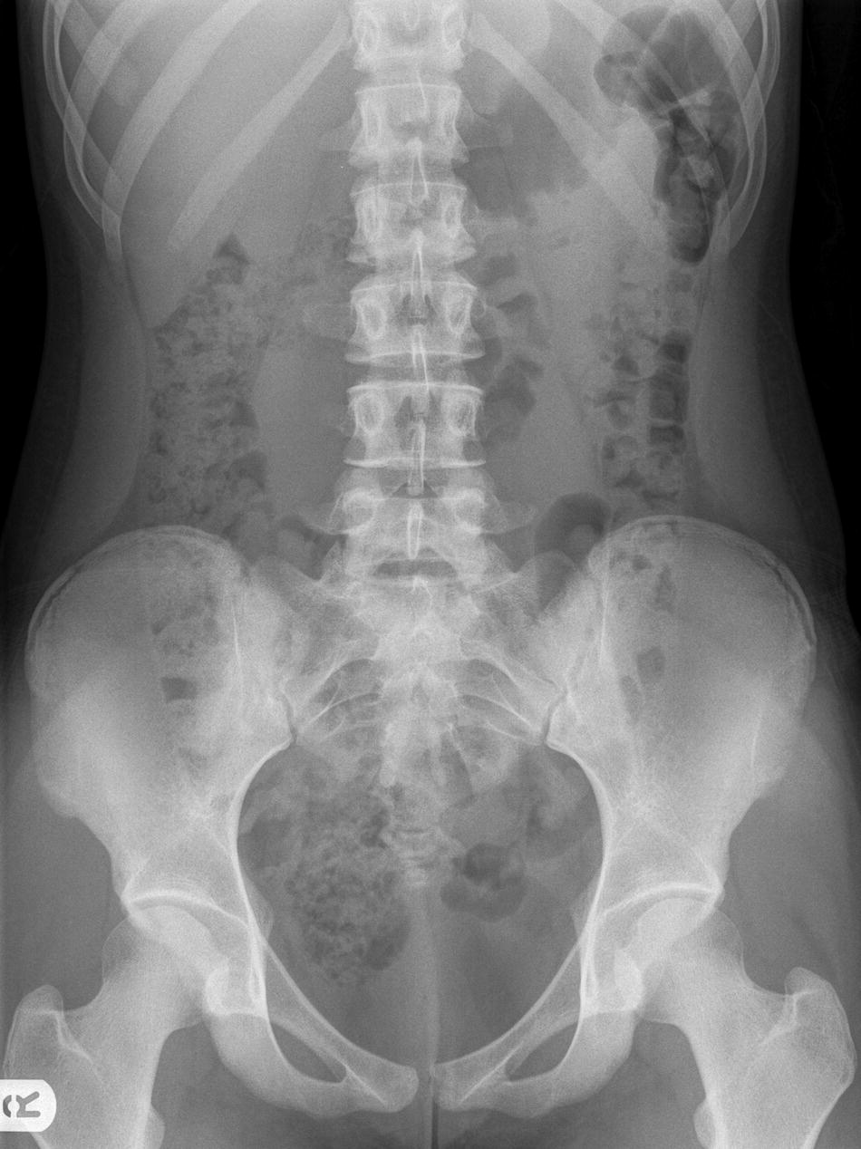
Question 1:
- Present this radiograph.
- What is the diagnosis?
- Give the most likely underlying cause.
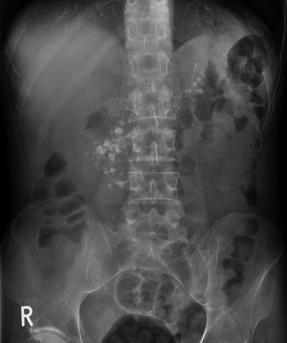
Question 2:
- Present this radiograph.
- Give two indications for the procedure the patient has undergone.
- Give two specific complications.
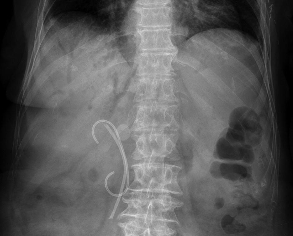
Question 3:
- Present this radiograph.
- Which age group does this condition commonly affect?
- What is the initial management?
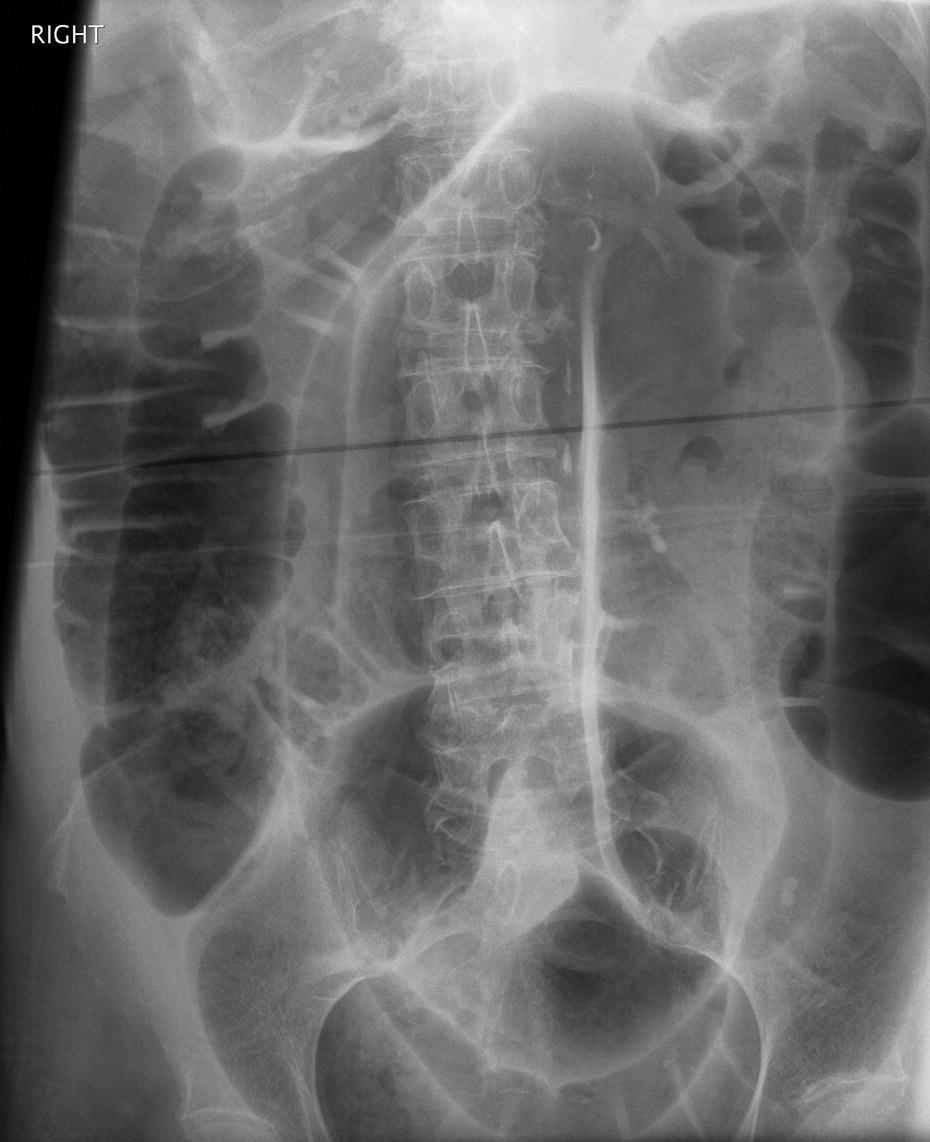
Question 4:
- Present this radiograph.
- What is the most likely diagnosis?
- Give a complication.
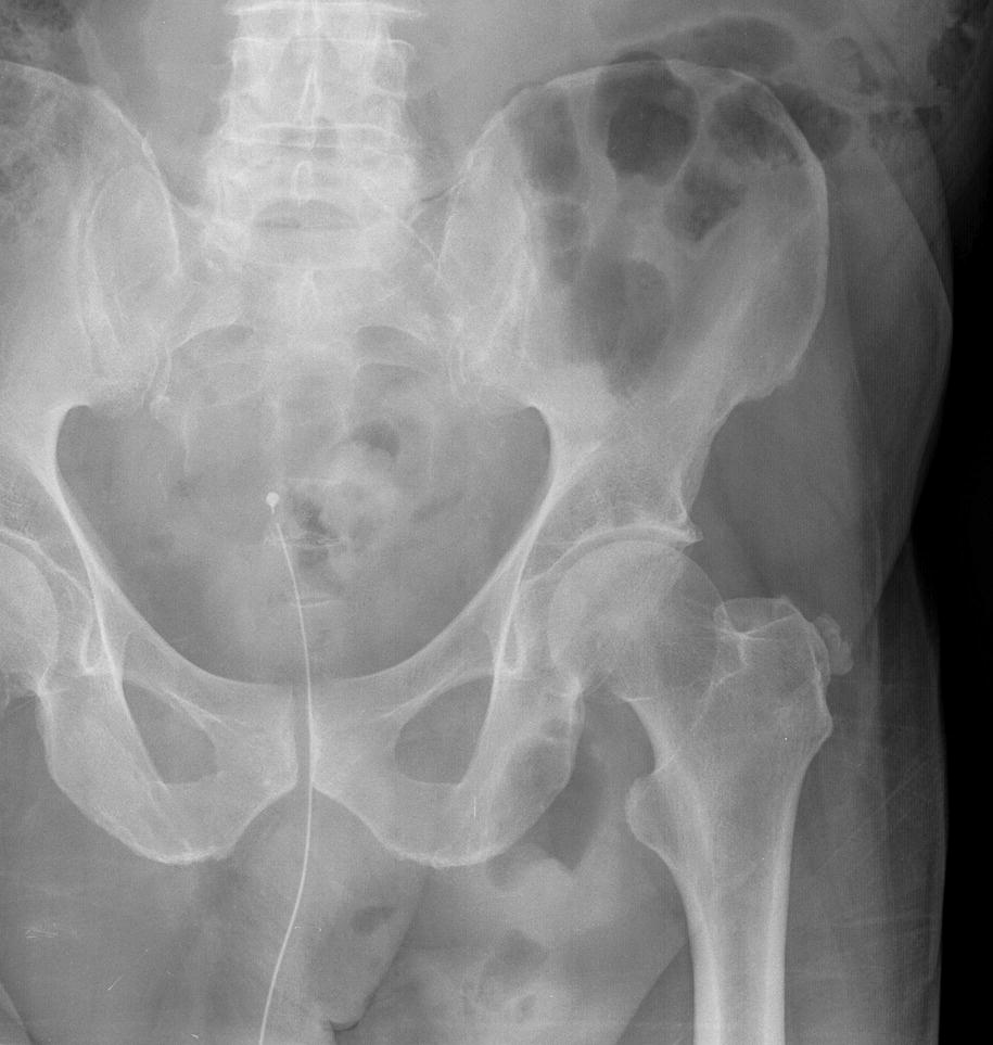
Question 5:
- Present this radiograph.
- What size does an abdominal aortic aneurysm have to be for the risk of spontaneous aneurysm rupture to be larger than the risk of operative management by open surgery or endovascular aneurysm repair (EVAR)?
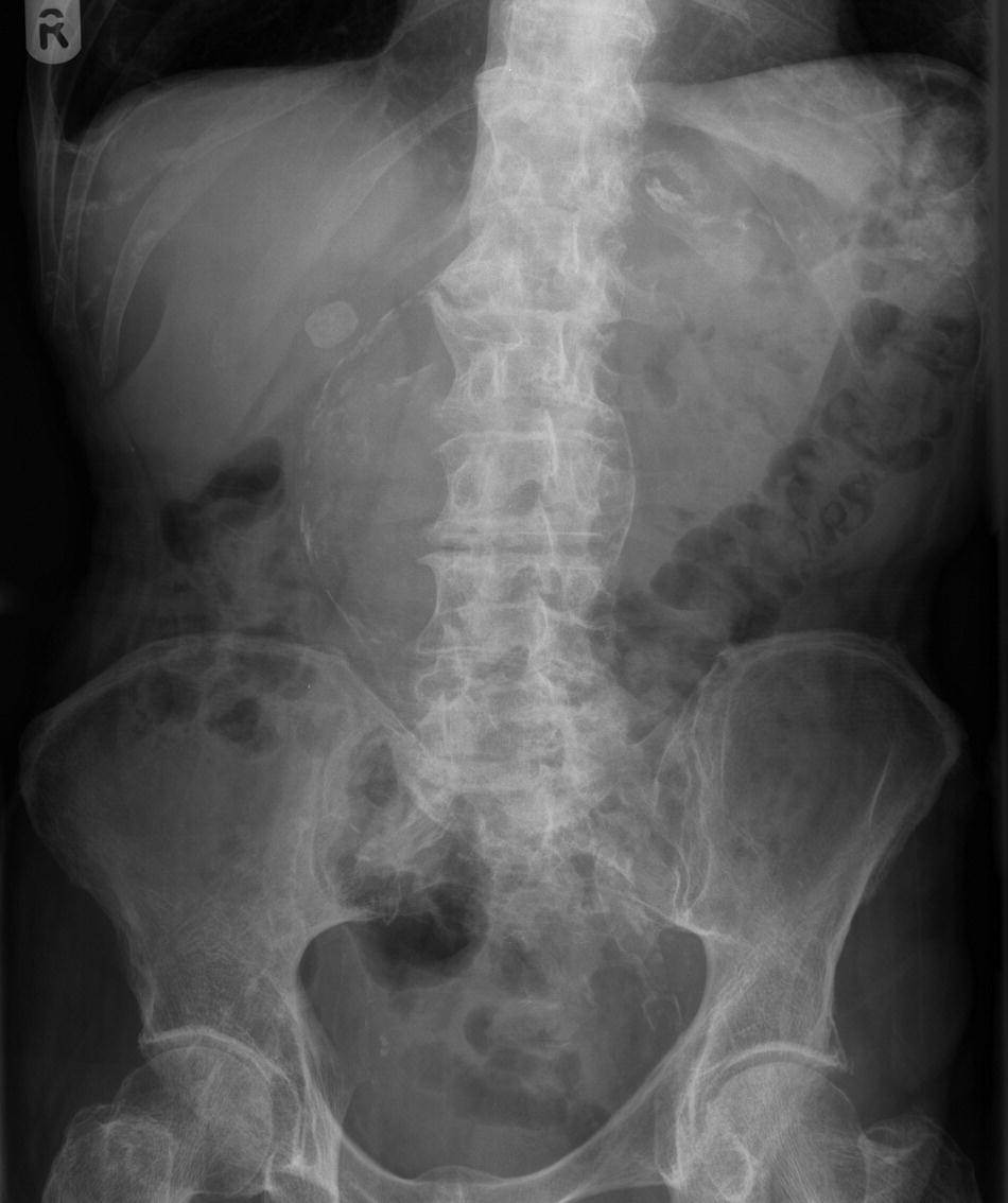
Question 6:
- Present this radiograph.
- What is the diagnosis?
- What is your initial management plan?
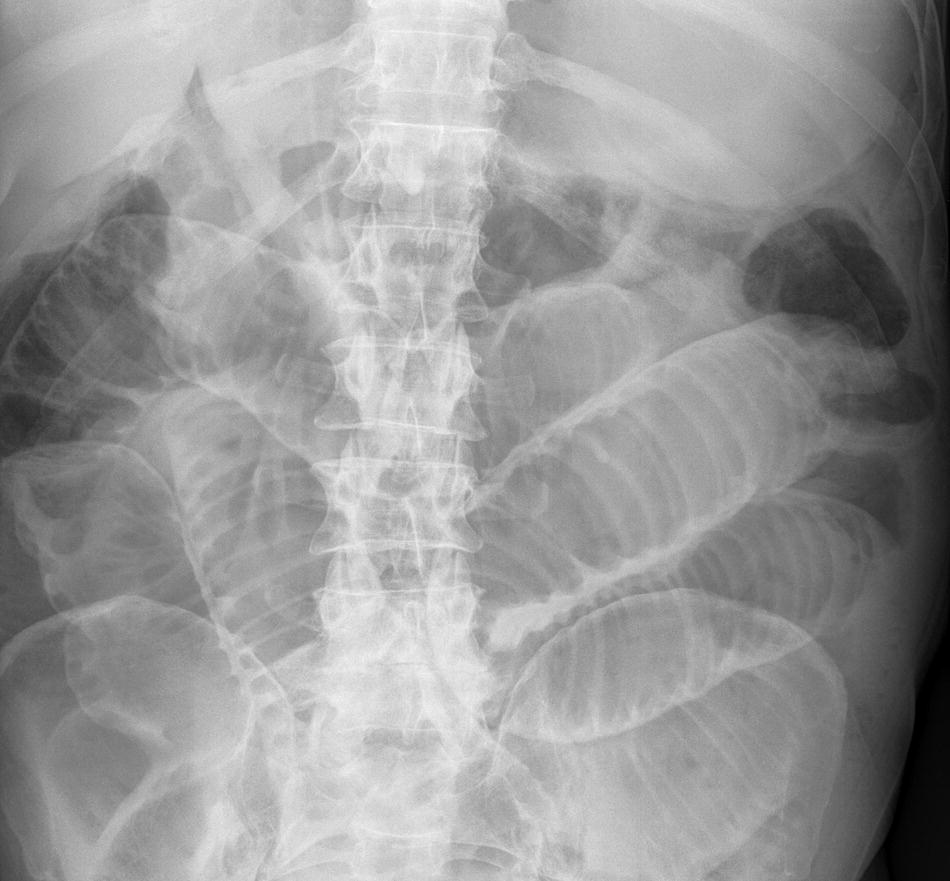
Question 7:
- Present this radiograph.
- What should have been done prior to this X-ray?
- The patient was unaware of the pregnancy. What is the next most appropriate investigation?
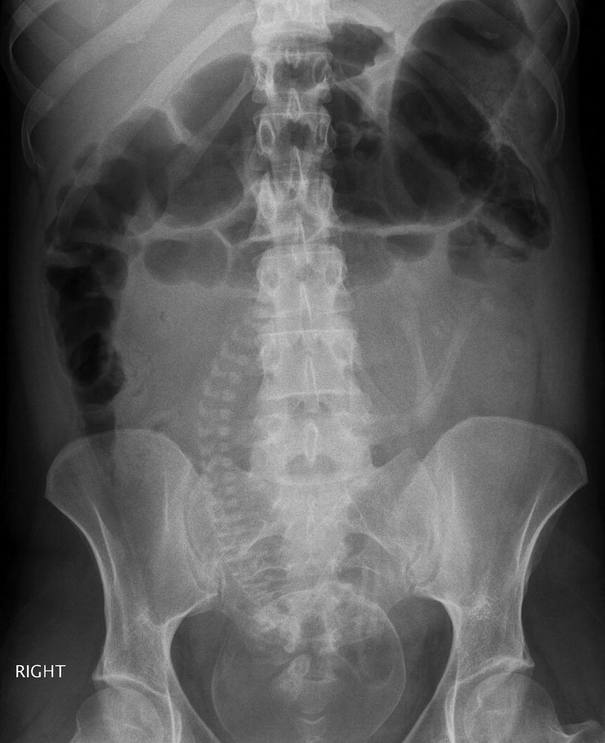
Figure 156: Mrs VN. Taken on unknown date. Severe abdominal pain for 8 h.
Question 8:
- Present this radiograph.
- Give three causes for the abnormality shown.
- List two possible complications.
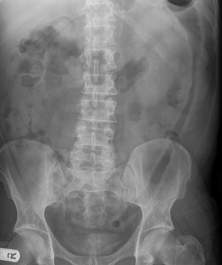
Question 9:
- Present this radiograph.
- What is demonstrated?
- In which ethnic group is it more common?
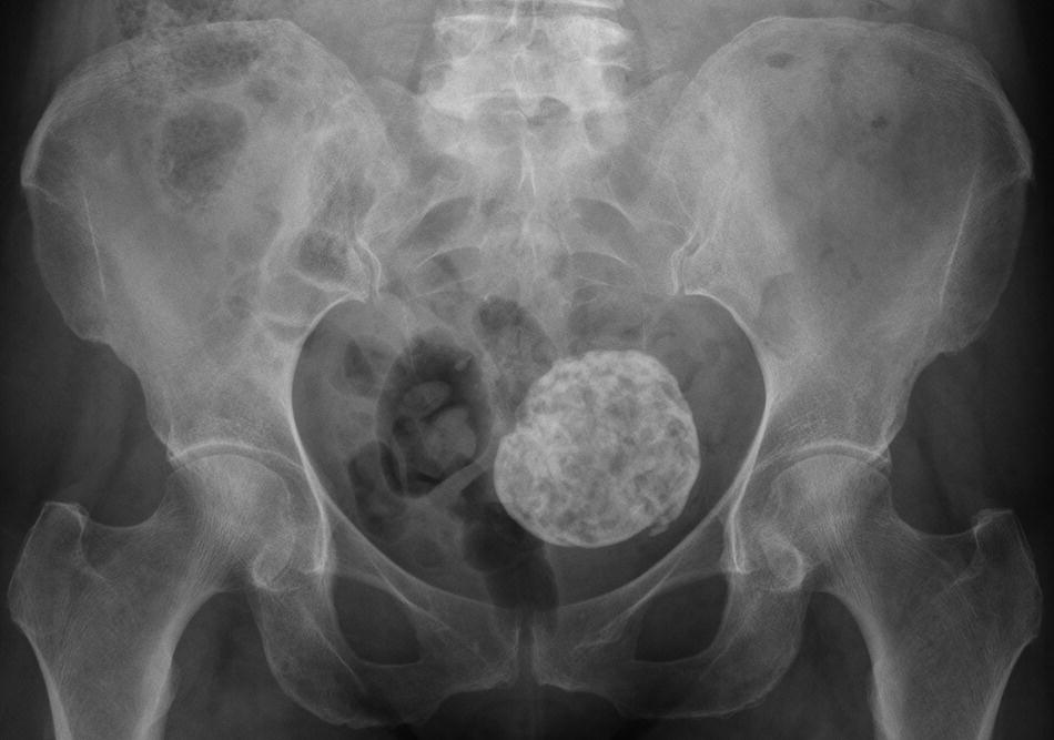
Question 10:
- Present this radiograph.
- Why might the patient present with diarrhoea?
- Give two possible complications.
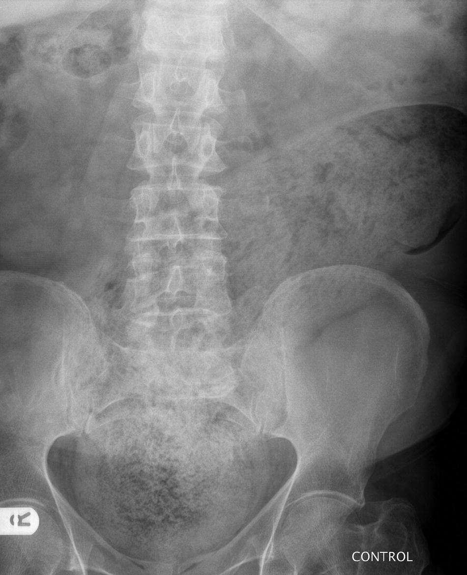
Question 11:
- Present this radiograph.
- Give two indications for the stent shown.
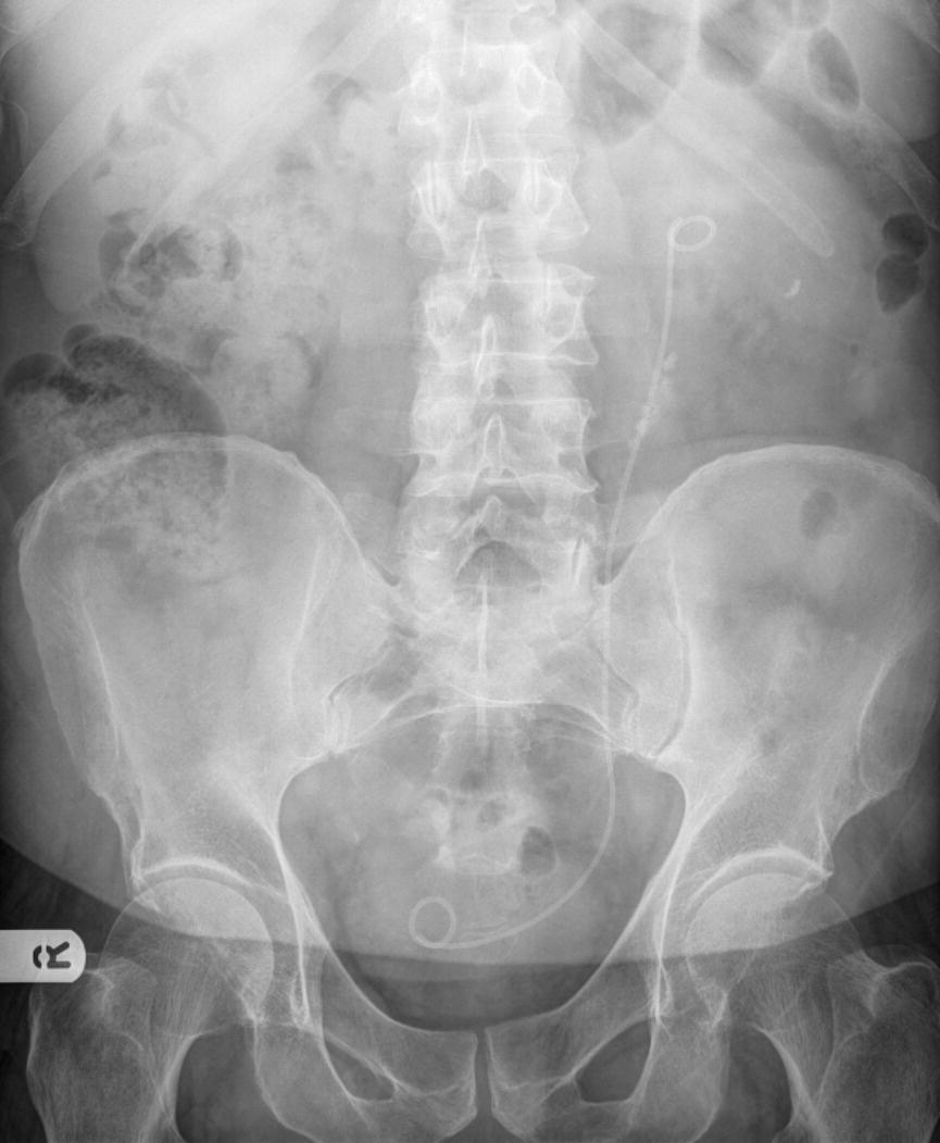
Question 12:
- Present this radiograph.
- Give two likely underlying causes for this appearance.
- What investigation would you like to do next?
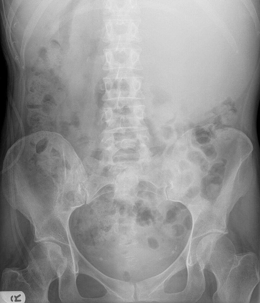
Question 13:
- Present this radiograph.
- Give two possible causes for this appearance.
- If the appearances are secondary to bowel obstruction, at what level is the obstruction likely to be?
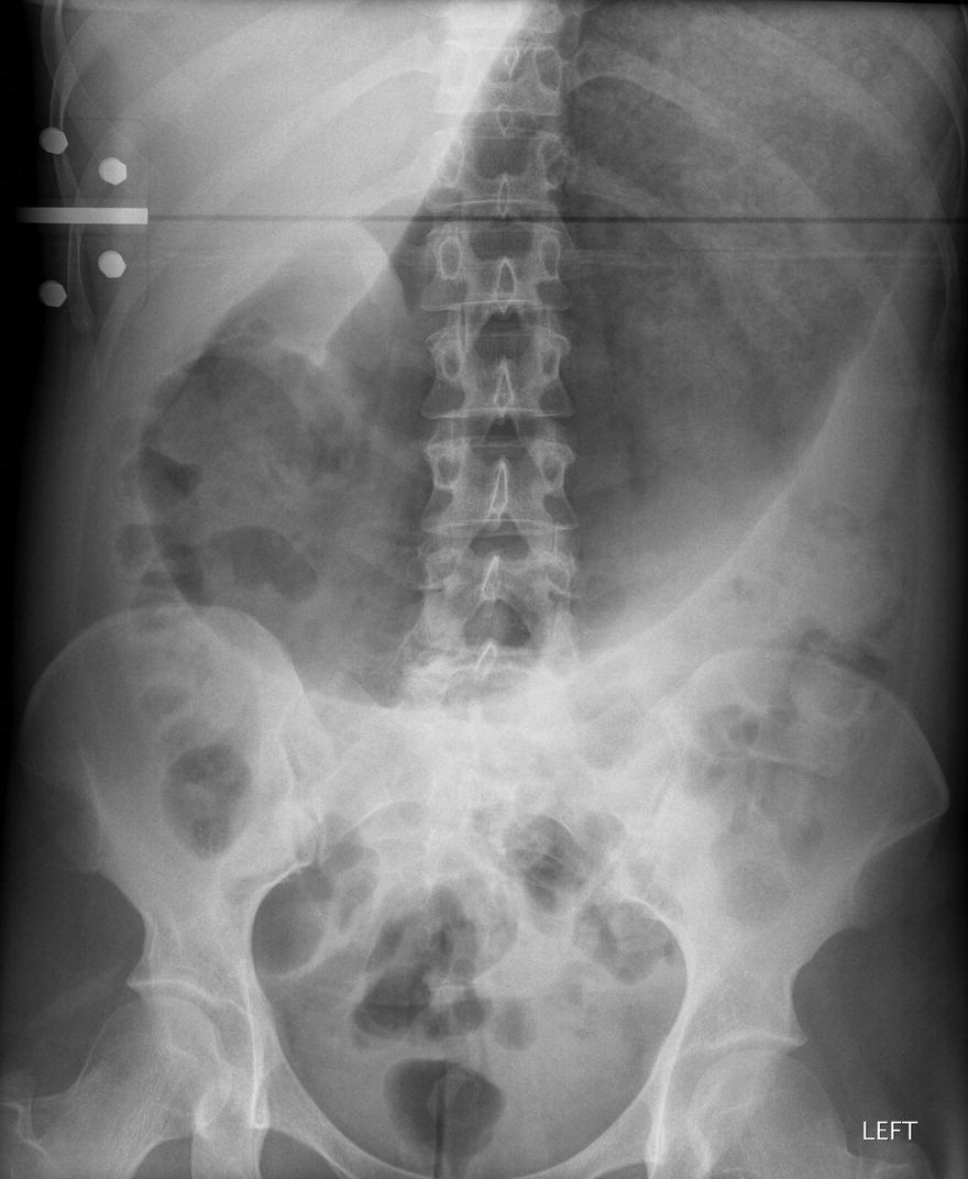
Question 14:
- Present this radiograph.
- Give two possible causes for this appearance.
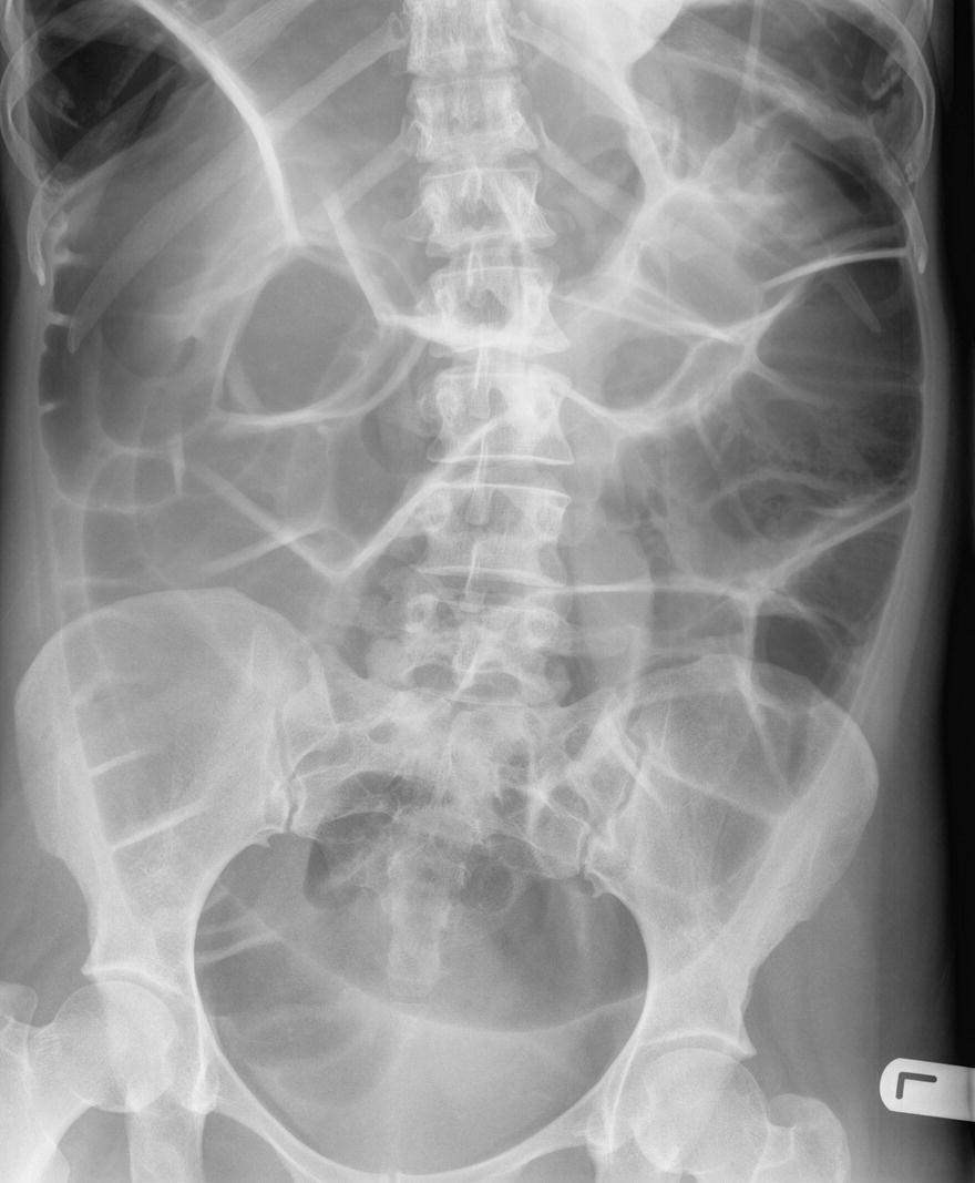
Question 15:
- Present this radiograph.
- What conditions may predispose to these findings?
- Give one method of treatment.
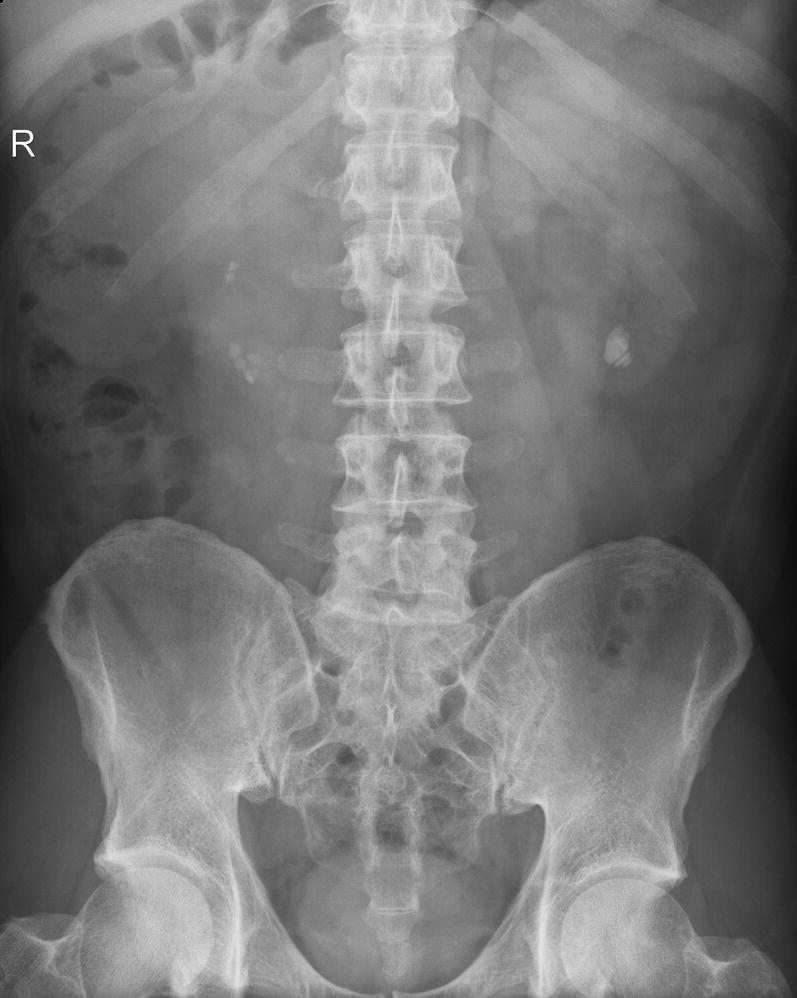
Question 16
- Present this radiograph.
- The patient has had previous abdominal surgery. What is the likely cause of the abnormality identified?
- What is the initial management?
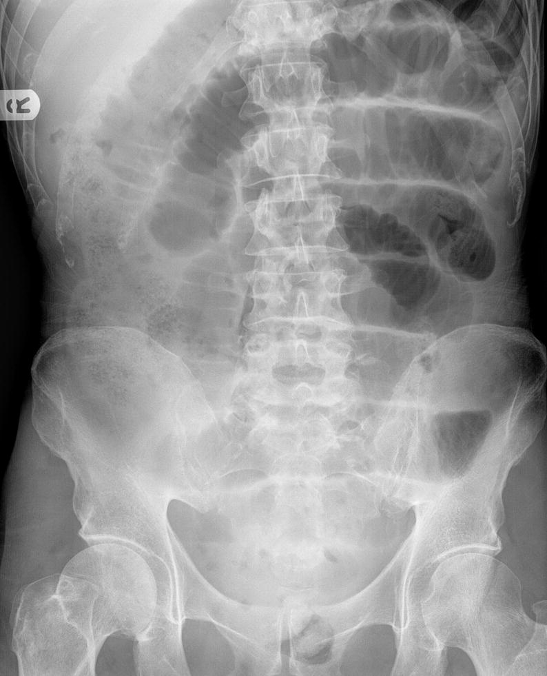
Figure 165: Mrs EA. Taken on unknown date. Abdominal pain and vomiting for 24 h.