(See FELINE ADVISORY BUREAU.)
An operation in which a prosthetic ligament is inserted around the patellar ligament and the fabella to restabilise a stifle in which the cruciate ligament has been ruptured. (See CRUCIATE LIGAMENTS.)
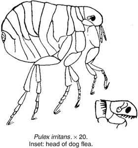
(See under FLIES.)
(See HOLOPROSENCEPHALY.)
A condition in the gerbil due to excessive secretion from the HARDERIAN GLAND. There is erythema, scabs and sometimes moist dermatitis around the nostrils that may become infected, leading to sinusitis.
Treatment: reduce humidity, provide a sand bath and, where groups are kept, reduce numbers. Topical treatment of the bacterial infection can be used, but systemic antibiotics may be required.
‘Facial eczema’ is a synonym for a form of dermatitis caused by light sensitisation in cattle and sheep. (See LIGHT SENSITISATION.)
The facial nerve is the seventh of the cranial nerves, a motor nerve that supplies the muscles of expression of the face.
In the case of unilateral ‘facial paralysis’, which very often follows accidents in which the side of the face has been badly bruised. The muscles on one side become paralysed but those on the opposite side are unaffected. This absence of antagonism between the two sides results in the upper and lower lips, and the muscles around the nostrils, becoming drawn over towards the unaffected side, and the animal presents an altered facial expression. The ear on the injured side of the head very often hangs loosely and flaps back and forward with every movement of the head, and the eyelids on the same side are held half-shut. MRI can be used to detect facial nerve abnormalities in dogs. (See also under GUTTURAL POUCH DISEASE; LISTERIOSIS.)
This is more correctly termed DERMATOGRAPHISM.
Smoke from these may contaminate pastures and cause disease in grazing animals. (See FLUOROSIS; MOLYBDENUM.)
‘Fading’ is the colloquial name for an illness of puppies, leading usually to their death within a few days of birth. Signs include: progressive weakness which soon makes sucking impossible; a falling body temperature; and ‘paddling’ movements. Affected puppies may be killed by their dams. Possible causes include canine viral hepatitis; another is a canine herpesvirus; a third may be a blood incompatibility; a fourth Bordetella; a fifth is hypothermia or ‘chilling’, in which the puppy’s body temperature falls. Another possible cause may be Clostridium perfringens infection.
Kittens A similar syndrome may be caused by the feline leukaemia virus.
A test used to detect ANTHELMINTIC RESISTANCE. It involves taking a faecal sample and counting the worm eggs, giving a dose of the anthelmintic and then taking a second faecal sample after a certain number of days depending on the anthelmintic used. The egg count remaining is counted.
(singular, Faex)
Excreta, excrement or the body waste removed from the ALIMENTARY CANAL via the RECTUM, moved by PERISTALSIS after its formation in the COLON. As the faeces build up in the rectum they stretch the rectal wall and stimulate nerves to signal the need to evacuate the contents. DEFAECATION occurs by relaxation of the sphincter muscles of the ANUS. In dogs, cats, some other animals and humans faeces are called STOOLS.
(See COPROPHAGY.)
The singular of FAECES.
Fainting fits (syncope) are generally due to cerebral anoxaemia occurring through compromised blood circulation, sudden shock, or severe injury. It is most commonly seen in dogs and cats, especially when old, but cases have been seen in all animals.
These are the smallest breed of horse. They must not be higher than 8.2 HANDS (82 cm) high, but they are usually between 7 hands (70 cms) and 8 hands (80 cms). They are mainly bay and black but spotted greys occur. They originate from Crillo stock from Argentina, with some SHETLAND PONY and WELSH PONY. There has been considerable INBREEDING to maintain the small size.
Falcons are the genus of birds Falco, a collective containing 37 species of wild bird. In Britain, the peregrine falcon (Falco peregrinus) is the fastest bird on earth at 20 miles (320 km) an hour. Male falcons are called tercel (American tiercel) and the chicks, whilst in down, are called eyas. The female is larger than the male. In Britain, many falcons are kept for displays but they may be used for hunting, especially abroad. The red-tailed hawk (Bueo jamaicensis) or the Harris hawk (Parabuteo unicinctus) are popular.
Avian pox has been found in imported peregrine falcons, giving rise to scab formation on feet and face and leading sometimes to blindness. Tuberculosis is not uncommon, and may be suspected when the bird loses weight. ‘FROUNCE’ and ‘inflammation of the crop’ are old names for a condition, caused by infestation with protozoan parasites (Tritrichomonas), which can be successfully treated. Frounce causes a bird to refuse food, or to pick up pieces of meat and flick them away again, swallowing apparently being too painful; there is also a sticky, white discharge at the corners of the beak and in the mouth. CAPILLARIA are found in the oropharynx or intestines of infected birds and can be treated with FENBENDAZOLE, ‘sour crop’ results in a swollen crop with foul smell containing meat and due to delay in emptying. Unless rapidly treated surgically under ANAESTHESIA there is rapid COLLAPSE, TOXAEMIA and death. ENTERITIS can be due to bacteria, viruses, fungi, endoparasites or toxins, COCCIDIOSIS (due to Caryospora spp.) occurs. TICKS feed on the birds.
ASPERGILLOSIS is a common cause of weight loss and even death in captive falcons. Bone fractures may arise as a result of calcium deficiency through birds being fed a bone-free all-meat diet. This may be prevented by sprinkling sterilised bone meal or oyster shell on the meat, or feeding the bird with small rodents. BATING, while tethered, can also cause leg fractures. BUMBLE-FOOT is another cause of lameness.
In the Middle East, dosing falcons with ammonium chloride – a common, if misguided, practice believed to enhance their hunting qualities – has caused sickness and fatalities.
Hawks experience similar problems but BUMBLE-FOOT is a more serious problem.
These, one on each side, run from the extremity of the horns of the uterus to the region of the ovary.
Cats fall from windows of high-rise buildings when chasing birds, insects, etc. and sustain fractures of the mandible and hard palate as the chin hits the ground.
Dogs fall less commonly, but when they do, they sustain injuries to face, chest and extremities, with spinal injuries occurring when they fall from over two storeys high.
(See under PSEUDO-PREGNANCY.)
A condition which can cover many types of kidney problems which have a familial basis. It occurs in FINNISH LANDRACE SHEEP and in some breeds of dogs such as COCKER SPANIELS.
A group of diseases that cause dysfunction in the proximal renal tubules and then affect fluid reabsorption. The affected animal’s urine contains glucose, amino acids, sodium, phosphorus and uric acid. There is also metabolic acidosis. It is seen as an inherited form in BASENJIS.
In buildings that are ventilated artificially, it is mandatory under the Welfare of Farmed Animals Regulations 2007 (England and Wales) to have an alarm and stand-by system in order to prevent heat-stroke or anoxia. (See CONTROLLED ENVIRONMENT HOUSING.)
(See FOOD AND AGRICULTURE ORGANIZATION.)
Local application of an electric current as a passive exercise which stimulates muscles and nerves.
The form of GLANDERS affecting the body and limb surfaces. It is usually chronic in horses.
In the UK it is illegal for the castration of horses, donkeys, mules, dogs or cats to be carried out without an anaesthetic. (See ANAESTHETICS, LEGAL REQUIREMENTS; CASTRATION.) Only a veterinary surgeon is permitted to castrate any farm animal more than two months old, with the exception of rams, for which the maximum age is three months.
Only veterinary surgeons are permitted to carry out a vasectomy or electro-ejaculation of any farm animal; likewise the de-snooding of turkeys over 21 days old, de-combing of domestic fowls over 72 hours old, and de-toeing of fowls and turkeys over 72 hours old. Nor can anyone but a veterinary surgeon remove supernumerary teats of calves over three months old, or disbud or dishorn sheep or goats.
Certain procedures legal overseas are prohibited in the UK, namely freeze-dagging of sheep, penis amputation and other operations on the penis, tongue amputation in calves, hot branding of cattle and the de-voicing of cockerels. Very short DOCKING of sheep is also prohibited in the UK.
(See VETERINARY FACILITIES ON THE FARM.)
This committee took over from the FARM ANIMAL WELFARE COUNCIL on 1 April 2011. It is an expert committee of the DEPARTMENT FOR ENVIRONMENT, FOOD AND RURAL AFFAIRS and the devolved administrations of Scotland and Wales. Address: FAWC Secretariat, Area 8B, 9 Millbank, c/o 17 Smith Square, London SW1P 3JR. Telephone: 0207 238 5124/ 6340/ 5016; email: fawcsecretariat@defra.gsi.gov.uk.
An independent body set up by the government in 1979 to keep under review the welfare of farmed animals. Farms, markets, abattoirs and vehicles were inspected and, where appropriate, recommendations made to government. Reports were issued from time to time on the welfare of particular species or aspects (transport, slaughter, etc.) of the use of farm animals. This Council closed in March 2011 and was superseded by the Farm Animal Welfare Committee.
A scheme undertaken by one of several independent organisations which periodically visit farms and provides an overview of the farm, its management, and welfare and disease standards. It is undertaken in the United Kingdom, Ireland, Australia, some parts of the USA and other countries. It can in some ways be considered an extension of the HACCP principle onto farms. It involves areas of BIOSECURITY, and TRACEABILITY. Most responsible traders including supermarkets will usually only purchase food from farms and processors where there is farm assurance.
(See SPRAYS USED ON CROPS; FERTILISERS; METALDEHYDE POISONING.)
A disease caused by the inhalation of dust, from mouldy hay, etc., containing spores of e.g. Thermopolyspora polyspora or Micropolyspora faeni. Localised histamine release in the lung produces oedema, resulting in poor oxygen uptake. It is classed as an acute extrinsic allergic alveolitis. Repeated exposure causes respiratory distress, even when the interval between exposures is several years. The condition has been recognised in humans, cattle, horses and turkeys. In chickens, a similar condition has been caused by inhalation of dust from dead mites in sugar cane bagasse (fibrous residues).
(See WORMS.)
A person who shoes horses. Farriery is a craft of great antiquity and the farrier has been described as the ancestor of the veterinarian. In the UK, farriery training is strictly controlled. Intending farriers must undergo a five-year apprenticeship, including a period at an authorised college, then take an examination for the diploma of the Worshipful Company of Farriers before they can practise independently. The training is controlled by the Farriers Training Council and a register of farriers kept by the Farriers Registration Council.
Address: Sefton House, Adam Court, Newark Road, Peterborough PE1 5PP; Telephone: 01733 319911; email: frc@farrier-reg.gov.uk; website: www.farrier-reg.gov.uk.
The act of parturition in the sow.
A rectangular box in which the sow gives birth. Their use is helpful in preventing overlying of piglets by the sow, and so in obviating one cause of piglet mortality; however, they are far from ideal. Farrowing rails serve the same purpose but perhaps the best arrangement is the circular one which originated in New Zealand. (See ROUNDHOUSE.)
Work at the University of Nebraska suggests that a round stall is better, because the conventional rectangular one does not allow the sow to obey her natural nesting instincts, and may give rise to stress, more stillbirths and agalactia.
(See MASTITIS METRITIS AGALACTIA SYNDROME.)
In the sow, the farrowing rate after one natural service appears to be in the region of 86 per cent. Following a first artificial insemination, the farrowing rate appears to be appreciably lower, but at the Lyndhurst, Hants AI Centre, a farrowing rate of about 83 per cent was obtained when only females which stood firmly to be mounted at insemination time were used. The national (British) average farrowing rate has been estimated at 65 per cent for a first insemination. Australian research has shown that the quality of feed given to gilts from about 10 weeks will affect the number of eggs shed as an adult. In the USA feeding more energy and less protein has increased the chances of a sow having more than four pregnancies.
Sheets or bands of fibrous tissue which enclose and connect the muscles.
Infestation with liver flukes.
Normal body fat is, chemically, an ester of three molecules of 1, 2 or 3 fatty acids, with 1 molecule of glycerol. Such fats are known as glycerides, to distinguish them from other fats and waxes in which an alcohol other than glycerol has formed the ester. (See also LIPIDS [which include fat]; FATTY ACIDS. For fat as a tissue, see ADIPOSE TISSUE). There are two types of fat – brown fat and white fat. Brown fat is seen particularly in young animals and is used to provide body heat on exposure to cold. White fat is metabolised following SYMPATHETIC NERVOUS SYSTEM stimulation releasing NORADRENALIN and is used for movement. White beige fat is an intermediate fat type derived from white fat with the properties of white and brown fat but is used less efficiently than either. A LIPOMA is a benign fatty tumour. For other diseases associated with fat see STEATITIS; FATTY LIVER/KIDNEY SYNDROME OF CHICKENS AND TURKEYS; OBESITY, DIET AND DIETETICS.
These accumulations of fat in the caudal COELOM of reptiles constitute their normal fat stores. Reptiles do not have subcutaneous fat deposits or an OMENTUM. Fat bodies should not be confused with fatty tumours (See LIPOMA).
Found in fat and also called ADIPOCYTES.
Seen in cats. (See EOSINOPHILIC GRANULOMA.)
(See EXERCISE; MUSCLE; NERVES.)
Fats protected from rumen degradation are digested lower in the gut and serve as a concentrated energy source. They can also improve milk and meat production and increase conjugated linoleic acid.
In poultry rations these can lead to ‘TOXIC FAT SYNDROME’. (See LIPIDS for cattle supplement; also ECZEMA in cats.)
These, with an alcohol, form FAT. Saturated fatty acids have twice as many hydrogen atoms as carbon atoms, and each molecule of fatty acid contains two atoms of oxygen. Unsaturated fatty acids contain less than twice as many hydrogen atoms as carbon items, and one or more pairs of adjacent atoms are connected by double bonds. Polyunsaturated fatty acids are those in which several pairs of adjacent carbon atoms contain double bonds. Certain fatty acids, e.g. linoleic acid, are known as ‘essential’ and are necessary for coat health. There may be species differences; for example, skin ulceration can be presented by fatty acid supplementation in the black rhinoceros, but they are not required in the white rhinoceros.
Omega-3 fatty acids appear to have a beneficial effect on heart muscle, but need to be balanced with omega-6 fatty acids as the latter can have a deleterious effect. In humans, conjugated linoleic acid has anti-carcinogennic effects, improves food digestibility and reduces body fat deposition.
Butyric acid can help inhibit infection of the mucous membranes by Salmonella and Campylobacter spp. and so reduce human food-borne illness.
A condition in which there is an excess of fat in the parenchyma cells of organs such as the liver, heart and kidneys.
This is a condition in laying hens which has to be differentiated from FLKS (see next entry) of high-carbohydrate broiler-chicks. Factors involved include high carbohydrate diets, high environmental temperatures, high oestrogen levels, and the particular strain of bird. FLHS in hens is improved by diets based on wheat as compared with maize; whereas FLKS is aggravated by diets based on wheat. Death is due to haemorrhage from the enlarged liver.
A condition in which excessive amounts of fat are present in the liver, kidneys and myocardium. The liver is pale and swollen, with haemorrhages sometimes present, and the kidneys vary from being slightly swollen and pale pink to being excessively enlarged and white. Morbidity is usually between 5 per cent and 30 per cent. It can be caused by many factors, including genetics, diet, management, hormones, environment and toxic substances. A similar condition is seen in cage birds that receive diets high in fats and carbohydrates and do not exercise.
Prevention FLKS has been shown to respond to biotin (see VITAMINS), and accordingly can be prevented by suitable modification of the diet.
Signs A number of the more forward birds (usually two to three weeks old) suddenly show signs of paralysis. They lie down on their breasts with their heads stretched forward; others lie on their sides with their heads bent over their backs. Death may occur within a few hours. Mortality seldom exceeds 1 per cent.
A ‘production disease’ which may occur in high-yielding dairy cows immediately after calving. It is then that they are subjected to ‘energy deficit’ and mobilise body reserves for milk production. This mobilisation results in the accumulation of fat in the liver, and also in muscle and kidney. In some cases the liver cells become so engorged with fat that they actually rupture.
An important consequence of this syndrome may be an adverse effect on fertility. Cows with a severe fatty liver syndrome were reported to have had a calving interval of 443 days, as compared with 376 days for those with a mild fatty liver syndrome.
Complications such as chronic ketosis, parturient paresis (recumbency after calving), and a greater susceptibility to infection have been also been reported.
The only sign may be wattles paler than normal; the birds remain apparently in good condition. The cause may be varied – genetic, nutritional, management, environmental, and presence of toxic substances. Adding choline, vitamins E and B12, and inositol to the diet can remedy the condition. Reducing the metabolisable energy level in the diet by about 14 per cent usually prevents it.
Fauces is the narrow opening which connects the mouth with the throat. It is bounded above by the soft palate, below by the base of the tongue, and the openings of the tonsils lies at either side. Faucitis is inflammation of the tissues around the opening and may occur in isolation or form part of a more generalised stomatitis.
(See ACETONAEMIA; ACIDOSIS; KETOSIS; NUTRITION, FAULTY; FEED BLOCKS; DIET AND DIETETICS; LAMENESS in cattle; BLINDNESS.)
Faulty wiring of farm equipment has led to cows refusing concentrates in the parlour, not because they were unpalatable (as at first thought), but because the container was live so that cows wanting to feed were deterred by a mild electric shock. (See also EARTHING; ELECTRIC SHOCK, ‘STRAY VOLTAGE’ AND ELECTROCUTION.)
A dual-purpose breed originating from France. They are large birds, good layers, and were considered as possibly contributing to broiler breeder genes. Cocks weigh 3.5 kg (8 lb) and hens 3 kg (6.5 lb). They have five toes and feathered shanks. The plumage of hens is mainly brown and creamy-white and cocks have black, brown and straw-coloured feathers. Their eggs are brown.
Favus is another name for ‘honeycomb ringworm’. (See RINGWORM.)
Abbreviation of FARM ANIMAL WELFARE COUNCIL. (See FARM ANIMAL WELFARE COMMITTEE.)
(Feathering; adjective, Feathered)
Long hairs on the fetlocks of draught horses and some ponies. In dogs, feathering is seen on the ventral surface of the body, the caudal aspect of the legs and the ventral tail of spaniels and setters.
An inherited problem particularly of budgerigars and parrots thought to be due to a single recessive gene. Abnormal feathering is seen as soon as they start to feather up and the flight, tail and contour feathers continue to grow. Most birds die at 10 to 12 weeks but they can survive two or more years. Affected birds should not be bred from. It is also known as ‘Chrysanthemum disease’.
Feather picking (feather pulling) in poultry and in cage birds, particularly parrots, may be due to boredom or insecurity.
Poultry In many cases it is due to the irritation caused by lice or to the ravages of the depluming mite. In such cases the necessary anti-parasitic measures must be taken. Insufficient animal protein in the diet of young growing chicks, especially when kept under intensive conditions, may cause the vice. Once the birds start pulling the feather they sooner or later draw blood, and an outbreak of cannibalism results. Treatment consists of isolating the culprit, if it can be found at the beginning, and of feeding the birds a balanced diet containing green food. The addition of blood meal in the mash is effective in many cases. The use of blue glass in intensive houses has stopped the habit in some cases. Use of trees in free-range pens reduces feather-pecking.
Parrots In parrots the syndrome is much more complex and triggered by many factors. Feather pecking is never seen in birds in the wild. The condition is diagnosed by the fact that the affected bird has a fully-feathered head and the wings, legs and body are deplumed as only they can be reached by the beak. The condition has to be thoroughly investigated: often there is an underlying disease, and response to appropriate treatment resolves the feather picking. Specific treatment varies from the application of an ELIZABETHAN COLLAR to the administration of psychotic drugs, but sometimes environmental changes have been effective – e.g. the transference of a caged bird to an aviary or the provision of a CONSPECIFIC individual for company.
An anthelmintic used for the treatment of parasitic gastroenteritis and parasitic bronchitis in cattle, sheep, pigs and horses and in combination with PYRANTEL and PRAZIQUANTEL for the control of intestinal worms in dogs. Chemically, febantel is a pro-benzimidazole which is converted in the body to benzimidazole.
An organisation representing veterinary organisations in 38 European countries and with four sections representing practitioners, hygienists, veterinary state officers and veterinarians in education, research and industry. Address: Avenue de Tervuren 12, B-1040 Brussels, Telephone: (00) 32 2533 70 20; E-mail: info@fve.org
The European Federation of Animal Health, an association of veterinary medicine manufacturers.
(See ADDITIVES.)
These ‘self-help’ lick blocks, placed out on pasture, are useful especially on hill farms for preventing loss of condition and even semi-starvation in the ewe.
Most feed blocks contain cereals as a source of carbohydrate, protein from natural sources supplemented by urea, minerals, trace elements and vitamins. In some blocks glucose or molasses is substituted for the cereals as the chief source of carbohydrate. A third type contains no protein or urea but provides glucose, minerals, trace elements, and vitamins; being especially useful in the context of hypomagnesaemia (and other metabolic ills) in ewes shortly before and after lambing.
Their effectiveness for providing specific ingredients is variable as animals differ in the extent to which they use feed blocks.
The gain in weight, in kg or lb, produced by 1 kg or 1 lb of feed; it is the reciprocal of the feed conversion ratio.
If FCRs are to be used as a basis of comparison as between one litter and another, or one farm’s pigs and another’s, it is essential that the same meal or other foods be used; otherwise the figures become meaningless.
The amount of feed in kg or lb necessary to produce 1 kg or 1 lb of weight gain.
(See DIET AND DIETETICS; FAULTY NUTRITION.)
Feed must be stored separately from fertilisers, or contamination and subsequent poisoning may occur.
The safe storage period on the farm of certain feeds is given under DIET AND DIETETICS.
Poultry and rats and mice must not be allowed to contaminate feeding-stuffs, or SALMONELLOSIS may result. If warfarin has been used, this may be contained in rodents’ urine and lead to poisoning of stock through contamination of feeding-stuffs. (See also TOXOPLASMOSIS.)
Unsterilised bone-meal is a potential source of salmonellosis and anthrax infections.
(See also ADDITIVES; CONCENTRATES; DIET AND DIETETICS; MITES, PARASITIC - OTHER MITES - FORAGE MITES; MOULDY FOOD; MYCOTOXICOSIS; CUBES AND PELLETS; SACKS; LUBRICANTS.)
Feeding-Stuffs Regulations 2000 control the constituents of animal feed including pet food. They specify, among other items, permitted additives, colourants, emulsifiers, stabilisers, maximum amounts of vitamins and trace elements, and permitted preservatives.
Feedlots involve the ZERO-GRAZING of beef cattle on a very large scale. In the USA there are some feedlots of 100,000 head each, and many more containing tens of thousands of cattle. Veterinary problems arise when these cattle are brought to the feedlot from range or pasture, and fed on grain. Shipping fever is a common ailment; likewise liver abscesses.
Related to, or of, the Family Felidae including the domestic cat (Felis catus).
A charity in the UK dedicated to promoting the health and welfare of cats through improved knowledge. It provides information for those involved in veterinary care, rescue work and boarding establishments as well as the general cat-owning public. After 55 years the name was changed in 2013 to INTERNATIONAL CAT CARE. (See also INTERNATIONAL SOCIETY OF FELINE MEDICINE.)
(See ANAEMIA; TOXOPLASMOSIS; HAEMOBARTONELLA; FELINE LEUKAEMIA; FELINE BABESIOSIS.)
(See FELINE DYSAUTONOMIA (KEY-GASKELL SYNDROME).)
An uncommon parasitic disease of cats due to infestation of erythrocytes with Babesia felis causing anaemia, lethargy and sometimes jaundice. Young cats may develop immunity to Babesia felis without prior signs of illness; older cats often have recurrent illness. Subclinical infections occur. The disease can prove fatal. (See also BABESIOSIS.)
A disease of the upper respiratory tract of cats involving Bordetella bronchiseptica. Clinical signs can be mild, but fatal pneumonia can develop especially in kittens. Some animals may become symptomless carriers of the organism (which is also responsible for respiratory disease in other species, including kennel cough in dogs). Treatment is by antibiotics.
Infection with calicivirus (of which there are several strains) is an important cause of FELINE INFLUENZA (‘CAT FLU’). Signs include fever, discharge from the eyes and nose and ulcers of the mouth and tongue. The virus is disseminated by sneezing cats, through contact with nasal secretions picked up on the hands and clothing of attendants, etc. Calicivirus is also associated with chronic stomatitis, a limping syndrome and a virulent systemic form associated with high mortality.
Cancer is an important disease of cats. Cancer of the lymph nodes is common, as is that of the bone marrow. Skin cancer and mammary gland cancers also occur. (See also under CANCER.)
Clinical signs of this heart condition include dyspnoea, weight loss and lethargy. Diagnosis is by radiography. Beta blockers, digitalis and diltiazem have been used in treatment. The cause is unknown.
An acute upper respiratory disease caused by Chlamydiophila felis; also known as feline pneumonitis.
Signs Severe swelling and redness, nasal discharge, sneezing and coughing. It commonly affects groups of animals, rarely single cats. Treatment includes topical and/or systemic antibiotics. Chlamydiosis vaccine (available as a combination product) protects against clinical disease but not infection.
It is seen as ocular hyperaemia and/or CHEMOSIS. Causative agents are feline herpes virus-1 (FHV-1), Chlamydophila felis, Mycoplasma felis or Gram-negative bacteria. Diagnosis is best confirmed by PCR. Treatment of FHV-1 involves the use of topical anti-viral agents such as acyclovir, or famciclovir given orally – these products are not licensed for cats in the UK. Treatment of Chlamydophila and Mycoplasma is best achieved with a tetracycline such as doxycycline.
This is a common infection in cats that usually causes mild, often asymptomatic, infections, but it may progress to FELINE INFECTIOUS PERITONITIS (FIP).
Gum and tooth diseases are quite common in cats. (See MOUTH, DISEASES OF; TEETH.)
Resorption of the teeth by the body is common in domestic cats. This may occur as (usually) painful cavities at the gum line (often hidden by a flap of gum) or painless root resorption. (See MOUTH, DISEASES OF.)
(See under DIABETES MELLITUS.)
A condition in cats first recognised at Bristol University’s department of veterinary medicine in 1981–2. It is also called feline autonomic polygangliopathy.
Signs include depression, loss of appetite, prominent nictitating membranes, dry and encrusted nostrils – suggesting a respiratory disease. Constipation and a transient diarrhoea have both been reported; also incontinence in some cases. The pupils are dilated and unresponsive to light and tear production is diminished. There may be difficulty in swallowing and food may be regurgitated. A common finding is enlargement of the oesophagus (MEGAOESOPHAGUS). Lesions include loss of nerve cells, and their replacement by fibrous tissue, in certain ganglia. The disease is similar to GRASS SICKNESS in horses. The cause is still unknown.
Treatment involves countering dehydration, maintaining nutritional status through assisted feeding, use of eye drops to encourage nasal and tear secretions and using laxatives to encourage bowel movements.
Prognosis The recovery rate is stated to be about 25 per cent, but recuperation may take weeks or months. Cats with a greatly enlarged oesophagus, persistent loss of appetite, or bladder paralysis are the least likely to survive.
(See also CANINE DYSAUTONOMIA.)
An uncommon infection affecting the blood cells of cats that is likely to be spread by ticks. Several Ehrlichia species have been implicated. The commonest findings are fever, malaise and gastroenteritis, but the respiratory system and joints may also be affected. Tick repellents help prevent infection; treatment is usually with TETRACYCLINES.
This has been reported in Sydney, Australia, and is characterised by non-fatal cases of hind-leg ataxia, and sometimes by side-to-side movements of head and neck. On post-mortem examination, demyelinating lesions and perivascular cuffing involving the brain and spinal cord were found. The cause is thought to be a virus, but efforts to transmit the disease have failed.
Hair loss that usually occurs on the posterior aspects of the hind-limbs and along the belly and back. Often associated with flea infestation (see ECZEMA), it may respond to control of the parasite and administration of steroids.
Conjunctival swabs from cats with conjunctivitis and from clinically normal cats were examined microbiologically. Non-haemolytic streptococci and Staphylococcus epidermis were isolated from both groups while beta-haemolytic streptococci, rhinotracheitis (feline herpes 1) virus, Mycoplasma felis and Chlamydophilia felis were isolated from cases with conjunctivitis.
This can be mild and transient. Sometimes the term is applied not to an inflammation of the gums but merely to a hyperaemia – an increased blood flow – which ‘may alarm the owner but does not hurt the (young) cat’.
Gingivitis can also be acute or chronic, easily treatable, or highly intractable.
One of the commonest causes of gingivitis in middle-aged or elderly cats is the accumulation of tartar on the surface of the teeth. If neglected, the tartar will gradually encroach on to the gums, causing these to become inflamed. Unless the tartar is removed, a shrinkage of the gums is likely to follow. As the gum recedes from the teeth it leaves pockets or spaces into which food particles and bacteria can lodge, exacerbating the inflammation, causing halitosis and leading to the roots of some teeth becoming infected.
The yellowish tartar deposits can become so thick and extensive that eventually they completely mask the teeth. A cat in this condition undoubtedly suffers much discomfort, finds eating a little difficult, and may have toothache. Health is further impaired by the persistent infection. The cat becomes dejected.
Even in such advanced cases, removal of the tartar (and of any loose teeth) can bring about almost a rejuvenation of the animal.
This form of chronic gingivitis can be successfully overcome by treatment and, indeed, prevented if an annual check of the teeth is carried out by a veterinary surgeon.
Intractable gingivitis Some cases of this are associated with a generalised illness rather than merely disease of the mouth. For example, chronic kidney disease, and possibly diabetes, may cause ulcers on the gums (as well as elsewhere in the mouth).
Some strains of the feline calicivirus may also cause gum and tongue ulceration. Bacterial secondary invaders are likely to worsen this, especially if the cat’s bodily defence systems have been impaired by, say, the feline leukaemia virus, some other infection, or even stress.
Antibiotics or sulphonamides are used to control the bacteria; vitamins prescribed to assist the repair of damaged tissue and to help restore appetite, and other supportive measures taken. However, some cases of feline gingivitis do not respond.
It is likely that all the causes of feline gingivitis have not yet been established. Further research will no doubt bridge the gaps in existing knowledge, and bring new methods of treatment and a better prognosis. (See also FELINE STOMATITIS.)
This is not a very common condition in cats, mainly seen in males but does involve haemophilia A, haemophilia B and haemophilia in MAINE COON cats. There are varying degrees of haemorrhage usually following trauma, HAEMATOMAS may develop, or bleeding into joints. Prolonged bleeding following any surgery may be the first indication that the animal is affected.
This is caused by an autosomal recessive gene; the signs are progressive ataxia in infancy, and the condition appears linked to an abnormal coat colour.
One of the causes of feline influenza. Infection may occur in combination with feline calicivirus. Clinical signs may be severe and include epiphora, coughing, dyspnoea and corneal ulcers. Secondary bacterial infection can lead to fatal pneumonia. Cats recovering from acute infection may be left with chronic nasal and ocular disorders; they will also become carriers of the virus. Infection is spread by sneezing, and may be carried on equipment, clothing, hands of attendants, etc. (See FELINE VIRAL RHINOTRACHEITIS; FELINE INFLUENZA.)
An uncommon condition reported in cats, mainly within Europe and initially described in Switzerland in 2000. Usually there are sudden behavioural changes with AGGRESSIVENESS and SEIZURES with no response to treatment. Diagnosis is indicated by the signs and history and used of MRI findings. The cause is not yet known and so the condition is often described as Feline Idiopathic Hippocampal Necrosis.
(See HYPERTHYROIDISM.)
The most commonly acquired heart condition in cats. It is seen at any age, either sex (although common in middle-aged males) and breed (common in Persians). Cats may show laboured breathing after exercise, look depressed, have low appetite, and may show tachycardia, heart murmur, or abnormal rhythm. If undetected it can lead to pulmonary oedema and arterial THROMBOSIS.
Cystitis of unknown cause but which can be improved by increasing water consumption, stress reduction, environmental enrichment and modification, and paying attention to an individual cat’s needs in a multi-cat household.
Formerly known as the feline T-lymphotropic lentivirus (FTLV). It was discovered in California by N. C. Pedersen and colleagues. Spread by the saliva of infected cats, or less often via the milk or placenta, it has a prolonged incubation period leading to permanent infection.
The virus establishes a permanent infection; the prognosis is poor. Clinical signs can be transient and mild – fever, depression, enlarged lymph glands. As the virus causes immunosuppression, secondary infections account for many of the clinical signs.
Diagnosis is confirmed by laboratory demonstration of antibodies. Treatment is aimed at control of secondary infection by antibiotics; many cases, however, end fatally.
This disease is caused by the bacterium Mycoplasma haemofelis (formerly classified as Haemobartonella felis). It is treated with antibiotics. Blood transfusions or fluid therapy may be required in severe, acute cases.
Adult cats may carry the parasite, the disease lying dormant until some debilitating condition (e.g. stress or immunosuppression) lowers the cat’s resistance. (See HAEMOTROPIC ANAEMIA.)
Signs are those associated with anaemia: loss of appetite, lethargy, weakness and loss of weight. Anaemia may be severe enough to cause panting.
Diagnosis may be confirmed by identifying the causal agent in blood smears or testing for bacterial DNA.
This was formerly called feline distemper. The disease is caused by a parvovirus. Cats of all ages are susceptible; survivors appear to acquire lifelong immunity. The disease is less common than it was, as a result of successful vaccination programmes.
Cause The virus is indistinguishable from mink enteritis virus, resistant to heat and many disinfectants, and can survive outside its host for over a year. It causes severe damage to the bowel and bone marrow.
Signs Kittens and young unvaccinated cats are most at risk although non-immune cats of any age are susceptible. In susceptible cats the only sign may be sudden death, but more usually the virus causes an acute enteritis associated with fever, loss of appetite, vomiting, bloody diarrhoea with rapid dehydration and death. In newborn kittens infected in the uterus, the CEREBELLUM may be affected giving rise to a staggering gait.
Disease may be less severe in cats that are immune due to previous vaccination or exposure to infection.
Diagnosis may be confirmed by laboratory tests – examination of bone marrow and blood smears. Post-mortem examination reveals inflammation of the ileum, oedema of the mesenteric lymph nodes and liquefaction of the bone marrow. The disease must be differentiated from poisoning, toxoplasmosis, intestinal foreign bodies and septicaemia.
Prevention Live and inactivated vaccines are available; live vaccines, however, are not suitable for use in pregnant queens.
Treatment Supportive measures to counter dehydration and shock and potentially overwhelming secondary bacterial infections. Even though the bowel is often severely compromised, nutritional support is essential. In a cattery, isolation of in-contact animals and rigid disinfection must be practised. (See also NURSING OF SICK ANIMALS.)
A slowly progressive and fatal disease of young cats, and sometimes of older ones also, caused by a coronavirus. Although the coronavirus is commonly found in cats, most do not develop the disease. Where FIP develops, it usually does so in a ‘wet’ form in which fluid accumulates in the body cavities. A ‘dry’ form also occurs. It is the cat’s own immune response to the virus that determines which form of the disease is likely to be present.
Clinical signs in the early stages are non-specific. Fever, depression, loss of appetite, gradual loss of weight, distension of the abdomen due to fluid. Occasionally, diarrhoea and vomiting occur.
There may be distressed breathing in the ‘dry’ form. Inflammation within tissues can cause damage to organs such as the liver, kidneys, eyes, and brain. Both forms are fatal although some individuals with the ‘dry’ form may survive for many months. Confirmation of a diagnosis of FIP can be strongly suggested on clinical examination but confirmation depends on tissue biopsy or post-mortem examination. Treatment with recombinant feline INTERFERON omega (rfIFN-ω) reduces viral infection.
Prevention is difficult where cats are grouped together as the virus is very infectious. Some catteries try to control infection by repeated antibody testing and isolation techniques combined with stringent hygiene. Where queens are known to be a possible source of infection to their kittens, isolating the queen before kittening and early weaning into a clean environment provides a good method of producing virus-free kittens. A vaccine is available in some countries but its use is controversial.
The name is loosely applied to respiratory infections involving more than one virus, known as the feline viral respiratory disease complex. It commonly occurs in cat-breeding and boarding establishments, the infection(s) being highly contagious. Feline calicivirus and feline herpes virus (feline rhinotracheitis) are commonly involved but other infectious agents such as Bordetella bronchiseptica and Chlamydophila can create very similar signs. Secondary bacterial invaders account for many of the more serious signs.
Signs Sneezing and coughing. The temperature is usually high at first; the appetite is depressed; the animal is dull; the eyes are kept half-shut, or the eyelids may be closed altogether; there is discharge from the nose; condition is rapidly lost. If pneumonia supervenes the breathing becomes very rapid and great distress is apparent; exhaustion and prostration follow. Diagnosis is confirmed by isolation of the virus from nasal swabs by a specialist laboratory.
Treatment Isolation of the sick cat under the best possible hygienic conditions is immediately necessary. There should be plenty of light and fresh air, and domesticated cats need to be kept fairly warm.
Antibiotics help to control secondary bacterial infection. Food should be highly palatable and easily digested. (See NURSING OF SICK ANIMALS; PROTEIN, HYDROLISED.)
Owing to the very highly contagious nature of the viruses causing feline influenza, disinfection after recovery must be very thorough before other cats are admitted to the premises.
Prevention Live attenuated combined vaccines against feline viral rhinotracheitis and feline calicivirus are available. Vaccines are generally effective, but as there are several strains of feline calicivirus, they may not protect against them all. Other controls include strict hygiene (of premises and attendants) and the segregation of carrier (infected) cats.
Feline juvenile osteodystrophy is a disease, of nutritional origin, in the growing kitten.
Cause A diet deficient in calcium and rich in phosphorus; kittens fed exclusively on minced beef or sheep heart have developed the disease within eight weeks. As the use of proprietary diets has increased, then the condition has become less common.
Signs The kitten becomes less playful and reluctant to jump down even from modest heights; it may become stranded when climbing curtains owing to being unable to disengage its claws. There may be lameness, sometimes due to a greenstick fracture; pain in the back may make the kitten bad-tempered and sometimes unable to stand. In kittens which survive, deformity of the skeleton may be shown in later life, with bowing of long bones, fractures, prominence of the spine of the shoulder blade, and abnormalities which together suggest a shortening of the back.
This is a cause of ulcers, and small, palpable swellings under the skin. The disease is transmissible to human beings. (See LEISHMANIASIS.)
Formerly thought to be caused by M. lepraemurium; now shown to be caused by several MYCOBACTERIA that cause leprosy in rats. It has been assumed that most infection of cats was from rats. The skin shows nodules with or without hair, mainly on the head and limbs and occasionally the body. Some reports include lesions of the mouth and the conjunctiva. Disease progress is related to the infective dose and the ability of the host to establish an immunity.
A disease of cats caused by a virus (FeLV) discovered by Professor W. F. H. Jarrett in 1964. The virus gives rise to cancer, especially lymphosarcoma involving the alimentary canal and thymus, and lymphatic leukaemia. Anaemia, glomerulonephritis, and an immunosuppressive syndrome may also result from this infection, which can be readily transmitted from cat to cat. Many cats are able to overcome the infection. The virus may infect not only the bone marrow, lymph nodes, etc., but also epithelial cells of mouth, nose, salivary glands, intestine and urinary bladder.
Kittens of up to four months of age are more likely to become permanently infected with FeLV than older cats, but many cases do occur in cats over five years old.
Many cats which have apparently recovered from natural exposure to the virus remain latently infected, but keep free from FeLV-associated diseases. Such cats may infect their kittens via the milk.
Significance of FeLV Most deaths of FeLV-positive cats are not directly attributable to this virus, but to other viral, bacterial or protozoal infections which are rendered far more serious because of the immunosuppression caused by the virus.
Signs These vary and may be vague, with a gradual loss of condition, poor appetite, depression and anaemia; or they may be related to immunosuppression such as stomatitis or chronic diarrhoea. The cancer induced by FeLV may create enlarged lymph nodes leading to breathing difficulties or enlarged abdominal organs.
Diagnosis FeLV infection can be confirmed by detecting viral protein or genetic material in blood samples using techniques such as ELISA tests, immunofluorescence and polymerase chain reaction (PCR) assays. Viral isolation can also be performed. Healthy cats that test positive should always have follow-up testing as false positives can occur with some of these tests.
Control It is possible to spread the FeLV infection from infected cats whether sick or currently healthy. This can be a problem if the infected cat lives with other cats or goes outside. This can be prevented by a ‘test-and-removal’ system. Infected cats are removed for euthanasia, and any other cats in the same household are then tested. If FeLV-positive, they too are removed, even if clinically healthy. Retesting of the FeLV-negative cats is necessary after three and six months. If still FeLV-negative, they can be considered clear, and new cats introduced on to the premises, if desired.
The virus may persist in the bone marrow of cats which have ostensibly recovered. Such a latent infection can be reactivated by large doses of corticosteroid; the virus can be recovered by cultivation of bone marrow cells. FeLV is not transmitted from cats with a latent infection.
Inactivated vaccines will not protect cats that are already infected and no vaccines are 100 per cent effective.
Formerly called FELINE UROLOGICAL SYNDROME (FUS). The name is given to the several conditions causing painful and difficult URINATION, often with blood-stained urine. It may also be associated with changes in behaviour and URINATION habits. In cases that lead to obstruction of the URETHRA it can, if untreated, lead to death. In many cases the cause is unknown and is referred to as FELINE IDIOPATHIC CYSTITIS (FIC). Less commonly, bladder stones (calculi), the formation of sand-like material, cancer and bacterial infections can cause the signs of FLUTD. In those cats that develop URETHRAL OBSTRUCTION, it is usually males as the URETHRA is longer and narrower than in females, the cause in IDIOPATHIC cases is due to the formation of urethral plugs. These are composed of varying proportions of STRUVITE and organic matter.
Cause Various theories have been advanced to account for FLUTD: possible contributory factors include as yet unidentified infectious agents such as a virus or viruses, dry diets and poor water intake, and a high level of magnesium in the diet. The effects of heredity and castration have also been mentioned. However, it is now recognised that STRESS can be an important factor. Certain types of cat are recognised to be predisposed on account of their inability to cope with stressful situations such as the presence of other cats in the same household or the outside environment. Stress can trigger inflammation of the bladder wall (CYSTITIS) via nerve pathways, but factors from within the bladder itself can also trigger this nerve-induced (NEUROGENIC) inflammation compounding the problem. Feline dry diets are now formulated to maintain urine at the correct degree of acidity to avoid the problem.
Signs The cat may be seen straining to pass urine, with only very little to be produced. The urine may be bloodstained. Cat-owners sometimes mistake FLUTD for constipation; straining may also be seen with CYSTITIS, when the bladder is usually empty, and when the URETHRA is blocked and so the bladder is full. Other signs include loss of appetite, dejection, and restlessness. The abdomen will be painful to the touch, owing to distension of the bladder.
NB: URETHRAL OBSTRUCTION is an emergency requiring immediate veterinary attention, in default of which there is a great risk of collapse and unconsciousness. The bladder may rupture, causing additional shock, and leading to PERITONITIS.
Treatment If a cause of FLUTD is identified it should be treated appropriately. Often, however, the disease is likely to be multifactorial. In acute cases anti-inflammatory medication and pain relief is indicated. In the case of a blocked urethra, skilled manipulation can sometimes free a urethral plug. If this fails, a catheter will have to be passed to empty the bladder; this is imperative to relieve the back pressure on the kidneys. If CATHETERISATION fails, it will be necessary to empty the bladder by means of direct aspiration (cystocentesis).
Prognosis There are cases in which, after removal of the urethral obstruction, it does not recur. However, FELINE IDIOPATHIC CYSTITIS cases are prone to relapse and in cats with obstructed urethras repeated blockages are common. In these cases specialised surgical procedures or ultimately EUTHANASIA may be advisable. (See also INTERSTITIAL CYSTITIS.)
Post-operative treatment includes antibiotics and urine acid-alkali balance control in an attempt to dissolve the remaining crystals. A low-magnesium, urine-acidifying diet, including TAURINE, is also recommended and proprietary preparations are on sale to meet this requirement. (See PRESCRIPTION DIETS.)
(See ECZEMA.)
A skin condition caused by Mycobacterium lepraemurium of mice and possibly M. tuberculosis. These are ACID-FAST ORGANISMS. Any skin lesion containing acid-fast bacteria should be considered a potential public heath hazard. If there is no public health risk, the lesion may be treated by incising the area and then dosing with enrofloxacin. Unlicensed dapsone and clofazimine have been used under the ‘CASCADE’. (See also MYCOBACTERIUM.)
A condition caused by stress and/or anxiety resulting in excessive licking of the skin. Any ectoparasites need to be eliminated, before megestrol acetate or anti-stress drugs such as phenobarbitone and diazepam are used.
(See FELINE INFECTIOUS ENTERITIS.)
(See TEETH, DISEASES OF, TEETH - NECK LESIONS IN CATS’ TEETH; FELINE INFECTIOUS ENTERITIS.)
(See FELINE CHLAMYDIAL INFECTION.)
A high-grade FIBROSARCOMA occurring at sites traditionally used for vaccination in cats and reports started to appear 1991. Cases still occur despite changes in vaccine formulation and in feline vaccination protocols.
This was formerly thought to be cowpox virus. It is characterised by moist reddened lesions of the skin that may become scabby and purulent. Treatment is with disinfectant shampoos, and by creams to areas not accessible to licking; corticosteroids are contraindicated.
(See PYOTHORAX.)
A painful sunburn caused by cats sleeping with their feet or ears pointing to the sun. Ultraviolet light barrier creams have been used to prevent it.
This is similar clinically to BOVINE SPONGIFORM ENCEPHALOPATHY (BSE). The first signs are hypersensitivity to noise and visual stimuli. Ataxia follows and eventually the cat will not be able to get up. The cause is believed to be the eating of material from cattle affected by BSE. In a zoo, two pumas and a stray cat which shared their food were fed on split bovine heads. Both pumas and the cat died from FSE. At the height of the BSE outbreak in the 1990s in the UK, one case of FSE was being reported every six weeks.
Inflammation of the cat’s mouth.
Causes Various, including several viruses.
Signs These include difficulty in swallowing, halitosis, excessive salivation, loss of appetite and sometimes bleeding.
Treatment The aim is to limit secondary bacterial infection by means of antibiotics. A supplement of vitamins A, B and C may help. If the cat will not eat, subcutaneous fluid therapy will be required.
Chronic stomatitis in elderly cats may be due to EOSINOPHILIC GRANULOMA, or malignant growths such as squamous cell CARCINOMA or FIBROSARCOMA. (See also MOUTH, DISEASES OF.)
(See FELINE IMMUNODEFICIENCY VIRUS.)
This is of unknown aetiology, but may be immune-mediated. It causes painless non-pruritic swellings of the soles of the feet. Often they resolve without treatment; otherwise corticosteroid creams are used.
(See FELINE LOWER URINARY TRACT DISEASE.)
The name given to a condition in which head-tilt, ataxia, nystagmus and, occasionally, vomiting were seen. Duration of signs was short, only up to 24 hours.
Feline viral rhinotracheitis is involved in the feline viral respiratory disease complex (FELINE INFLUENZA). The disease was discovered in the USA to be caused by a HERPESVIRUS, and first recorded in Britain in 1966. Severe signs are usually confined to kittens of up to six months old. Sneezing, conjunctivitis with discharge, coughing and ulcerated tongue may be seen. Bronchopneumonia and chronic sinusitis are possible complications.
Cause A herpesvirus. Infection may occur in a latent form, and a possible link has been suggested between this virus and feline syncytia-forming virus.
Live and inactivated vaccines are available against feline calicivirus and feline herpesvirus which may be implicated in the infection.
Treatment May include the use of a steam vaporiser, lactated Ringer’s solution to overcome dehydration and antibiotics. Vitamins and baby foods may help.
An odourless sulphur-containing amino acid present in cat urine which degrades to chemicals producing the characteristic ‘catty smell’.
These strong, useful ponies originated from Cumbria, England. They are normally black, with no white markings, and have FEATHERED legs. They are susceptible to FOAL IMMUNODEFICIENCY SYNDROME (originally called FELL PONY SYNDROME).
(See FOAL IMMUNODEFICIENCY SYNDROME.)
A country term used to describe ‘SUMMER MASTITIS’. As the quarter affected is usually non-productive after infection, it probably refers to “stealing the quarter”.
(See FELINE LEUKAEMIA.)
In the male dog this may occur as the result of a SERTOLI-CELL TUMOUR of a testicle.
Femur is the bone of the thigh, reaching from the hip-joint above to the stifle-joint below. It is the largest, strongest, and longest individual bone of the body. The bone lies at a slope of about 45 degrees to the horizontal in most animals when they are at rest, articulating at its upper end with the ACETABULUM of the pelvis, and at its lower end with the tibia. Just above the joint surface for the tibia is the patellar surface, upon which slides the patella, or ‘knee cap’.
Fractures of the head of the femur are common. Surgical repair may be possible.
A benzimidazole anthelmintic used in cattle, sheep, camelids, horses, pigs, poultry, dogs and cats. (See WORMS, FARM TREATMENT AGAINST.)
An analgesic for use in small mammals (rabbits, ferrets, guinea pigs, rats and mice). In horses long-term relief can be provided by application of fentanyl patches. It is usually combined with FLUANISONE for use as a neuroleptoanalgesic.
A herb used mainly in dog and cat remedies to stimulate appetite. In the USA it was used in a calf feed produced by one company and became habit forming so the animals would only eat feeds containing it. It was ruled in court that the herb had to be withdrawn.
Carbohydrates in the feed of pigs are sometimes fermented with lactobacilli to produce lactic acid and thereby suppress salmonellae and coliform multiplication.
Ferns other than bracken occasionally cause poisoning in cattle. For example, Dryopteris filix-mas (male fern) and D. borreri (rusty male fern) give rise to blindness, drowsiness and a desire to stand or lie in water. Poisoning is occasionally fatal. (See also BRACKEN POISONING.)
(Mustela putorius furo) These attractive creatures are increasingly popular as pets and about 20 per cent are working ferrets. They need careful and expert handling – a bite to the finger can penetrate to the bone. In the UK the breeding season begins in March and continues until the end of August. There is seasonal weight variation with weight loss in the spring and weight gain in the autumn. It is preferable that females (‘jills’ – males are ‘hobs’) not used for breeding are spayed. Unmated jills may be in oestrus for the whole of the breeding season, with the occurrence of persistently high levels of oestrogen. This can cause severe health problems, including a possibly fatal PANCYTOPENIA. Surgical neutering is a major cause of adrenal gland disease with progressive body ALOPECIA and PRURITUS. The alternatives to spaying are injections of proligestone, given via the scruff of the neck, or mating with a vasectomised male. The latter will result in a pseudo-pregnancy lasting about 42 days; the jill may need to be mated again if she returns to oestrus.
Other diseases of ferrets include HYPOCALCAEMIA, three to four weeks after giving birth; MASTITIS; ALEUTIAN DISEASE; CANINE DISTEMPER (often fatal with respiratory and gastrointestinal signs, vaccination prevents this); BOTULISM (type C); abscesses; enteritis due to E. coli or campylobacter. A CROHN’S DISEASE-like condition occurs. Ferret systemic CORONAVIRUS is an emerging fatal disease first recorded in USA in 2002. Skin tumours are not uncommon. Periodontal disease is often caused by the accumulation of dental calculus. UROLITHIASIS can occur; the ferret can be fed the cat food formulated to prevent this condition. Ferrets are susceptible to zinc poisoning and any galvanised material can be a risk.
Common external parasites include FLEAS, OTODECTES, SARCOPTIC MANGE (see MITES, PARASITIC), and TICKS. IMIDACLOPRID can be used to treat. Internal parasites include COCCIDIOSIS (Eimeria furo, E. ictidea, Isospora laidlawii), CRYPTOSPORIDIOSIS, GIARDIASIS (Giardia duodenalis), TOXOPLASMOSIS. Worm (HELMINTH) infestations include HEARTWORMS (Dirofilaria immitis), HOOKWORMS (Ancylostoma spp.), TAPEWORMS (Dipylidium caninum) and ROUNDWORMS (Toxocara spp.; Toxascaris leonina). Laboratory parameter reference ranges can be found in Veterinary Record (2012) 171 218, and there is a website dealing with health and management of ferrets at www.wildlifeinformation.org.
Ferritin is a form in which iron is stored in the body. Ferritin concentrations in serum are closely related to total body iron stores, and ferritin immunoassays can be used to assess the clinical iron status of human beings, horses, cattle, dogs, and pigs.
(See REPRODUCTION.)
Fertilisers should not be stored near feeding-stuffs, as contamination of the latter, leading to poisoning, may occur. Deaths can occur after cattle gain access to the remains of a fertiliser dump, often caused by a crust of superphosphate and ammonium sulphate that remained on the ground.
For the risk associated with unsterilised bone-meal, see under ANTHRAX and SALMONELLOSIS.
Hypomagnesaemia is frequently encountered in animals grazing pasture which has received a recent dressing with potash. (See also BASIC SLAG; FOG FEVER.)
(See CONCEPTION RATES; FARROWING RATES; INFERTILITY; CALVING INDEX.)
In New Zealand and the USA, a severe hind-foot lameness of cattle has been attributed to the grazing of Festuca arundinacea, a coarse grass which grows on poorly drained land or on the banks of ditches, and being tall stands out above the snow. In typical cases, the left hind-foot is affected first, and becomes cold, the skin being dry and necrotic. Signs appear 10 to 14 days after the cattle go on to the tall-fescue-dominated pasture. Ergot may be present, but is not invariably so.
It has been suggested that ‘fescue foot’ may be associated with a potent toxin, butenolide, produced by the fungus Fusarium tricinctum.
Adjective derived from FETUS, which is the unborn developing offspring after the embryonic stage.
Examples of these are TOXOCARA infection in bitches and TOXOPLASMOSIS in utero of cows, ewes, sows, bitches and cats.
(See CHORION; AMNION; ALLANTOIS; also UTERUS, DISEASES OF and EMBRYOLOGY.)
(See MUMMIFICATION OF FETUS.)
The joint in the horse’s limb between the metacarpus or metatarsus (cannon bones) and the 1st phalanx (long pastern bone). At the back of this joint are situated the sesamoids of the 1st phalanx. (See BONES.)
(See EMBRYOTOMY.)
The young growing in the uterus after the organs have been formed. The word is from the Latin fetus, meaning offspring; the spelling ‘foetus’ is common, but incorrect. For an outline description of the development of the fetus, see under EMBRYOLOGY. For fetal circulation, see the diagram under CIRCULATION OF BLOOD. (See also FREEMARTIN.)
Fever is one of the commonest signs of infectious disease, and serves to make the distinction between febrile and non-febrile ailments. In true fever the body’s own in-built thermostat changes to a higher setting and the core temperature is raised accordingly. It is thought to be beneficial in many cases of infectious disease in countering the infective agent. Fever is a common sign in problems other than infectious disease, such as immune-mediated disease, pain and cancer especially in dogs and cats.
Examples of specific fevers are BLACK-QUARTER, BRAXY, DISTEMPER, EQUINE INFLUENZA and SWINE FEVER.
When fever reaches an excessively high stage, e.g. 41.5° C (107° F), in the horse or dog, the term ‘hyperpyrexia’ (excessive fever) is applied, and it is regarded as indicating a condition of danger; while if it exceeds 42° C or 42.5° C (108° F or 109° F) for any length of time, death almost always results. Occasionally, in certain fevers or febrile conditions, such as severe heat-stroke, the temperature may reach 44.5° C (112° F). (See also under TEMPERATURE, BODY.)
Signs Animals with a fever are usually depressed, lethargic and have a reduced appetite. They may appear stiff and reluctant to move and have mildly elevated respiratory and/or heart rates. There is usually a certain amount of shivering, to which the term ‘rigor’ is applied, but this is very often not noticed by the owner. Later, there may be PERSPIRATION, rapid breathing, a fast, full, bounding pulse, and a greater elevation of temperature are exhibited. Thirst is usually marked; the appetite disappears; the urine is scanty and of a high specific gravity; the bowels are generally constipated, although diarrhoea may follow later; oedema of all the visible mucous membranes, i.e. those of the eyes, nostrils, mouth, occurs. (See also HYPERTHERMIA.)
Fever may sometimes have a beneficial effect.
Experiments with newborn mice show that fatal infection with Coxsackie B1 virus can be modified to a subclinical infection if the animals are kept in an incubator at 34° C (93° F) and thus attain the same body temperature as mice of eight to nine days old. Similarly, puppies infected with canine herpesvirus survive longer and have diminished replication of virus in their organs if their body temperature is artificially raised to that of adult dogs. It is also said to assist in treating MYXOMATOSIS in rabbits.
The term used for the structural parts of plants which are commonly called DIETARY FIBRE or ROUGHAGE. It forms an important part of nutrition and feeding animals. Much of this cannot be digested by CARNIVORES or man, but is able to be partially utilised by HERBIVORES. (See also DIETARY FIBRE; DIET AND DIETETICS; HORSES, FEEDING OF; RATIONS FOR LIVESTOCK.)
Animals that require fibre as a major part of their diet. RABBITS, GUINEA PIGS and CHINCHILLAS in the wild live on grass, plants, twigs and bark.
An involuntary contraction of individual bundles of muscle fibres.
Fibrin is a substance upon which depends the formation of blood clots. (See CLOTTING OF BLOOD; PLASMA.)
Fibrin is found not only in coagulated blood, but also in many inflammatory conditions. Later it is either dissolved again by, and taken up into, the blood, or is ‘organised’ into fibrous tissue.
A high molecular weight protein in solution in the blood plasma also known as Clotting Factor 1, which following the action of thrombin is converted to fibrin and this is precipitated to form part of the process of clotting. Concentration of this is increased in inflammatory conditions, especially lesions of serous surfaces and in ENDOCARDITIS. (See also CLOTTING OF BLOOD.)
It breaks down FIBRIN by enzymatic action such as blood clots.
An irregularly-shaped cell responsible for the synthesis of CONNECTIVE TISSUE.
It is a mixture in varying proportions of white FIBROUS TISSUE and CARTILAGE. It tends to be white in colour, and the fibrous tissue provides strength and toughness and cartilage provides elasticity.
This is the protrusion of an intervertebral disc which has degenerated and enters the spinal canal, causing damage of the blood vessels and resulting in restriction of the blood supply (ISCHAEMIA) and damage of the SPINAL CORD (MYELOPATHY). The signs are sudden in onset, do not progress and are often asymmetrical and involve varying degrees of paralysis and loss of, or poor pulse in, the limbs. It is particularly seen in middle-aged dogs of large breeds, and also is common in horses.
(See TUMOURS.)
A tumour arising from fibrous or CONNECTIVE TISSUE; always malignant and often highly invasive.
The formation of fibrous tissue, which may replace other tissue. (See also CIRRHOSIS.)
Fibrous tissue is one of the most abundant tissues of the body, being found in quantity below the skin, around muscles and to a lesser extent between them, and forming tendons to a great extent; quantities are associated with bone when it is being calcified and afterwards, and fibrous tissue is always laid down where healing or inflammatory processes are at work. There are two varieties: white fibrous tissue and yellow elastic fibrous tissue.
White fibrous tissue consists of a substance called ‘collagen’ which yields gelatin on boiling, and is arranged in bundles of fibres between which lie flattened, star-shaped cells. It is very unyielding and forms tendons and ligaments; it binds the bundles of muscle fibres together, is laid down during the repair of wounds, and forms the scars which result; it may form the basis of cartilage; and it has the property of contracting as time goes on so may cause puckering of the tissues around.
Yellow fibrous tissue is not so plentiful as the former. It consists of bundles of long yellow fibres, formed from a substance called ‘elastin’, and is very elastic. It is found in the walls of arteries, in certain ligaments which are elastic, and the bundles are present in some varieties of elastic cartilage. (See ADHESIONS; WOUNDS.)
One of the bones of the hind-limb, running from the stifle to the hock. It appears to become less and less important in direct proportion as the number of the digits of the limb decreases. In the horse and ox it is a very small and slim bone which does not take any part in the bearing of weight; while in the dog it is quite large, and with the tibia, takes its share in supporting the weight of the body.
(See FELINE IDIOPATHIC CYSTITIS.)
A vulnerable breed. They are black coated and popular for hunting. They were bred into a taller type; all are single coloured, including black, dark brown (liver) or roan.
Ficus carica contain cellulase, xylanase and gluconase and dried fig mean is used in the tropics to improve digestibility of some feeds.
(See FILARIASIS.)
Filariasis is a group of diseases caused by the presence in the body of certain small thread-like nematode worms, called filariae, which are often found in the bloodstream. Biting insects act as vectors. (See HEARTWORM and TRACHEAL WORMS for canine filariasis; also EQUINE FILARIASIS; BRAIN DISEASES.) Infection is generally not considered a ZOONOSIS, although a case of conjunctivitis has been reported.
Parafilaria bovicola causes bovine filariasis in Africa, the Far East, and parts of Europe. The female worm penetrates the skin, causing subcutaneous haemorrhagic lesions that resemble bruising. Eggs are laid in the blood there. Downgrading of carcases at meat inspection is a cause of significant loss. MACROCYCLIC LACTONES (DORAMECTIN, IVERMECTIN, MOXIDECTIN) by injection are useful for control.
(See MONKEYS, DISEASES OF.)
These are minute filaments with specific antigenic properties attached to the surface of bacteria. They can be used in vaccines against E. coli, for example. (See also under GENETIC ENGINEERING.)
A technique in which a fine hypodermic needle is used to aspirate a tissue sample for cytological examination. (See CYTOLOGY; LABORATORY TESTS; PARACENTESIS.)
Finnish Landrace sheep are remarkable for high prolificacy, triplets being common, and four or five lambs not rare.
Not a common dog in Britain but is seen at shows. Progressive retinal atrophy may develop as a recessive trait.
(See FELINE INFECTIOUS PERITONITIS.)
A member of the phenylpyrazole family of non-systemic insecticides/acaricides applied topically for the treatment and prevention of flea and tick infestation in cats and dogs. It acts by blocking the invertebrate neurotransmitter γ-aminobutyric acid, causing uncontrolled activity of the nervous system and death. In cats, one application is active for up to five weeks against fleas and for one month against ticks. In dogs, it is active for two months against fleas and for one month against ticks. It is not recommended for use on cats under 12 weeks or dogs under 10 weeks old, nor for animals suckling young. In view of the risk of animals becoming infected with tick-borne diseases abroad (see CANINE BABESIOSIS), it may be beneficial to treat them with such long-acting products before travelling. Fipronil is toxic to rabbits, GALLINACEOUS BIRDS and fish.
These are required in commercial kennels under the terms of the Animal Boarding Establishment Act 1963.
Fire is used as a management tool when working with heather and moorland. It also removes non-flying ARTHROPODS such as TICKS and thereby the diseases that they carry. Many fires are accidental or malicious and can result in deaths of animals, BURNS or SMOKE INHALATION. Fires involving animals, particularly farm animals, are included in the WELFARE CODES FOR ANIMALS.
Animals can react adversely to fireworks. All types should be kept away from them. Farm animals and horses should be in field far from bonfires or activity. If used to housing, it is probably best to house them and also try and cover windows etc. Similarly, placing pet or caged animals in a quiet place and away from the noise and flashes is helpful. Staying with pets and horses can be helpful provided the attendant is calm. Sedatives can be used but it is better to try and familiarise the pet by means of specific CDs which allow the animals to slowly become accustomed to loud noises. (See also THUNDERSTORMS.)
An old-fashioned treatment of thermocautery for locomotory injuries, used mainly in horses and sometimes dogs. It is a form of counter-irritation undertaken with a red hot iron under local anaesthesia, and usually involving the lower limb. It is mainly used in ligament injuries, and is thought to aid healing by resulting in enforced rest from the pain produced. There are doubts about its efficacy and the RCVS proposed a ban in the 1990s. However it was accepted that a veterinary surgeon must justify the need to fire based on the failure or absence of an alternative treatment. It is used commonly in some parts of the world but is not listed in the MUTILATIONS (PERMITTED PROCEDURES) REGULATIONS.
Bar firing involves burning the skin over the and around an area of tendon strain; pin firing involves localised burning directly into an affected tendon or other inflammatory lesion such as a CURB or a bony EXOSTOSIS.
A non-steroidal anti-inflammatory drug used as a painkiller in canine osteoarthritis and to control inflammatory responses and post operative pain; it is also licensed for horses. It acts by inhibiting prostaglandin synthesis.
(See FOAL IMMUNODEFICIENCY SYNDROME.)
These are covered by the Diseases of Fish Act, and all are NOTIFIABLE in Britain: furunculosis and columnaris (bacterial); infectious pancreatic necrosis, viral haemorrhagic septicaemia, infectious haematopoietic necrosis and spring viraemia (viral); whirling disease (protozoan); ulcerative dermal necrosis and erythrodermatitis (of unknown cause).
Yersinia ruckeri infection causes the death of trout and is called ENTERIC REDMOUTH DISEASE. The fish become lethargic, dark and swim near the surface and show exophthalmos and small haemorrhages particularly on the pectoral fin. It appears to affect the heart.
On a fish-farm in England, 4,900 rainbow trout died from CEROIDOSIS over a four-month period. Affected fish swam on their sides or upside down, and often rapidly in circles. A few were seen with their heads out of water, swimming like porpoises.
Aquarium fish may be affected with fish tuberculosis, caused by Mycobacterium piscium, M. platypoecilus, or M. fortuitum. These cause a granulomatous condition which can prove fatal. Skin infection may develop in people handling diseased fish. (See also PETS, CHILDREN’S AND EXOTIC; WHIRLING DISEASE; SPRING VIRAEMIA OF CARP.)
This may occur through liquor from silage clamps seeping into streams, etc. The following, in very small concentrations, are lethal to fish: DDT, Derris, BHC (Gammexane), Aldrin. Many agricultural sprays may kill fish, as will snail-killers used in fluke control. In one case, many thousands of trout died. The owner of the trout farm reported that they had been leaping out of the water on to the banks. The Devon VI Centre’s findings suggested that the inadvertent contamination by excessively chlorinated water, into the stream supplying the trout farm, was to blame. In Hampshire the flushing of drains with a chlorine preparation led to similar trouble in river trout. The autopsy findings were ‘scalding of the flanks, fins, and gills’. (See also AFLATOXINS.)
Mortality among young salmon in cages was found to be caused by heavy colonisation of gills by Trichophyra species protozoa.
Toxins from CYANOBACTERIA in plankton can cause a high fish mortality; though clams, oysters, scallops, and mussels can absorb the alkaloids without harm.
In people, paralytic shellfish poisoning can occur within 30 minutes; deaths from respiratory paralysis within 24 hours have been recorded.
Fish-farming is a rapidly expanding industry, especially (in the UK) in Western Scotland. Rainbow trout and Atlantic salmon are the main species farmed. As the salmon cages are floated in sea lochs, the fishes come into close contact with wild fish attracted by the feed which may pass out of the cage. Thus disease may be spread from the wild fish to the farmed, with results that can be devastating. Fish lice are the greatest problem; they literally eat the fish alive.
In mainland Europe, carp and eels are farmed. Tilapia is an African fish which is farmed in various countries; it can be farmed in the warm water effluent from power stations. Sea bream and turbot are also farmed. In the USA, channel catfish are farmed in the southern states. The world’s largest producer of farmed fish, however, is China, where more than 20 species are produced.
The FARM ANIMAL WELFARE COUNCIL issued a report on the welfare of farmed fish in 1996.
This very popular hobby mainly concentrates on tropical fish. Many of these are imported and may have travelled a considerable distance before arriving in the UK. The methods used for their capture in some countries may cause injury. The result of this and of subsequent mishandling may not be apparent until the fish are in the possession of the hobbyist. Deaths even then can still be due to the method of capture.
(See ARGULUS.)
Fish-meal is largely used for feeding pigs and poultry, although it is also added to the rations for dairy cows, calves and other farm livestock. It is composed of the dried and ground residue from fish, the edible portions of which are used for human consumption. The best variety is that made from ‘white’ fish – known in the trade as white fish-meal. When prepared with a large admixture of herring or mackerel offal it is liable to have a strong odour, which may taint the flesh of pigs and the eggs of hens receiving it.
Fish-meal is rich in digestible, undegradable protein, calcium, and phosphorus; it has smaller amounts of iodine and other elements useful to animals. It contains a variable amount of oil. It forms a useful means of maintaining the amount of protein in the ration for all breeding females and for young animals during their period of active growth. From 3 per cent to 10 per cent of the weight of food may consist of white fish-meal. When pigs are being fattened for bacon and ‘fattening-off’ rations are fed, the amount of fish-meal is reduced; during the last four to six weeks it is customary to discontinue it entirely.
Many investigations have emphasised the very great economic value of fish-meal for animals fed largely upon cereal by-products. It serves to correct the protein and mineral deficiencies of these and thus enable a balanced ration to be fed. It serves a very useful purpose by enabling more home-grown cereals to be fed and largely replaces protein-rich imported vegetable products. (See also AMINO ACIDS; DIET AND DIETETICS.)
Livestock owners should beware of feeding inferior fish oils, which often cause illness owing to their quickly becoming rancid, in place of good-quality cod-liver oil. (See RANCIDITY.)
(See ICHTHYOSIS.)
Concentrated and purified stickwater; the liquid which is pressed out of fish during oil-extraction and meal-making processes.
(plural, Fistulae)
An unnatural narrow channel leading from some natural cavity, such as a duct of the mammary gland, or the interior of the rectum or ANAL SAC, to the surface. A fistula may result from a congenital abnormality or, occasionally, may be created artificially by a surgeon. In cows, treads by neighbours, tears by barbed wire, bites or other injury to the teats sometimes result in a fistula through which milk escapes from the side of the teat. In a dental fistula, which occurs in cats and dogs most commonly but is also seen in the horse, an abscess develops at the root of a molar tooth, and the pus burrows upwards and bursts through the skin on to the surface of the face (See MALAR ABSCESS).
Occasionally a fistula heals, but often it is extremely hard to close, especially if it has persisted for some time. Surgery may be necessary.
Fistulous withers is a condition in which a sinus develops in connection with the withers of the horse. It may follow an external injury such as from an ill-fitting saddle and infection with bacteria, when, on account of the poor blood supply, local necrosis (death) of the ligaments above the vertebrae, or of the summits of the spinous processes, with suppuration, sets in. Actinomyces organisms are sometimes found (also Brucella in areas where it has not been eradicated). In other cases, filarial worms have been found embedded in the ligament, and are responsible for those cases which arise without any previous history of injury to this part of the body.
Signs There is pain and swelling over the withers, perhaps more obvious on one side than the other, and working horses resent the application of the collar, or may be reluctant to work. Later on the swelling usually bursts, but it may appear to subside in a few cases. The openings which are left when the purulent material is discharged may heal over in time, but other swellings form and burst as before. In many cases one or two openings remain permanently and a thin stream of pus is constantly discharged.
Treatment Fistulous withers is always a serious condition which should be treated before great and perhaps irreparable damage has been done to the tissues involved. Old-standing cases are notoriously difficult to treat, and many animals have to be destroyed.
The application of poultices and blisters to the outside is absolutely useless. Antibiotics may be effective; otherwise, extensive surgery may be necessary.
The treatment generally takes from two weeks (in very slight cases) to as long as three months or more, where the sinuses are deep and bone is involved.
Fits is another name for convulsive seizures accompanied usually by at least a few seconds of unconsciousness. Epilepsy is the commonest cause of fits in the adult dog. The animal may be relaxed or even asleep at the time when the fit occurs, and the first phase consists of a tonic spasm of voluntary muscle with arrest of respiration; this lasts 30 to 40 seconds and is succeeded by clonic contractions of limb muscles (‘galloping’). After this the dog usually appears to be exhausted for a period varying from a few seconds to a few minutes, with a gasping form of respiration. Some dogs then get up and appear normal almost immediately, while others wander restlessly for half an hour or more, bump into furniture and eat greedily if they find any food. The pattern of the fit is reasonably consistent in any individual dog, but varies considerably from one dog to another. In between these fits, the dog appears to be entirely normal.
Fits may also: occur during the course of a generalised illness such as canine distemper or rabies; follow a head injury; be associated with a brain tumour; accompany hypocalcaemia; or follow some types of poisoning. In puppies, hydrocephalus is a cause of fits, but more commonly cutting of the teeth or infestation with parasitic worms.
Deprivation of drinking-water may cause convulsions in dogs as in pigs. (See SALT POISONING.)
Fits are also encountered in cage birds, especially African Grey and Timneh Grey parrots (Psittacus erithacus erithacus and P. e. s. timneh). The cause is nearly always dietary (predominantly hypocalcaemia due to feeding a calcium-deficient diet, but sometimes hypoglycaemia is recognised). Treatment is, usually, simply by adjusting the diet. Epilepsy is occasionally encountered in Amazon parrots (Amazona species); phenobarbitone is usually effective.
Treatment Anti-convulsant drugs, such as phenobarbitone or bromides, may be successful; the dose being the lowest found to control fits over a period. In dogs in which these drugs produce side-effects, phenobarbitone may be tried, though it may cause whining in some dogs. Diazepam is useful, given rectally during a convulsion. (See CONVULSIONS; EPILEPSY; HYSTERIA, CANINE.)
(See FELINE IMMUNODEFICIENCY VIRUS.)
These are used as a basis for establishing the parameters of welfare for farm animals. It implies both a fitness and a sense of well-being for the animal. The “freedoms” are: 1. Freedom from hunger and thirst. 2. Freedom from discomfort. 3. Freedom from pain, injury and disease. 4. Freedom to express normal behaviour. 5. Freedom from fear and distress. They evolved from a previous set of five freedoms produced by the Report of the Technical Committee to Enquire into the Welfare of Animals Kept Under Intensive Livestock Husbandry Systems, the Brambell Report, December 1965. The so-called Brambell’s Five Freedoms were freedom to stand up, lie down, turn around, groom themselves and stretch their legs. (See FARM ANIMAL WELFARE COMMITTEE.)
5-hydroxytryptamine. (See SEROTONIN.)
(See 5-HYDROXYTRYPTOPHAN.)
(See SEROTONIN.)
A naturally occurring amino acid also known as OXYTRIPTAN and it is a chemical precursor and intermediate stage in the biosynthesis from TRYPTOPHAN of the neurotransmitters SEROTONIN and MELATONIN.
NB. 5-Hydroxytryptophan is used as a dietary supplement for the treatment of depression in humans. If eaten by dogs it can result in sudden onset of nervous signs with vocalisation, twitching, fixed dilated pupils, vomiting, diarrhoea, muscle rigidity, hyperaesthesia and disorientation. Treatment with cyproheptadine and DIAZEPAM may result in recovery over a period of about four to five days.
A name often used for MILK-DROP SYNDROME in dairy cows or one form of LEPTOSPIROSIS in cattle caused by Leptospira hardjo.
Soft, weak; often applied to muscles or organs when lacking muscular tone.
(plural, Flagellae)
Whip-like processes possessed by certain bacteria and protozoan parasites and used for purposes of movement.
A condition which may result when one or more ribs are fractured in two places; the damaged area moves slightly inwards on inspiration, and outwards on expiration, rather than as normal.
A term used to describe the behaviour of fish when suffering from skin irritation caused by parasites or other conditions. In trying to rub against stones or other objects in an attempt to relieve the condition, they often have to lie on their sides. When turning from the normal position to their side, and back again, ‘flashing’ is noticed by the observer.
Not a common breed these days. Inherited conditions by unknown mechanisms that are seen include ENTROPION, DISTICHIASIS and GLAUCOMA.
‘Flat pup’ syndrome is a condition in two to three-week-old puppies, in which the hind-legs and sometimes also the forelegs are splayed out sideways. The condition usually corrects itself, although physiotherapy may be helpful. (See ‘SWIMMERS’.)
Among these are acriflavine, euflavine, and proflavine, derivatives of aniline. Acriflavine, the hydrochloride of diamino-methyl-acridinium, is an orange-red crystalline powder, soluble in water and forming a powerful antiseptic solution in strengths of 1 in 1000. It stains horn and skin tissues bright yellow. It has been used to control bacterial infection, and stimulate healing, in wounds.
Viruses of the family Flaviviridae are single-stranded RNA viruses which are arthropod-borne. They cause YELLOW FEVER, hence the name (flavi, yellow). They cause ENCEPHALITIS and include JAPANESE B ENCEPHALITIS, LOUPING-ILL, MURRAY VALLEY ENCEPHALITIS, USUTU VIRUS, WESSELSBRON DISEASE, WEST NILE VIRUS, etc.
An organism reported as a cause of gill rot in koi carp.
Proprietary name for the antibiotic flavophospholipol (bambermycin) formerly marketed as an in-feed growth promoter. It is not licensed in the EU. (See ADDITIVES.)
Flea-collars for dogs and cats are used to assist in flea control. They are impregnated with a parasiticide, or insect growth inhibitor. Carbaril, propoxur and diazinon are among the insecticides used. All will kill fleas when used as directed; most are active for several months. They should be loosely fastened and the animal should not be allowed to chew them. Animal-owners should select a reliable make, for sometimes ineffective collars appear on the market; they should also watch for any signs of skin inflammation as a few animals are allergic to some of the chemicals used. Children should not be allowed to play with the collar.
Fleas are members of the order Siphonaptera.
They produce non-sticky eggs that drop to the floor or bedding. They appear as white specks, like small sugar grains, and pop when burst. Hatching is dependent on environmental temperature and humidity and takes from two days (in summer) to 12 days or much longer in cold weather.
When fully grown, the legless larva spins a cocoon, in which the pupa develops. It can remain alive in the cocoon for several years.
If infestation is suspected, but adult fleas cannot be seen, combing may gather some black or dark-brown flea faeces. These will form a reddish halo if placed on moistened cotton wool.
Pulex irritans is the human flea, but it is frequently found on dogs and cats, and occasionally on pigs and goats.
Ctenocephalides canis is the dog flea, but is often found on man and cat. It can transmit Dipylidium caninum, as also may the cat flea, C. felis, and the human flea, P. irritans. Recent surveys indicate that C. felis is the most common species found in UK homes. All these fleas cause severe irritation, and in young or debilitated animals may cause anaemia if numerous. Sensitisation to flea-bites is an important cause of ECZEMA.
Spilopsyllus cuniculi, the European rabbit flea, a vector of MYXOMATOSIS, is now considered to be second in importance to the mosquito in this respect; it also infests cats and occasionally dogs. It was introduced in 1966 into Australia, as a vector of myxomatosis, in order to reduce the rabbit population.
Reproduction of the flea is partly dependent on the reproductive hormones of the rabbit, and so the greatest numbers are present during the rabbit’s pregnancy.
In cats S. cuniculi attach to the ear pinna causing an itchy dermatitis, but do not breed even on pregnant cats.
Archaoppsylla erinacei, the hedgehog flea, only occasionally and temporarily infests dogs, but may cause an allergic dermatitis in them. Cats might become infested too.
Echidnophaga gallinacea, the ‘stick-tight’ or chicken flea, is usually found attached in dense masses to the head of a fowl or the ear of a dog or cat. Man, horses and cattle are occasionally infected. It is a common parasite throughout the tropics and is frequently the cause of death in poultry. The female flea, after fertilisation, inserts its mouth parts into the cuticle of the host, and remains there. Ulcers may form; and in any case the flea is difficult to move.
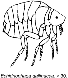
Tunga penetrans, the true jigger flea, differs only in slight details from the latter species. The female, however, penetrates the skin, and lying in an inflammatory pocket with an opening to the exterior, becomes as large as a pea. It is found in Africa and America in man and all the domestic mammals, especially the pig. The eggs are laid in the ulcers; and the larvae crawl out and pupate on the ground.
Flea infestation may be of veterinary importance in intensively-reared ruminants.
Destruction of fleas (See INSECTICIDES). Bedding must be destroyed or disinfected and the surrounding floorboards and cracks cleaned thoroughly using a vacuum cleaner or the animal will shortly be reinfested. This is even more important than ridding the host of fleas. There are sprays for use on bedding and soft furnishings and can help kill fleas and stop eggs hatching.
Powders and aerosol sprays, applied externally; ‘pour on’ or ‘spot on’ formulations applied to the skin under the fur or coat; shampoos; and tablets to be taken internally are all available for the control of fleas. There are many preparations marketed: natural pyrethrins and their synthetic derivatives; organophosphorus compounds; carbamates and amidines and avermectins are all used in topical applications. Imidacloprid can be used in dogs, cats and rabbits, but fipronil can be used only in dogs and cats, being toxic in rabbits. Collars impregnated with carbamates or pyrethroids are popular among owners, but their effectiveness appears to be limited to prevention of reinfestation.
Cythioate, an organophosphorus compound, and lufenuron, a benzoyl urea derivative, are given as tablets or oral suspension.
Permethrin, a pyrethrin derivative, is formulated as a powder, ‘pour on’ and shampoo. Aerosol sprays often contain a mixture of piperonyl butoxide and pyrethrins. All are effective, properly used, but the manufacturers’ directions must be followed carefully, with regard both to handling and to the suitability of the particular product for cats or dogs. Puppies and kittens must only be treated with products recommended for use in young animals.
(See also FLEA-COLLARS; INSECT GROWTH REGULATOR.)
A breed formed around 1600 in Germany from the Simmental cattle of Switzerland. They have a white face and are used for milk, meat and as draught animals.
Sheep fleece covers the whole sheep’s body and requires shearing annually before the summer except for a few breeds that naturally shed their wool. It is removed from the sheep in one piece. In the Middle Ages, sheep were mainly kept for their fleece, mainly spun into wool for clothing and this resulted in owners becoming very wealthy. In the 20th century the creation of semi-synthetic materials meant that the fleece has had little value and was mainly undertaken for welfare reasons. In the 2010s wool prices have increased. The average sheep fleece is 2.3 kg to 4.5 kg (5 lb to 10 lb), although in sheep bred for wool the weight might be about 13.5 kg (30 lb). Value of the fleece partly depends on its CRIMP.
A breed of domestic rabbit weighing up to 10 kg. It has a long, muscular body and broad hindquarters. Bucks have larger heads than does.
A disease cause by gliding bacteria Flexibacter maritimus, it results in erosive lesions on the body surface due to necrosis of the body, head, fins and occasionally the gills. They can be called ‘eroded mouth syndrome’ or ‘black patch necrosis’. It is seen in marine fish including Dover sole (Solea solea), sea bass (Dicentrarchus labrax), turbot (Scophthalmus maximus) and in wild and farmed salmon (Salmo salar).
Flies are mostly, but not exclusively, members of the order Diptera – the 2-winged flies.
Even the common housefly can transmit infection such as ANTHRAX and TUBERCULOSIS, and also various species of parasitic worms. The stablefly’s role in the production of summer mastitis is well known, and other flies, such as the sheep headfly, may be responsible for cases of this disease too. The autumnfly (and almost certainly others) can transmit an eye worm of cattle, and also the infective agent Moraxella bovis which causes the more commonly recognised contagious keratitis or New Forest disease.
The approach of a cloud of flies, such as the headfly, will cause cattle to cease grazing and huddle together. The movement or presence of a mass of even non-biting flies over the animal’s body represents a further cause of ‘worry’ or restlessness; and both the headfly and the autumnfly feed on secretions from eyes, nose, etc., and on the serum exuding from small wounds.
Cattle may become sensitised to the secretions poured into the bite wound, so that an allergy arises with sometimes the production of serious skin lesions which, in turn, may attract other flies.
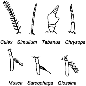
Antennae of various flies. The small hair in the lower row is the ‘arista’.
Sawfly poisoning was first reported in Denmark in 1974, but it has been recognised in Australian cattle and sheep since 1955, and heavy losses have occurred. Goats are also susceptible.
The sawfly larva (Arge pullata) is bright yellow with black dashed lines on the back. It defoliates birch trees, and then drops to the ground to pupate or search for more food.
Order Diptera Insects which have one pair of wings.
Simulium (buffalo gnats) The flies of this genus are small, black or reddish-brown, thick-set hump-back flies – hence their name. The females at certain times appear in swarms and attack cattle, horses and other animals.
The eggs are laid in water. The larvae, which are aquatic and creep about like leeches, can only live in well-aerated running water; in still water they are asphyxiated. The larva when mature spins a silky cocoon which is attached to water weeds. In this the pupa lies loosely, breathing by means of extruded gill-tufts. The fly is very active in Central Europe, where cattle may die in two hours after an attack. They show laboured breathing, stumbling gait, rapid pulse, and swellings in pendulous places. In less severe cases loss of appetite, abortion, depression, and temporary or permanent blindness may result.

Simulium. Adult larva, and pupa. The adult fly is magnified × about 10.
Sandflies Two-winged flies, of which the blood-sucking females transmit infections, including that of LEISHMANIASIS, which is reported in dogs.
Mosquito The mosquito, the carrier of malaria and yellow fever to man, is also of importance in tropical veterinary medicine, transmitting diseases such as avian malaria (see Plasmodium gallinaceum), HEARTWORM of dogs, BLUETONGUE, EQUINE ENCEPHALITIS, HORSE-SICKNESS, AFRICAN and RIFT VALLEY FEVER). In temperate climates, too, mosquitoes are important disease vectors.
Four genera of mosquitoes are of veterinary importance: Aedes, Anopheles, Culex and Mansonia.
Eggs are laid on the surface of water or floating vegetation, either singly (Aedes and Anopheles) or as ‘rafts’ of eggs.
Larvae undergo three moults, and develop only in water, in which they are highly mobile.
Culex modestus is the main vector of WEST NILE FEVER in Europe transmitting infection from birds to humans, the mosquito is found in southern England.
Larvae-eating fish, such as Alphanus dispar, are being used in the Nile Delta and elsewhere for mosquito control. (See also DDT and DIELDRIN.)
Midges, biting (culicoid) (see under this heading)
Gadflies (tabanidae) The family of the gadflies is a large and important one, as the females are blood-suckers and are found in many parts of the world. In the UK the name ‘gadfly’ is usually restricted to WARBLES. It is eradicated in the UK but GADDING still occurs.
Gadfly eggs are laid in masses on leaves and plants near water. The larvae are more or less aquatic, but towards maturity they live in damp earth or decaying vegetation. The larva is cylindrical, pointed at both ends, and with most of the segments carrying pseudo-pods or false feet. The pupa resembles that of a moth. In temperate climates, development takes nearly a year. The males feed on plant juices, but the females are blood-suckers, and in addition carriers of various diseases – for example, trypanosomiasis, swamp fever in horses, and filariasis in man.
The bite is painful, and causes much irritation to horses and cattle, resulting in gadding, decrease in milk yield, and so on. No remedies are really satisfactory, although nets have been used with some success on horses.
If the pools most commonly frequented by these flies are covered with a thick layer of paraffin oil, the flies are killed. If this plan is adopted early in the season the numbers can be kept under control.
Tabanidae These are large, stout flies, the females of which suck blood and transmit diseases such as SURRA (mechanically) and other blood diseases such as ANTHRAX. Another species transmits swamp fever in horses (see EQUINE INFECTIOUS ANAEMIA).
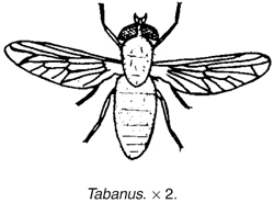
Haematopota This is also a worldwide genus. The species has smoky wings, and include the British clegg or horse-fly which, in addition to being a veritable pest to horses and other domestic animals, inflicts a very painful bite in man.
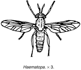
Chrysops is distinguished by its long slender antennae, and its green or golden eyes spotted with purple. It is found all over the world, including Britain. This genus is the carrier of the parasite of Calabar swelling in man. It also can inflict a very painful bite.
The non-biting two-winged flies have an even greater significance to man and his animals than the biting flies.
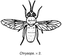
Muscidae The flies belonging to this family are smallish to medium-sized flies. The type of this family is Musca domestica.

Musca domestica The great majority of flies found in houses belong to this species. It is a medium-sized fly with four black stripes on its back, and a sharp elbow in the fourth wing vein. The eggs are laid; about 120 in a batch, preferably in horse manure, but occasionally in human or other excreta. They hatch rapidly (depending on the environment), and the issuing larva (or maggot) feeds and moults and finally becomes full grown in four to five days. It leaves the manure at this stage, and crawls to a dry spot where it pupates. The puparia are more or less barrel-shaped and dark brown in colour. In another four or five days in summer the adult fly emerges. The shortest time on record between the laying of the egg and the appearance of the adult is eight days; 10 to 12 days is more normal. In three to four days the female is ready to lay eggs. The fly lives over the winter in the pupal stage, although in kitchens and warm places adults may be seen at every season of the year.
The house-fly can transmit disease by swallowing bacterial spores, and either bringing them up in their vomit or passing them out in their faeces; or by carrying them about on its hairs and legs. Two species of stomach worm are carried by this fly, in which they pass part of their life-cycle. Among other organisms known to be carried by this fly are anthrax, tuberculosis, and many species of worm eggs. (See FLY CONTROL MEASURES, below.)

Headfly This is a non-biting fly which, as its name Hydrotaea irritans suggests, is a cause of great irritation to cattle, sheep, etc., especially since so many headflies often settle on the same animal. The fly will take advantage of any abrasion on the sheep’s skin. Both fly-repellents and head-caps have been used for control. Pine-tar oil is a useful repellent.
The headfly is responsible for carrying bacteria to cows’ teats (especially when already damaged by biting flies or other causes), and appears to have an important role in producing SUMMER MASTITIS. It is also involved in the spread of New Forest eye infection caused by Morexalla bovis.
The headfly. (Crown copyright photograph.)
Face flies These ‘autumn flies’ (Musca autumnalis) plague beef and dairy cattle, and horses, at pasture, feeding on watery secretions from nostrils and eyes.
Dipterous larvae or maggots – Myiasis The most important to the veterinary surgeon and the agriculturist are those non-biting muscid flies which have taken on a parasitic existence in their larval stages. Myiasis means the presence of dipterous larvae (or other stages) in organs and tissues of the living animal and the disorders and destruction of tissue caused thereby. (See STRIKE.)
The myiasis-producing flies can be divided into three groups: specific, semi-specific and accidental.
Specific: This group consists of flies which need to breed in living tissue. It includes Chrysomyia bezziana, Cordylobia anthropophaga, Wohlfahrtia magnifica, Booponus intonsus, and all the Oestridae.
Semi-specific: This group consists of flies which, normally breeding in carcases, may live in the living animal. It includes the blow-flies, the sheep-maggot flies, and some of the flesh-flies.
Accidental: This group includes all flies the larvae of which, accidentally swallowed with the food, may live in the intestine.
The more important of the above flies are considered below.
‘Blow-flies’ Calliphoridae are largish muscids of a metallic or yellow colour.
‘Common blow-fly’ or ‘Blue-Bottle’ (Calliphora sp.) has reddish palps, black legs, and a bristly thorax. The general colour is dark blue with lighter patches on the abdomen. The colour, however, is not lustrous. The ova are usually deposited in decaying animal matter, but occasionally in living tissue.
‘Green-bottle fly’ (Lucilia sericata) is the British sheep-maggot fly. It is also found in Australia and America.
L. caesar, a common species in Europe, does not ‘blow’ sheep in this country, but does so in countries such as Russia, where other species are absent. Other species of Lucilia in India and Australia occasionally are also implicated.
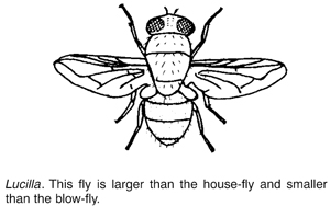
‘Copper-bottle fly’ (Lucilia cuprina) is the strike fly which attacks sheep in Australia and South Africa.
These are of a bright metallic or bluish-green colour, with many strong bristles on the thorax arranged in two parallel rows. There are no stripes on the thorax or abdomen. The cheeks are not hairy as in Calliphora.
This genus blows wool, but occasionally infects wounds.
Chrysomyia bezziana, found in India, Africa and the Philippines, is a metallic greenish-blue blow-fly, closely related to Lucilia, but with dark transverse abdominal bands and with fewer and less-developed thoracic bristles. The metallic sheen is more brassy than in Lucilia. This fly breeds only in living tissue – it discharges from natural orifices, or in sores and cuts. Up to 500 eggs may be laid at one time. They can hatch in about 30 hours, and the larvae rapidly reach maturity, crawl out and pupate on the ground. Several other species of this genus are semi-specific myiasis flies, normally breeding in decaying matter. These include C. albiceps, a notorious sheep-maggot fly in Australia.
‘Screw-worm fly’ (Callitroga americana) in America can be distinguished from the old-world species by the three well-marked blue dorsal stripes on the thorax and dark hairs on the abdomen. It is of a dark bluish-green colour, with a well-marked yellowish-red face. (See also FLY CONTROL MEASURES, below.)
This species will lay eggs in decaying animal or vegetable matter, but will also oviposit in any diseased tissue, in wounds in the vulvae of freshly calved cows, the umbilical cord of calves, and so on. The ova can hatch in 24 hours, and the maggots in four to six days. The pupal stage on the ground lasts three to 10 days. The maggot resembles a blue-bottle maggot, but the deeply cut constrictions between segments and the prominent rings of spines give it its popular name.
As soon as the egg hatches, the larva starts burrowing into the flesh. It can penetrate the sound tissue of living animals, and may even lay bare the bones.
‘Tumbu fly’ (Cordylobia anthropophaga) is a specific myiasis fly in Africa, attacking many hosts. It is a dirty brownish-yellow blow-fly with blackish markings. Eggs are laid in dust and rubbish on which the host, usually a dog, is accustomed to lie. The small larva may live apart from the host for 10 days, but it may eventually burrow into the epidermis or die. It moults in this position, and forms a ‘tumbu’ below the skin with an opening to the exterior through which it breathes. The ‘tumbu’ does not suppurate unless the larva dies. The larva emerges in about seven or eight days, and two or three days later it pupates. The adults emerge in about 20 days. This fly does not burrow into the deeper tissues. The scrotum is a common site of the maggot. Putting a drop of oil or Vaseline over the breathing hole will force the larva to protrude, when it can be removed.
Booponus intonsus is a light yellow specific myiasis fly found in the Philippines, which is somewhat allied to Cordylobia. It infects bovines and goats.
The eggs are laid on the hairs on the lower parts of the legs; and the larvae make their way to the coronet and bury themselves in the flesh. The larvae resemble the screw-worm. The larval period seems to last two or three weeks, when it leaves the host and pupates in the ground. The pupal life is 10 days.
The larvae, called the ‘foot maggot’, cause a considerable lameness with numerous superficial wounds and distortion of the horn.
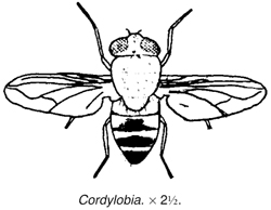
‘Flesh flies’ (Sarcophagidae) are closely related to the Muscidae. The body is more elongated than that of the blow-flies, and they are usually grey in colour, with a mottled abdomen and a striped thorax. They generally produce living larvae instead of laying eggs. Two genera are important.
Sarcophaga spp. These are large grey flies with red eyes and square chequered markings on the abdomen. The third segment of the antenna is long. All the species normally breed in decaying animal matter, but may be found in old festering wounds. They are found throughout the world.
Wohlfahrtia magnifica resembles the preceding genus, but has well-defined round spots on the abdomen. The third segment of the antenna is short and the arista is without bristles. It is widely distributed in Russia, Asia Minor and Egypt. The larvae never attack carcases, but are always found in wounds and natural cavities of living animals. The fly deposits living larvae on sores and discharges.
In Australia the most important sheep-maggot flies are Calliphora augur, a large orange-coloured fly; C. stygia, the common sheep-maggot fly, often called the ‘golden-haired blow-fly’; and Chrysomyia albiceps var. putoni, the larva of which is known as the ‘hairy maggot’.

Injuries due to maggots The injuries due to maggots may be roughly divided into two classes – larvae attacking wounds and discharges, and larvae attacking the wool of sheep. The former type of injury is found on any animal, including man. The flies usually, but not always, select old sores. Some, such as Chrysomyia americana (the ‘screw-worm’) will penetrate into the sound tissue, and prefer fresh wounds or carcases. The infected wound usually has a watery discharge. Prevention is obviously most important. (See also under MYIASIS.)
Blood-sucking muscid flies These flies, which resemble the house-fly in general appearance, are responsible for an enormous amount of damage to farm animals. When one considers that they include such flies as the tsetse fly, the stable-fly and the horn-fly, this is easily understood.
Stomoxys This genus is mainly confined to Africa and Asia, but one species, S. calcitrans, the stable-fly, is worldwide in its distribution.
Stomoxys breeds in stable manure and in other places where moisture and organic material are found. The eggs hatch in about two to three days, and the larva, which is similar to but smaller than Musca, becomes full-grown in two to three weeks. The pupal stage lasts nine to 13 days. Development is more rapid in the tropics, where the time between egg and adult may be reduced to 12 days.
This fly is a serious pest to horses and other animals. It will also bite man. Apart from the extreme irritation of its bite, it can transmit anthrax, surra and other diseases. It is also the intermediate host of Habronema microstomum, a worm parasite of horses.

Haematobia H. stimulans is a common blood-sucking parasite of cattle, and occasionally of horses and man, in Europe. It resembles Stomoxys, but has spatulate palps as long as the proboscis, and hairs on both sides of the arista. It breeds in fresh cattle dung. The larva becomes full-grown in about six to nine days, while the pupal stage lasts five to eight days.
Lyperosia L. irritans is very closely related to Haematobia, but can be distinguished from it by the absence of bristles from the underside of the arista. It is found in Europe (including the UK) and America. It is a very serious pest to cattle, clustering round the base of the horns, a habit which gives the fly its popular name of horn-fly. The irritation caused by their bites is estimated to cause a drop in milk yield amounting in some cases to 50 per cent. The flies breed in fresh cow dung. Flies emerge in about 15 days after the egg is deposited. The maggots must have moisture, and can be destroyed by any means which will dry the manure quickly. The horn-fly seldom goes far from its host, and may be destroyed by attaching splash-boards to ordinary dippers. The fly leaves the cattle at the moment of entering the bath, but the dip, caught and flung back by the splash-board, drenches and destroys the flies. The hotter and more excited the cattle, the closer the flies stick and the greater the number killed. Any oily dip is suitable. (See also FLY CONTROL MEASURES, below.)
Tsetse flies (glossina) The flies of this genus are, with one exception found in Arabia, confined to Africa. They are the notorious carriers of trypanosomiasis in man and animals. Glossina resembles a large stable-fly but has a feathered arista, long slender palps, a slender shaft to the proboscis, and a peculiar wing venation. The life-cycle is unusual: the female produces a single living larva at a time and deposits it when full-grown. It immediately pupates. One female produces only about a dozen larvae in her life.
More than a dozen species of Glossina are known. The most important are: G. palpalis; G. morsitans; G. brevipalpis; G. longipalpis; G. pallidipes; G. tachinoides.

Bot and warble flies The bot family Oestridae consists of hairy, heavy flies with rudimentary mouth parts. The female attaches the egg, or, in the case of the nostril flies, places the larva on a suitable host, and the remainder of the larval life is parasitic. When mature the larvae leave the host and pupate on the ground.
These flies may be placed in three groups according to the habitat of the larva:
1. In the alimentary canal –
Gastrophilus, the horse bot;
Cobboldia, the elephant bot.
2. In the head sinuses –
Oestrus, the sheep nostril fly;
Rhinoestrus, the horse nostril fly;
Cephalomyia, the camel nostril fly; and others.
3. In the subcutaneous tissue –
Hypoderma, the warble-fly (See WARBLES);
Dermatobia, the macaw worm fly; and others.
Bot flies The flies of the genus Gastrophilus are large and hairy, with large compound eyes and three ocelli. The females have an elongated ovipositor which is bent under the body when at rest. Four species are of importance.
G. intestinalis (G. equi), the common horse bot, has cloudy wings; it deposits its eggs on any part of the horse, but especially on the distal ends of the hairs. The eggs require moisture and friction (supplied by licking) before they will hatch.
G. nasalis (G. veterinus) is smaller, more hairy, and has a rusty-coloured thorax. It oviposits usually at the proximal ends of hairs under the jaw. It lays one egg and flies to a distance, returning later to lay another.
G. haemorrhoidalis has a bright orange-red tip to the abdomen. It deposits its eggs only at the base of the small hairs on the lips of the horse. The eggs may hatch without moisture or friction.
G. pecorum resembles G. intestinalis. In colour it is yellowish-brown to nearly black, with brownish-clouded wings. Its habits are similar to that species.
The distribution of the first three is universal, but the last seems to be restricted to Europe and South Africa.
The life-cycle of the species of this genus is not yet fully understood. Some of the newly hatched larvae may pierce the skin or buccal mucous membrane; in any case the larvae are found in various parts of the alimentary tract. Each species has its own special preference. G. intestinalis is usually found in the stomach, occasionally the duodenum; G. nasalis prefers the duodenum, but has been found in the pharynx and stomach; G. haemorrhoidalis is found in the stomach, duodenum, rectum, and even in the anus; while G. pecorum usually occurs in the pharynx or stomach, but may be recovered from any part.
Bots when present in large numbers in the stomach or intestine, or even in small numbers about the pharynx and anus, may cause a considerable suffering to their host by mere mechanical obstruction. The adult fly worries the horse considerably, especially the species G. nasalis and G. haemorrhoidalis, and may cause loss of condition.
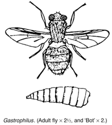
Treatment Formerly, carbon disulphide, administered in autumn and early winter by stomach tube and followed by warm saline. This has been replaced by a haloxon formulation given in the feed and by ivermectin paste, which have both proved effective (also against roundworms). Withholding water four hours before and after dosing is recommended when treating against bots. (See AVERMECTINS; IVERMECTIN.)
Some control is possible by regular removal of the ‘nits’ from the lower limbs of grazing horses during summer.
Oestrus O. ovis, the sheep nostril fly, is somewhat larger than the house-fly and is greyish-yellow to brown in colour. It is found practically all over the world. It deposits eggs, or larvae. The hovering female ‘strikes’ at the nostrils, and the young larva crawls up the nose, and may lodge in one of the sinuses of the skull. It remains there until fully grown, when it is sneezed out and pupates in the ground.
Prevention is by applying tar to the nostrils. This may be done by means of a salt lick, access to which may only be obtained by smallish holes (5 cm or 2 in) smeared with tar. Ploughing a single furrow across a sheep pasture allows the sheep to protect their nostrils from the flies, ‘strike’, and gives some measure of protection. Some anthelmintics are effective.
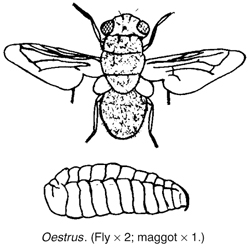
Rhinoestrus R. purpureus (R. nasalis), the horse nostril fly, is common in Central Europe and North Africa. It is a smallish fly with the body covered with small tubercles, and is closely related to Oestrus. The female deposits a number of living larvae at one time in the eyes or the nose of the horse (and occasionally man). The larvae may be found about the cranial cavities or even in the pharynx or larynx. Russian gadfly is a synonym.
Hypoderma Two species of warble-fly, H. bovis and H. lineatum, are found in cattle (and occasionally in the horse). Both are very extensively found in Europe and America but have been eradicated from the UK and the disease is NOTIFIABLE.
H. bovis is a largish fly with yellow hair just behind the head. The underpart of the abdomen is nearly black, while the tail end is orange-yellow. The legs have few hairs.
H. lineatum is rather smaller with a reddish-orange tail and rough hairy legs.
H. bovis lays its eggs one on the base of each hair at a time. The fly has a most terrifying effect on cattle, and causes them to gallop madly in all directions. H. lineatum irritates animals less than does H. bovis. The ova are generally deposited while the animal is lying in the shade. A number of eggs – up to 14 – are laid on the same hair, and are often in full view.
In both cases larvae emerge in several days and pierce the skin. They travel up through the connective tissue and finally reach the back. Under the skin the larvae form a small swelling (about the middle of winter), which moves about at first, but gradually becomes still and enlarges. A small opening appears in the centre through which the larva breathes. In spring the larva falls to the ground and pupates. Several weeks later the adult fly emerges.
The presence of the larvae may decrease the milk yield by 10 to 20 per cent, cause a considerable depreciation in flesh near the points where the larvae are, and enormously reduce the value of the hide. The adult fly also causes loss through the mad chasing about of cattle. (See also under WARBLES.)
H. diana is a warble-fly affecting deer in the UK and elsewhere.

Dermatobia D. hominis, the macaw worm-fly, is a parasite of cattle and other domesticated animals (and occasionally man) in tropical America. It is a medium-sized fly, grey or steel-blue in colour, with pale brown wings. The female lays its eggs on the body of some blood-sucking arthropod, usually a mosquito. This carrier attacks an animal five or six days later, and the larvae, rapidly escaping from their shells, pierce the skin of the host, and form a local tumour near where they were deposited. In a month or so they emerge and pupate.

Pupipara This family, which includes the sheep ked and the horse ked (New Forest fly), was so called because live larvae are produced which pupate at once. The adults in this case are blood-sucking parasites with a hard integument with a broad neckless head and very stout legs ending in grasping claws. Wings are present or absent.
Hippobosca equina, the New Forest fly or horse ked, has wings which, however, are seldom used, the fly preferring to run swiftly between the hairs of the host.
Paragle fly Paragle redicum, an anthomyiid fly, lays its eggs in canine faeces, and the white specks have been mistaken for tapeworm segments. Larvae may also be passed alive through the canine (and also the human) gut.
Hymenoptera Insects which have two pairs of wings.
Sawflies These have four wings and a saw-like ovipositor. The larvae are said to be poisonous if swallowed.
Fly control measures Perceptive farmers have for many years realised the harmful effects of fly infestation on livestock. Controlled field trials, comparing the productivity of treated and untreated cattle, have convincingly demonstrated the advantages that can be gained by modern fly-control methods.
Flies interfere with normal rest and feeding. The approach of a cloud of flies often causes cattle to cease grazing and huddle together.
The horse fly (Tabanus), like the stable fly (Stomoxys), has a painful bite, and the wounds inflicted attract other flies; this exacerbates the ‘worry’ situation and often transmits even more infection. Some animals become sensitised to the secretions of biting flies, so that an allergy results. One example of this is the ‘SWEET ITCH’ of horses caused by biting (Culicoid) midges.
Flies are notorious for spreading livestock diseases. Even the common housefly (Musca domestica) can transmit anthrax, tuberculosis, and the larvae of some parasitic worms. The headfly (Hydrotaea irritans) is among several species that may transmit the bacteria that cause ‘SUMMER MASTITIS’. This can lead to gangrene of part of the udder, usually with the permanent loss of use of one-quarter – and to great pain and occasionally death of the cow.
Besides interfering with grazing, the headfly causes infested sheep to scratch, rub and knock their heads, often breaking the skin. Open wounds then attract other flies, increasing the tendency to self-mutilation. Sometimes the whole poll region becomes raw. Pine-tar oil has been used as a repellent, and head caps for protection, but they are inconvenient to use.
Plastic tags impregnated with synthetic pyrethroids have been effective in the reduction of ‘fly-worry’ in cattle. For sheep too, tags containing CYPERMETHRIN and PERMETHRIN can be effective in controlling the severity of damage caused by headflies. Cyromazine, in a ‘pour-on’ formulation, is useful against blowfly larvae on sheep and lambs.
The activities of the autumn fly (Musca autumnalis) are very similar to those of the headfly. Both flies feed on secretions from nose, mouth, eyes and wounds and are among several species that transmit the various pathogens causing New Forest disease. This involves an acute and painful conjunctivitis, with inflammation also of the cornea, which becomes opaque, so that cattle are often rendered temporarily blind. Complications may result in permanent eye defects and impaired sight; but even without them the disease causes stress and, since it interferes with feeding, loss of condition can be appreciable.
Other bacterial diseases spread by flies include SALMONELLOSIS and BRUCELLOSIS; while viral diseases such as swine fever and foot-and-mouth have in the past been similarly transmitted in the UK. The larvae of some flies also parasitise animals, for example, the ‘green-bottle’ fly causing ‘strike’ in sheep, the sheep nostril fly, and the horse bot flies. The mere approach of warble flies causes cattle to stampede, and the larvae undoubtedly cause pain during their migration through the cow’s body, and probably irritation while present in the skin swellings, or warbles. In a few instances, cows have died following the accidental crushing of larvae in the warbles, and larvae occasionally seriously damaged the spinal cord. In the UK, control measures have virtually eliminated warble infestation in cattle.
Deterring and killing flies The number of flies entering a milking parlour can be reduced by a spray-boom erected over the doorways, and a plain water mist, produced by ordinary sprinklers or misters, has been recommended for use in collecting yards where a spray-boom over the parlour entrance is impracticable. Such measures, however, do not reduce the total fly population of the farm – they do not kill. Electric fly traps do, and they can be useful when installed in piggeries and dairy cattle buildings. Which flies are electrocuted will obviously depend on the feeding and resting habits of the various species.
Of far wider application, and the most effective weapon against farm flies, is the insecticidal spray. This can be used to convert a livestock building into one big fly trap. A wide variety of effective insecticides is available.
For housed stock, spraying walls may suffice; but beef and dairy cattle at grass will be the target of flies coming from their resting places among trees. Fly control, if it is to benefit grazing animals, therefore requires application of an officially approved insecticide direct to their backs. For this purpose, the synthetic pyrethroids are effective.
These insecticides are chemically allied to the active ingredients of pyrethrum, but are more potent as fly-killers and are also light-stable so that they stay effective for longer in the sun.
Fly control for horses and livestock Numerous formulations based on cypermethrin and other synthetic pyrethroids are available, as both a spray and as pour-on applications, which are convenient to use.
A PVC fly band impregnated with cypermethrin, for threading on to either the browband or the crownpiece of a head collar, and an ear tag impregnated with 8 per cent cypermethrin, are also available for cattle at pasture.
Whether measured in terms of reduced animal suffering, or farmers’ incomes, or a lowered incidence of diseases – some of which are of public health as well as economic importance – fly control is very worthwhile. Any doubter should note the success of the UK government’s five-year plan to eradicate warble flies from the UK. Now, only sporadic outbreaks of warble infestation occur. Since horses, as well as cattle, suffer from warble-fly larvae (although to a much lesser extent), eradication has also benefited them and their owners.
Overseas Similar methods to those described above, suitably adapted for tropical climates, are in use. The spraying of ground with DDT and dieldrin has been very effective for the control of tsetse flies and human trypanosomiasis, but the practice was discontinued because of the development of resistance and the toxicity of those preparations to other species (see also under TROPICS, LIVESTOCK PRODUCTION IN THE).
The release of sterile male flies from aircraft has been used on a large scale in Puerto Rico to control the screw-worm fly.
Genetically engineered blow-fly maggots have been used in attempts to eradicate sheep blow-flies. The maggots were altered so that females which mate produce blind or sterile offspring – a hereditary characteristic which will ‘confer genetic death on future generations’.
(See also DIPS AND DIPPING.)
(See FATTY LIVER/KIDNEY SYNDROME OF CHICKENS AND TURKEYS; LIVER/KIDNEY SYNDROME OF POULTRY.)
(plural, Flocculi)
A flocculus is a small mass of fibrous material – examples include tufts of wool, coagulated protein in areas such as the aqueous humour of the eye. (See EYE, DISEASES AND INJURIES OF - IRITIS) A flocculus is also a small mass attached to each normal cerebellar hemisphere.
(See PASTURE, CONTAMINATION OF; SALMONELLOSIS; WATER-DROPWORT.)
(See BEDDING AND BEDDING MATERIALS Pigs; HOUSING OF ANIMALS.)
This practice is attractive to the pig farmer since it eliminates the cost of troughs and also saves space – the normal feeding passage becoming a catwalk over the pigs’ sleeping quarters.
From a health point of view, the precise com-position of the concrete floor may prove important. The constituents of sand used in concrete, such as ‘pit sand’, with a high iron content can cause iron poisoning once the surface layer has been licked off.
More important is the fact that, with floor feeding, loss of appetite in pigs – a common early sign of many diseases – may not be noticed. With trough-feeding, it is easy to see which pigs are uninterested in food.
Feeding pellets on floors instead of meal in troughs may also cause digestive upsets. The method may involve more stress than conventional systems.
As a rough guide, the following minimum figures may be given: bacon pig, 2 m2 (6 ft2); veal calf, 3.5 m2 (12 ft2); laying hen on deep litter, 1 m2 (2½ ft2).
From 1 January 2013 EU minimum floor space standards for pigs were specified:
Less than 10 kg - 0.15m2 (less than 22 lb - 1.5 ft2); 10 kg to 20 kg - 0.2 m2 (22 ft2 to 44 lb - 2 ft2); 20 kg to 30 kg - 0.3 m2 (44 lb to 66 lb - 3 ft2); 30 kg to 50 kg - 0.4 m2 (66 lb to 110 lb - 4 ft2); 50 kg to 85 kg - 0.55 m2 (110 lb to 185 lb - 5.5 ft2); 85 kg to 110 kg (185 lb to 220 lb - 6.5 ft2); more than 110 kg – 1 m2; gilts - 1.64 m2 (17.5 ft2); sows 2.25 m2 (24 ft2)
Floor sweepings in mill or barn have been added to feed and caused fatal poisoning. For example, pigs have died in this way from nitrate poisoning, and cattle from mustard seed poisoning.
The colloquial name for an inherited muscle disease of both Black and Yellow Labrador Retrievers, correctly called muscular dystrophy of Labrador Retrievers. The condition has been seen in both the UK and the USA. Inheritance is associated with an autosomal recessive gene, leading to atrophy of type II muscle fibres.
Signs Poor exercise tolerance, especially in cold weather, a stiff hopping gait, with sometimes collapse. Signs have been shown as early as eight weeks of age, but in other cases after several months.
Prognosis There is no cure but, if not severely affected, with care such dogs can make acceptable pets. They should not be used for breeding.
A chloramphenicol-like antibiotic which is used in cattle to treat infections caused by Pasteurella, Mannheimia and Haemophilus spp. It is also used in the treatment of fish, particularly in the treatment of furunculosis (Aeromonas salmonicida infection) in farmed Atlantic salmon. It is not recommended for use in breeding stock. The antibiotic is administered by injection in cattle and in the feed of fish. In the USA it has been used to successfully treat enteric septicaemia in Channel catfish (Ictalurus punctatus).
(See AGENE PROCESS.)
Infestation of animal feeds by flour mites (Acarus farinae; A. siro) can cause a significant loss of nutrient value, as explained under DIET AND DIETETICS. Like forage mites of various species, flour mites can also cause an irritating parasitic skin disease of animals. In one incident, 36 police horses were stabled in a building which was cleaned and whitewashed before their arrival. Unfortunately a feed barrow was overlooked and still contained oats left over from the previous year. New oats of high quality were delivered in sacks, and the delivery man opened one sack and topped up the barrow. After he had gone, new oats continued to be put on top of what was left in the large feed barrow, which was never completely emptied. A fortnight after the horses’ arrival, the last four in the line showed signs of head and neck irritation. One horse had rubbed the side of its neck bare; two others had dermatitis on the poll and alongside the mane.
Examination of the bottom layer of the barrow’s contents revealed an enormous number of flour mites, and these were also isolated from the skin lesions.
An unsuspected cause of diarrhoea in dogs may be dog biscuits or meal, stored in large bins, and heavily infested with forage mites. As flies may carry nymphal forage mites, fly control is important in reducing such infestations.
Forage mites and/or their eggs may be found in dog faeces, where they may have been mistaken for the eggs of strongyle worms. However, the mite’s egg is nearly twice the size of the worm’s.
Flour mites (A. siro and A. farinae), the house/furniture mite (Glycyphagus domesticus), and the mould mites (Tyrophagus putrescentiae and T. longior) may also be involved.
A neuroleptic drug of the butyrophenome group used in small animals (ferrets. rabbits, guinea pigs, rats and mice). It is usually mixed with FENTANYL) and the combination can be used as a neuroleptanalgesic for rabbits and small rodents.
A broad spectrum benzimidazole anthelmintic used in pigs and poultry and the only anthelmintic currently registered for use in UK poultry.
Often abbreviated to FDG or F-FDG, fludoxyglucose is a radioactive chemical injected into animals and man and is used in imaging by the PET SCAN.
A hormone preparation supplied in impregnated sponges for the synchronisation of oestrus in ewes. It may also be used to induce oestrus and ovulation in the non-breeding season (see CONTROLLED BREEDING).
(See under DEHYDRATION.)
(See LIVER-FLUKES; LUNG-FLUKES; SCHISTOSOMIASIS for blood flukes; PANCREAS, DISEASES OF; and RUMEN FLUKES.)
A medicine that kills flukes, usually referring to LIVER-FLUKES.
A synthetic pyrethroid compound used for the treatment of sheep scab and tick infestation. Impregnated in plastic strips, it is hung in beehives to treat varroasis in honey bees.
A non-steroidal anti-inflammatory drug used for relief of pain and inflammation in horses, dogs and cattle; formulated as granules, tablets or injection. Proprietary preparations are Finadyne (Schering-Plough), Cronyxin (Bimeda), and Binixin (Bayer).
(See under TETRACYCLINES which make bone fluoresce, and under WOOD’S LAMP which shows ringworm-affected hairs fluorescing. For the fluorescent antibody test, see under RABIES and IMMUNOFLUORESCENT MICROSCOPY.)
Fluorescin (Fluorescein) is a useful diagnostic agent in injuries and ulcers on the cornea of the eye. A weak solution is dropped into the eye and the injured area can be seen clearly demarcated from the surrounding healthy cornea. It can be bound to an antigen to provide rapid identification of an infectious agent.
This element occurs in body tissues and in some natural water supplies. Excess of fluorine causes mottling of the teeth. (For fluorine poisoning, see FLUOROSIS.)
Sodium mono-fluoroacetate is used to kill rats and mice, and it is in this connection that poisoning in domestic animals and man may arise. The drug causes distress, yelping, sometimes vomiting, and convulsions in the dog. A dose as small as 0.66 mg per kg body-weight is fatal. Treatment consists in the administration of PENTOBARBITONE SODIUM.
In 1963, two outbreaks of fatal poisoning involving numerous dogs, cats, cattle and a pony were attributed to the agricultural insecticide fluoroacetamide, a closely related compound.
BACTERIOSTATIC antimicrobials which inhibit the replication of DNA. The group includes danofloxacin (Advocin), enrofloxacin (Baytril), marbofloxacin (Marbocyl, Zenequin) and others. The increased use of these products has caused concern over the production of antibiotic resistance especially in Staphylococcus and Streptococcus species worldwide. Use of fluoroquinolones in juvenile animals has been linked with growing cartilage malformation.
An imaging technique using X-RAYS to provide real-time images of the internal structures of the animal using a fluoroscope.
Fluorosis, or chronic fluorine poisoning, is of economic importance in cattle, sheep, etc., grazing pastures contaminated by fluorine compounds emanating from iron and steel works and other industrial plant. It has also been reported in dairy cattle receiving mineral supplements with a high fluorine content, the result of incorporation of rock phosphate. This is something which animal feed manufacturers should guard against, and they should offer guarantees concerning maximum fluorine content in their products.
Signs Acute: sudden death, convulsions, cardiac and respiratory arrest, and burns can occur. Chronic: there is severe lameness, and a resulting loss of condition; milk yield is greatly reduced. The teeth may become mottled, and the bones particularly liable to fracture. Cows may stand with their legs crossed in cases of fracture of the pedal bones. Hip lameness is probably more common.
Antidote Calcium aluminate is of some limited value as an antidote to fluorine poisoning.
Fluothane is a trade name for HALOTHANE. It is a volatile anaesthetic used in most species but now has been superseded by the less toxic ISOFLURANE and sevoflurane.
Fluriprofen, Flurbiprofen and Ibuprofen are non-steroidal anti-inflammatory drugs used in human medicine, and sometimes given to dogs by their owners, or eaten by dogs with access to the tablets, with resultant poisoning (sometimes fatal). Stomach ulceration and kidney failure have been caused.
Eye drops containing flurbiprofen are used as pre-operative treatment for cataract extraction.
Used in pig liquid feeding systems to move any feed particles lodged in the pipes. Water at pH 3.5, produced by organic acids, is circulated every night to disinfect the system and can be reused for this purpose. Weekly, the mixing tanks are flushed with alkaline water at pH 12.8, and once every two weeks the entire system is treated with this alkaline water, which once used is pumped into the slurry store.
Flushing of ewes aims for rising metabolism in breeding ewes some six to three weeks before service, by putting them on to protein-rich feed. The purpose is to intensify subsequent oestrus and thereby ensure that each ewe is in fit condition to breed. Some trials, however, have failed to demonstrate the effectiveness of the practice.
(See FELINE LOWER URINARY TRACT DISEASE.)
This is a form of EPILEPSY seen in CAVALIER KING CHARLES SPANIELS. The dog behaves as if it were trying to catch flies when none is present.
Blow-fly myiasis is the condition resulting from infestation of the living skin of sheep by the larvae of blow-flies which, in certain circumstances, lay their eggs in the wool. It can be prevented by INSECT GROWTH REGULATORS such as cyromazine and DICYCLANIL. (See BLOW-FLY STRIKE; FLIES - DIPTEROUS LARVAE OR MAGGOTS - MYIASIS; STRIKE.)
(See FINE NEEDLE ASPIRATION BIOPSY.)
Seen as an inherited disease affecting FELL and DALES PONIES. It was originally called FELL PONY SYNDROME. Affected foals are born normal and healthy but become ill within four weeks of birth with depression, weight loss, diarrhoea, coughing and nasal discharge. There is anaemia, ß lymphocyte deficiency and reduced antibody production. All die. There are FIS DNA-based carriers, which maintain the disease and when mated with other carriers can produce affected foals.
(See PARTURITION.)
Foals are young horses of either sex until the time they are one year old. Male foals are known as ‘colt foals’, and female foals are called ‘filly foals’. Most foals are born between March and June in Britain, although quite a number (especially thoroughbreds) are dropped earlier than this. Thoroughbreds are conventionally aged as from 1 January of the year in which they are born, and all other horses from 1 May, irrespective of whether they were actually foaled before or after these dates.
Generally speaking, foals run with their dams at grass during the summer, and are weaned at four to six months of age. With weakly foals, however, and in the case of highly-bred pedigree animals, it is not uncommon to allow them to run with their dams until nearly Christmas, so that they may get an exceptionally good start in life.
As a rule, foals will begin to eat grass when they are between three weeks and a month old, although some start earlier and some later than this. At about six weeks to 2½ months they will begin to eat dry corn from mangers along with their mothers.
Diarrhoea may occur as a result of changes in the mare’s milk, or as a result of the dam grazing avidly upon rich spring grass, etc.
Salmonella typhimurium may cause a subclinical infection; alternatively acute and severe diarrhoea or septicaemia may occur, the latter often following the former. (See SALMONELLOSIS.)
E. coli is another cause of acute diarrhoea in the foal. (See E. Coli.)
Corynebacterium equi is probably a more common cause of pneumonia than of diarrhoea; nevertheless the latter can be severe. Clostridial enterotoxaemia occurs; likewise campylobacter infections.
Viruses causing diarrhoea in foals include a CORONAVIRUS and a ROTAVIRUS. The latter may be associated with a profuse, watery diarrhoea and lymph-node enlargement, sometimes followed by death. (See also GLOBIDIOSIS.)
Navel-ill and joint-ill These are colloquial terms for dangerous infections which may attack the foal within the first fortnight of its life. Pain and swelling at the navel, with sometimes abscesses along the umbilical vein, may occur; but joint-ill (polyarthritis) signs may be noticed first. Often the hock and stifle joints are painful and swollen, there is fever, and the foal is obviously ill.
Treatment includes the use of appropriate antibiotics (but not corticosteroids, which have an immunosuppressive effect).
Septicaemia is always a danger likely to arise with or after those two conditions. Septicaemia due to Actinobacillus equuli infection (‘sleepy foal disease’) may occur within the first three days of life. The foal becomes dull, disinclined to suck, has stupor and diarrhoea, and prostration and death quickly follow. Polyarthritis may occur in foals which survive a little longer.
Organisms causing septicaemia and joint-ill include E. coli, Actinobacillus equi, Strep. zooepidemicus and Klebsiella pneumonia.
Haemolytic disease This results from an incompatibility between the blood of sire and dam, and the consequent production of antibodies which reach the foal in the colostrum and break down the foal’s red blood cells. These become so reduced in number that not only is there jaundice, but often also a fatal anaemia. If trouble from this cause were anticipated, the use of a foster-mother might save the situation but, obviously, this is seldom practicable. Moreover, unless the mare has previously had a ‘jaundiced foal’, there will be no indication of the problem. If she has previously had such a foal, however, it is possible to test her blood against the sire’s during pregnancy and obtain a fairly good idea as to their incompatibility or otherwise. Treatment must be undertaken quickly and consists in exchange transfusion – the removal of up to 2.8 litres (5 pints) of the foal’s blood and the simultaneous injection of up to 3.5 litres (6 pints) of a compatible donor’s blood, previously collected. The transfusion takes about three hours to complete. Recoveries following this treatment have been spectacular.
Worms Strongyles (red worms), Strongyloides westeri and ascarids may cause trouble in foals. The former give rise to malaise, cough, unthriftiness, tiredness and sometimes abdominal pain; the latter to diarrhoea and intermittent colic, among other signs. The Animal Health Trust recommends that foals be dosed every four weeks, alternately for ascarids and strongyles, until the age of one year.
Bone diseases in foals include valgus (see under BONE, DISEASES OF); and RICKETS which can render the growth-plate more vulnerable to injury.
Skin diseases Congenital or inherited conditions include Epitheliogenesis imperfecta and ICHTHYOSIS. ALOPECIA is sometimes a complication of STRANGLES; to which URTICARIA is an occasional sequel.
Pneumonia Rhodococcus equi causes pneumonia in foals, as does Corynebacterium equi, which occurs sporadically in the UK. A suppurative broncho-pneumonia, with abscesses in the lungs and pulmonary lymph nodes, it may be associated with ‘joint-ill’ and osteomyelitis.
Rhinopneumonitis A congenital infection with EHV 1 virus is a cause of early death in foals. (See EQUINE VIRAL RHINOPNEUMONITIS.)
Muscular dystrophy (See under MUSCLES, DISEASES OF.)
Pervious urachus, or ‘leaking at the navel’, is a condition in which the communication between the urinary bladder and the umbilicus or navel outside, which should close at the time of birth, remains patent and allows urine to dribble from it. The urine blisters the skin around, and ultimately results in considerable swelling and suppuration around the navel. The condition should be corrected by surgery.
Combined immunodeficiency (CID) occurs in some Arab foals. (See IMMUNODEFICIENCY; also under HERNIA; VALGUS; TYZZER’S DISEASE.)
This is seen in chickens in the USA and is thought to be due to disturbance in the intestinal flora composition and proliferation of Gram-negative pathogens.
An infection of sheep caused by the epsilon toxin of Clostridium perfringens type D (see Clostridium SPP.). Any problems that result in reduced ruminal or intestinal movements allow the toxin to build up. It also occurs in animals have often moved. Signs usually include depression and then recumbency.
(See POISONING.)
(See FLOUR-MITE INFESTATION; FORAGE MITES.)
(noun, Foetus)
(See FETAL; FETUS.)
(See EMBRYOTOMY.)
(See under SMOG and FOG FEVER.)
The colloquial name derived from the word ‘fog’, meaning the second crop of grass taken from pasture already cut once that season for hay or silage, has caused much confusion, since it has been applied to several different syndromes in cattle.
True fog fever has been defined as acute pulmonary emphysema occurring in adult cattle (typically single suckler beef cows) which have been moved from poor to lush grazing in the autumn. It should be differentiated from parasitic bronchitis, and also from extrinsic allergic alveolitis caused by micro-organisms in mouldy hay.
It may be identical with the acute bovine pulmonary emphysema encountered in North American adult cattle moved from range to lush pasture.
Cause The probable cause is the conversion in the cow’s lumen of the amino acid L-tryptophan in grass to 3-methylindole, a toxic substance.
Two clinical forms of fog fever may be seen. The mild form affects up to 50 or 60 per cent within affected groups of cattle, the animal remaining bright though breathing rather rapidly. In the severe form, the degree of respiratory distress varies greatly, is often severe, and results in a 30 per cent mortality rate, especially if animals are moved or stressed. Therefore, move animals very quietly to prevent death or pen them in the field and offer hay or silage. FLUNIXIN MEGLUMINE is used as treatment although 10 per cent methylene blue solution given intravenously may be effective.
Post-mortem Lesions include pulmonary congestion, oedema, and hyaline membranes, interstitial emphysema and diffuse alveolar epithelial hyperplasia.
Aftermath, ‘fog’. Grass grown for winter grazing.
The name for FOLIC ACID and its derivatives.
One of the vitamins of the B-complex. Storage in the liver is dependent on adequate dietary zinc. (See VITAMINS.)
This is seen as nucleated red blood cells (MEGALOBLASTS), it may also result in SPINA BIFIDA in offspring of deficient pregnant dams.
(See SKIN; OVARIES.)
This stimulates the development of the Graafian follicles in the ovary, and controls the secretion of oestrogens from the ovary. It is secreted by the anterior lobe of the pituitary gland. In the male animal, FSH stimulates sperm production in the testicle.
Follicular mange is another name for DEMODECTIC MANGE due to mites of the genus Demodex canis, which lives in the hair follicles of the skin. (See MITES, PARASITIC.)
(See POULTICES AND FOMENTATIONS.)
Fomites is a term used to include all articles that have been in actual contact with a sick animal, so as to retain some of the infective material and be capable of spreading the disease. Bedding material, fodder, mangers, stable or byre utensils, clothing, grooming tools, the clothes of an attendant, or even the attendant him or herself, may all be fomites.
These are a relatively common form of allergy in dogs and cats. There are many allergens containing proteins and can be part of foods such as milk, wheat and meat. They can result in diarrhoea, and/or skin lesions. OTITIS EXTERNA is common in dogs but less common in cats. (See ALLERGY; FATTY ACIDS.)
The United Nations organisation whose aim is to prevent hunger. It is a source of data and produces a wide range of statistics and information. It assists developing countries to modernise and improve their agriculture, fisheries and forestry and aims to ensure good nutrition for all, and food security. Headquarters address: Viale delle Terme Caracalla, 00153 Rome, Italy. Telephone: +39 06 57051; email: FAO-HQ@fao.org.
(See FEED CONVERSION RATIO.)
In countries such as Denmark and the USA, the inspection of meat and meat-derived products has long been carried out entirely by members of the veterinary profession. In the UK this has been only partly so, but to conform with EU regulations meat inspection, including pre-slaughter inspection of food animals, and the examination of organs and tissues as well as inspection of the dressed carcase are carried out under the supervision or direction of official veterinary surgeons under the centralised control of the Meat Hygiene Service. For conditions which render meat dangerous as food, see TUBERCULOSIS; SALMONELLOSIS; ANTHRAX; TRICHINOSIS; HYDATID DISEASE, etc.
(See E. Coli; SALMONELLOSIS; Campylobacter SPP. INFECTIONS; ROTAVIRUS; also BOTULISM). In the UK, Campylobacter spp. infection is the most frequent cause of food poisoning, followed by SALMONELLOSIS, but E. coli is becoming more common. Clostridium perfringens, Staphylococcus aureus and Bacillus cereus are also found. Yersiniosis and listeriosis, from infected milk or cheese, also occur.
Where meals are prepared for a number of people, as in homes for elderly people whose resistance is lowered, the risk of food poisoning is increased. The very young and immunologically comprised people are also more at risk. Following serious outbreaks of poisoning by E. coli O157 in Scotland in 1997, a government report by Professor Hugh Pennington recommended a series of measures to raise hygiene standards in food shops, etc.
This sets out regulations covering the whole of the food chain from retailers back to primary producers.
(See DIET AND DIETETICS; NURSING OF SICK ANIMALS; RATIONS FOR LIVESTOCK; also CAT FOODS.)
An independent body established in 2000 by the UK government with a brief to ‘protect public health from risks which may arise with the consumption of food, and otherwise to protect the interests of consumers in relation to food’. The agency reports to government but can publish its advice independently. The Meat Hygiene Service, the agency responsible for meat inspection in all licensed abattoirs, reports to the FSA.
This is affected by PERISTALSIS. In healthy humans, transit time is from eight to 24 to 36 hours. In healthy dogs using wireless motility capsule gastric emptying, time varied between about 400 and 900 minutes; small bowel emptying time was about 95 to 225 minutes and large bowel transit ranged from about 425 to 2,575 minutes, making a total transit time of about 1,290 to 3,445 minutes (21.5 to 57.4 hours). Although there was no complete relationship between body weight and gastrointestinal transit times, it appeared that low body weight dogs had longer gastric and small intestinal transit times than large and giant breed dogs. In African grey parrots (Psittacus erithacus), transit time was 120 to 130 minutes as measured by radiograph and 262 minutes by fluoroscopy. (See also INTESTINES.)
Although a member of the natural order Umbellifera – very many of the members of which are poisonous (e.g. water hemlock, water dropwort, and hemlock) – the extremely common fool’s parsley (Aethusa cynapium) is not a frequent cause of poisoning in animals. It is dangerous when fed to rabbits, if it is pulled in the early green succulent stage before the flowering tops are formed.
Under ordinary circumstances herbivorous animals do not readily eat fool’s parsley, for at the time when its growth is most luxuriant (i.e. in spring) there is generally an abundance of grass, which they prefer.
Signs In cows, there have been seen a loss of appetite, salivation, fever, uncertain gait, and paralysis of the hindquarters. In horses, an instance has been recorded in which a number of animals ate the plant in quantity; those which had white muzzles and feet became attacked with diarrhoea and all white areas of the body became severely inflamed, but other horses of a whole-colour remained unaffected. (See LIGHT SENSITISATION.) In other cases stupor, paralysis, and convulsions have been noticed.
First-aid Drenches of strong black tea or coffee should be given so that the tannic acid in them may combine with the alkaloids of the plant and form inert substances.
A very highly contagious NOTIFIABLE DISEASE that can affect all cloven-hooved species; other names are aphthous fever or epizootic aphtha. It is characterised by the formation of small vesicles (fluid-filled blisters) in the mouth and on the feet or, in the female, on the skin of the udder or teat. Economically, it can be devastating, particularly in cattle, pigs and sheep. The costs of eradication or control, involving diagnostic services, slaughter of infected or at-risk animals, compensation to farmers, disposal of carcases, or adoption of a vaccination policy, and loss of trade can be enormous. Where the disease is not dealt rapidly, it can become endemic. Then chronic signs develop such as failure to thrive and consequent loss of production. In such circumstances, a total ban on export of live animals and, to a very large extent, animal products can be very serious. FMD has been considered the single most important constraint to trade in animals and animal products.
Foot-and-mouth disease has occurred in virtually every country in the world in which cattle, sheep or pigs are kept. It has been endemic in parts of South America, continental Europe, Asia and Africa. Except in young animals, where the death rate may be up to 50 per cent, the disease is not characterised by high mortality. Usually fewer than 5 per cent of infected adult animals die.
The disease is transmissible to humans but the infection is usually mild and transient. It is not the same as (human) HAND, FOOT AND MOUTH DISEASE.
Cause Foot-and-mouth disease is caused by an aphthovirus of which seven types are recognised – including the three known as O (now named the PanAsia strain), A and C, which have caused outbreaks of the disease in Britain, and four more types which so far have been confined to Asia and Africa – Asia and Sat 1, Sat 2 and Sat 3. It is the PanAsia strain of type O, now dominant across much of Asia and the Middle East, that was responsible for the major outbreak in 2001. No cross-immunity is exhibited between types, and only partial cross-immunity between subtypes within a type. The virus has a high genetic variability.
The virus is present in the vesicles and in the fluid which comes from them when they burst; and since there is nearly always an excessive secretion of saliva from an animal affected with lesions in the mouth, it is through the medium of contamination with saliva that the disease is perhaps most readily spread. As well as this, however, the urine, faeces, and small amounts of serum from lesions in the feet, are factors in the spread to other animals; livestock markets and transport vehicles are particularly important in causing widening of an outbreak. But as the virus can survive for very considerable lengths of time, it may be picked up and spread by almost any object or animal that has been in contact with affected animals. Migratory birds in their flights from one country to another may act as carriers. Spread can occur by wind, watercourses, people and vehicles.
Bulk collection of milk has also been implicated. FMD virus may be excreted in milk before signs in the cow have appeared or become obvious.
The virus can survive in frozen liver or kidney for four months or more, and in bull semen stored at low temperatures.
The use of swill containing scraps of meat, bones, or other animal tissue for feeding to pigs is a very important factor in the spread of foot-and-mouth disease, and because of the number of outbreaks traced to swill, the (then) Ministry of Agriculture specified that swill shall have been boiled for at least one hour before being fed to pigs. It may have been neglect of this procedure that led to the 2001 UK outbreak. The feeding of swill is now completely banned.
The incubation period before signs of the disease appear after infection varies from 1 to 15 days, but the majority of cases show signs between the second and the sixth day after having been exposed to infection. An important feature of the disease in relation to its infectivity is that virus may be excreted before signs become evident; thus the infection may spread before the farmer is aware that his stock are infected.
With pigs, 10 days may elapse between excretion of virus and the development of lesions. With cattle and sheep, the figure may be five days; or an average of 2½ days.
Signs
Cattle At first, animals become dull, refuse their food, lie about in a sluggish manner, and cows suddenly give a lessened flow of milk. Their temperatures rapidly rise to 40° C or 40.5° C (104° F or 105° F), and fever is maintained until the crop of vesicles form, after which it subsides. A few hours after the initial dullness has been noticed, affected animals usually commence to salivate profusely – long ropes of stringy saliva hanging from the mouth. Lameness may be the first sign of the disease. Foot lesions generally appear four or five days after the vesicles form in the mouth. In these, blisters form around the coronets, between the claws.
The animal frequently smacks its lips in a characteristic manner, yawns, and protrudes its tongue. Blisters are found in all stages of development: the most common locations are on the dental pad and in the upper incisor region; on the tongue, especially around its tip; and on the insides of the cheeks and gums. The blisters each run a similar course; for a few hours they gradually rise, then they burst, liberating a small amount of yellowish, straw-coloured serum (which should be regarded as highly infective and as containing the virus). There remains behind a shallow, eroded, red, raw, ulcer-like area, to the edges of which little pieces of mucous membrane adhere for a short time until they are removed by the movements of the mouth. Adjacent affected areas merge, and in bad cases large irregular, ragged, red patches form, from the surfaces of which the mucous membrane has disappeared. The lesions are always extremely painful, and in consequence the animal is prevented from feeding. Generally, it can still drink, and it will often take liquid or very soft food, but it refuses dry food entirely. In from six days to a fortnight or so, healing begins, the lesions disappear and the animal appears to have recovered. It is, however, still carrying the virus.
Sheep Foot-and-mouth disease may be difficult to detect. Onset of lameness is variable – between two days and a week – and may pass off quickly. Vesicles in the mouth rupture and heal quickly, leaving no sign of lesions. The sheep may not appear obviously sick and feet and mouth must be examined closely to detect signs of infection.
Pigs Foot lesions usually begin either at the coronet or at the heels instead of between the claws as in cattle. The muzzle and end of the snout may show lesions. Young piglets may squeal with pain when moving.
In all affected species, the illness may cause animals to lose much weight, or to cease to grow. Abortion, infertility and diabetes are occasional complications. Foot-and-mouth disease may be the cause of sudden death in pigs, cattle and young sheep.
Animals in milk – cows, ewes and sows – may develop characteristic lesions on their teats or on the skin of the udder. The lesions are similar to those forming in the mouth, but they take longer to mature. In some cases the whole of the tip of the teat shows a single large blister, which is soon burst by milking or sucking. Subsequently an eroded appearance remains, until healing is established. Milk secretion rapidly diminishes. Permanent udder damage may result from the disease. The pain is usually acute, and the milk – contaminated with the exudate and with discharges from the lesions – is highly infective to young animals.
Differential diagnosis It is necessary to distinguish between foot-and-mouth disease, SWINE VESICULAR DISEASE and VESICULAR STOMATITIS by laboratory tests. In the UK, tissue and blood samples from suspect animals are sent to the Institute of Animal Health (IAH), Pirbright laboratory for testing. If large quantities of virus are present, the test can give a positive result in four hours. In some cases, it may be necessary to multiply the virus by culture in cells and it may take up to four days to confirm that virus is not present. The cell culture technique is also used to provide material for identifying the precise type and strain of the virus.
Control A policy of slaughtering affected and in-contact animals is operated in the UK, Canada, the USA, Norway and countries throughout the European Union where the disease is not endemic. Such a policy, involving compensation to owners of compulsorily slaughtered animals, is normally far less costly than a long-term vaccination policy. Slaughtered animals are disposed of by burning or burial; affected premises are thoroughly disinfected and the holding and surrounding area subject to a period of quarantine. Vaccination is practised in countries where a slaughter policy is unworkable because the disease is endemic and its incidence high; not vice versa, as might be thought by those who condemn the slaughter policy without having studied the reasons for it. ‘Overall’ vaccination is seldom practicable in such areas for reasons of cost, so ‘frontier’, or ‘ring’, vaccination (of all susceptible animals within a given radius of an outbreak) is usually practised. Israel is an exception; the high-yielding diary herds are vaccinated annually.
As a temporary measure, animals in areas surrounding outbreaks may be vaccinated to provide a ‘ring fence’ against infection. Contrary to its general policy, to protect Greece, the EU operates a vaccination policy in European Turkey and part of Turkey-in-Asia. The use of vaccination was seriously considered in the 2001 UK outbreak when the disease threatened to run out of control and the enormous numbers of animals slaughtered, and disposal of their carcases, created serious difficulties. In 2013 a synthetic vaccine was developed experimentally.
Vaccine bank In 1985, the United Kingdom, Australia, New Zealand, Finland, Ireland, Norway and Sweden formally established a foot-and-mouth disease vaccine bank. The participating countries are all free from the disease and do not normally vaccinate against it, but if an outbreak occurs, supplies of vaccine are held at the international vaccine bank at IAH Pirbright: 0.5 million doses of each of the seven main strains of FMD are kept in store. Any decision to initiate vaccination will depend on epidemiological findings.
Foot-and-mouth disease in Britain A serious epidemic in 1967–8 involved 2,397 outbreaks; the slaughter of more than 211,000 head of cattle, 108,000 sheep, 113,000 pigs, and 50 goats; and payments in compensation to owners of about £27 million. The policy and arrangements for dealing with the disease were subsequently reviewed by the Northumberland Committee, which recommended continuation of the slaughter policy with the option of ring vaccination.
Britain was free of FMD except for a minor, easily eradicated, outbreak on the Isle of Wight in 1981, until February 2001, when the worst outbreak ever broke out. Believed to have originated from a pig farm in Durham, where infected swill was fed, the disease spread to sheep which were mixed with others at markets in Hexham and Longtown. Transport of animals by dealers to Devon and other parts of the country caused rapid and widespread outbreaks; export to France and Holland caused further outbreaks which were rapidly contained. By the middle of the year, when the disease was subsiding, more than 3 million animals had been slaughtered. Cumbria, south west Scotland, Durham, Northumberland and Devon were the most seriously affected areas. Large parts of the countryside were out of bounds and the effects for the whole of the UK livestock farming industry, and for tourism, were devastating. In 2008, an outbreak occurred locally in Surrey when infection escaped from the Pirbright Institute.
A foot-bath with 3.75 cm (1½ in) pipes laid horizontally 5 cm (2 in) apart, even if filled with plain water, will help to detach mud; the pipes forcing the claws apart.
Caution A 5 per cent formalin foot-bath is often recommended for the control or prevention of foul-in-the-foot, but it is important not to exceed that strength or to put the cows through it too often. One veterinary investigation centre reported that on one farm, 90 out of 100 cows developed severe inflammation at their heels because they were walked through a 4.7 per cent formalin foot-bath twice daily for two weeks. Fifty of those cows developed further lesions, a few of which had not healed a month later.
Foot of the Horse. Diagram of the underside of a horse’s foot. (With acknowledgements to The UFAW Handbook on the Care and Management of Farm Animals, Churchill Livingstone.)
Provision must be made in the planning stage for ease of filling, cleaning, and disposal of the formalin solution. It is convenient to have the foot-bath installed at the parlour exit, so that cows become completely familiar with it and readily walk through it, whether filled or empty. (It would not be desirable to have the foot-bath at the entry to the parlour, owing to fumes from the formalin.)
Suggested dimensions for the foot-bath are as follows: length, about 3 m (10 ft); width 1 m (3 ft 6 in); depth 23 cm (9 in). The ideal is to have two successive foot-baths, the first containing plain water, and the second a solution of 5 per cent formalin.
A more recent recommendation is a 1 per cent solution for routine use as an aid to reducing herd lameness.
Foot-baths for sheep are used for the purpose of treating or preventing foot-rot and the foot lesions of orf.
The solutions most often used for foot-baths are 3 per cent formalin solution; or copper sulphate, 4 to 8 per cent. As a preventative of contagious foot-rot, a three-weekly run through a foot-bath gives excellent results. (See FOOT-ROT OF SHEEP.)
Caution A striking example of overdoing foot-bath use was the disastrous use of formalin in a foot-bath to treat lameness in a flock of 150 ewes. ‘As the lameness increased,’ MAFF stated, ‘so did the frequency and strength of the formalin liquid until the entire flock was crippled and had to be slaughtered.’
(See illustration on previous page; also CORNS; QUITTOR; LAMINITIS; SANDCRACK; SEEDY TOE; BRUISED SOLE; INJURIES FROM SHOEING.)
Skeleton of the foot consists of the lower part of the second phalanx, the whole of the third phalanx, and the sesamoid of the third phalanx or navicular bone. (See under BONES.) From the posterior angles of the third phalanx (coffin-bone) project two roughly quadrilateral plates of cartilage, one on either side, which are known as the ‘lateral cartilages’. These are important structures in the absorption of shock and in preserving the elasticity of the foot as a whole. Under certain conditions they become ossified, when the name ‘side-bones’ is applied. The three bones mentioned above are bound together by a series of ligaments which, while they allow free mobility in normal directions, prevent unnatural movements which might rupture the capsules of the coffin-joint. Lying between the two lateral cartilages and behind the third phalanx there is a fibro-elastic structure known as the ‘plantar cushion’ or digital torus, which, although strictly speaking it is not part of the skeleton of the foot, will be considered here for convenience. This plantar cushion is composed of extremely elastic, dense, fibrous tissue, poorly supplied with blood vessels and not greatly sensitive, and is one of the chief shock-absorbing structures of the foot. From above it is pressed upon by the descending deep flexor tendon, when the foot comes to the ground; from below it is pressed upwards by the horny frog. It cannot expand forwards to any great extent, because of the presence of the coffin-bone; and since it is practically a rubber-like buffer, it expands backwards and sideways. On either side of it, however, are the lateral cartilages, and these are pressed outwards in the process and carry with them the horny wall at the heels.
Sensitive structures Covering the parts described above and accurately moulded to them are the sensitive parts which nourish the horny hoof. These are: around the hoof-head above the coronary band, a perioplic matrix, the periople, which prevents undue evaporation from the wall; around the coronet, from one heel to the other, a structure about four-fifths of an inch wide, the coronary band, or coronary cushion, which nourishes and from which grows the horn of the wall; running down the inside of the wall all the way round and turning inwards and forwards at the heels, a laminar matrix, which is provided with laminae or ‘leaves’ which interdigitate with corresponding laminae on the inside of the wall; covering the lower surface of the coffin-bone, and nourishing the sole of the hoof, a solar matrix, or sensitive sole; and covering the lower surface of the plantar cushion and nourishing the frog, a furcal matrix, or sensitive frog. The term ‘pododerm’ is applied collectively to these sensitive structures. The pododermic tissues are in reality modified skin, and produce numerous minute tubular horn fibres which are firmly united to each other.
The hoof is composed of the wall, the sole, and the frog.
The wall is all that portion which can be seen when the foot rests upon the ground. It gives the foot its form. Its horn is hard, solid, only slightly elastic, and affords protection to the sensitive laminar matrix below it.
The inner surface of the wall has about 600 horny leaves or laminae, which dovetail with the sensitive laminae forming a firm union between wall and matrix. The upper edge of the wall is thin, flexible, and grooved for the lodgement of the coronary cushion. The lower edge is called the ‘bearing surface’, and is the part to which the shoe is fitted.
The sole is that part of the hoof which is nourished by the sensitive tissue covering the solar surface of the coffin-bone. It is divided into a body and two branches, and is roughly crescent-shaped. The sole is markedly vaulted in normal feet, especially in hind-feet, but in very many old horses it becomes flat or even convex; when excessively convex it is called a ‘dropped sole’. The white line of soft horn acts as a kind of cementing substance between the wall and the sole. This line is of great importance in shoeing, as it indicates the thickness of the wall, and is used as a guiding line through which the nails can be driven with safety. In the posterior part of the sole there is a V-shaped notch, between the branches of which lie the bars and the frog.
The frog is an exact mould of the lower surface of the plantar cushion which it protects. It is a roughly triangular wedge-shaped mass filling up the space between the bars and the V-shaped notch of the sole. It projects downwards more than the sole, and receives the greatest amount of the concussion in the normal foot; it is only seldom injured, however, for its horn is of very elastic consistency. The ground surface presents a well-marked median cleft, which corresponds to an elevation in its upper (inner) surface.
This name is used in the USA for what in Britain is called FOUL-IN-THE-FOOT caused by Fusobacterium necrophorum. Bacteroides nodosus has been isolated from some foot lesions of cattle in Britain, but its role has not been established.
Abscesses on the sensitive parts of the foot, often seen in pigs housed on rough concrete; this causes abrasions which become infected.
30 per cent of casualty pigs at one UK slaughterhouse had abscesses (a common reason for condemnation of meat) and 12 per cent of these abscesses were on the feet.
In a survey covering more than 6000 pigs, 30 per cent of the lesions were erosion of the heel, 24 per cent of the toe, and 21 per cent of the sole. Fine cracks to deep fissures constituted another 2.7 per cent of the lesions. (See diagram under BIOTIN.)
Causes These include excessively rough concrete, which can be abrasive. Softening of the horn under damp, dirty conditions is another factor; and nutrition may be involved in some instances.
Foot-rot of sheep is a disease of the horny parts and of the adjacent soft structures of the feet. The organism primarily responsible is Dichelobacter nodosus (Bacteroides nodosus, Fusiformis nodosus). The disease is commonly prevalent on wet, marshy, badly drained pastures, in old folds or sheep pens. Wet soil, however, does not cause foot-rot but merely facilitates infection. This is a mixed one, with B. necrophorus causing sufficient damage to permit the entry of D. nodosus.
In Australia two forms of foot-rot are recognised, in both of which D. nodosus is always present. The type of foot-rot which develops depends upon the proteolyptic capacity of the infecting strain of D. nodosus. In benign foot-rot the infecting strain is of low proteolytic activity; the resultant disease is limited and does not spread under the hard horn, although it might cause lifting of the sole of the foot. In virulent foot-rot the infecting strain is of high proteolytic activity and results in extensive separation of the hard horn, with uneven horn growth giving the clinical appearance of classical foot-rot.
It appears that transmission of foot-rot infection from cattle to sheep is possible.
D. nodosus cannot survive in the soil or on pasture for more than a fortnight.
Signs Lameness is the first noticeable feature. At first the sheep manages to put the foot to the ground, but after a time it goes on three legs only, the pain having greatly increased.
When the foot is examined either there will be found a swelling over the coronet, or an area of the horn of the hoof is found to be soft, painful on pressure, ‘rotten-looking’, with a variable amount of foul-smelling discharge present.
If neglected, the horn will begin to separate from the underlying sensitive tissues, and will eventually be shed. Sometimes the disease penetrates into the foot, affecting the ligaments or even the bone. One, two, three, or all four feet may be affected. If the two fore-feet are attacked, the sheep very often assumes the kneeling position for feeding. If the two hind-feet, any three feet, or all four feet are affected, standing becomes an impossibility, and the sheep, still retaining its appetite, will feed from the sitting position, crawling forward a few inches at a time to a new piece of grazing.
Prevention Foot-rot can be eradicated. Leave contaminated pasture free of sheep for three weeks. Isolate and treat all infected or suspected sheep. The feet of heavy sheep should not be allowed to get overgrown during wet weather; turning on to a bare fallow or stubble field, or walking along a hard road, is advocated by some to wear away the feet, but is not a very practicable proposition. The better way is to round up the sheep and trim each foot individually once every six to eight weeks.
Where the disease has not yet taken hold, the use of foot-rot vaccine may obviate much time-consuming work treating diseased feet; the manufacturer’s directions must be followed if the vaccine is to be effective. Vaccines contain inactivated strains of D. nodosus. The heritability of resistance to foot-rot is about 10 to 20 per cent.
Treatment It is advisable to separate the infected animals from the healthy, passing the latter through a foot-bath and changing the pasture to as high a ground as possible. If the lame sheep can be shut up in a dry, strawed yard, in pig-courts or in pens, and given handfeeding and individual attention daily, they recover much better than if they are left out in the open and only attended to occasionally. The feet should be carefully, but not excessively, trimmed to remove all necrotic material. When all the ‘rotten’ substance has been removed, the sheep should be passed through a foot-bath. In severe cases, zinc sulphate solution is preferable to formalin, which can cause severe pain; proprietary formulations based on zinc sulphate heptahydrate are available.
The shepherd should take care not to spread the disease to other sheep through the medium of hands or knife; both should be washed after dealing with each case, and all parings, diseased tissue, and infected swabs collected in a pail and burned. Neglect of these precautions often results in a continuance of new cases in a flock.
Aerosol sprays containing the antibiotic oxytetracycline, and a purple dye as marker, are popular. Injection of a long-acting antibiotic can be highly effective.

The old and the new in foot-rot treatment. Two injections with foot-rot vaccine at an interval of six weeks can help minimise the paring and cutting of feet necessary with traditional methods of control.
The economic and welfare consequences of foot-rot can be severe. Losses of up to 15 per cent in weight can be shown in affected ewes with reduced growth rate in lambs.
(See also FOOT-BATHS FOR SHEEP; CONTAGIOUS OVINE DIGITAL DERMATITIS.)
(See MITES, PARASITIC.)
A hole or opening. The word is applied particularly to holes in bones through which pass nerves or blood vessels. The foramen magnum is the large opening in the posterior aspect of the skull through which passes the spinal cord to enter the foramina in each of the vertebrae of the spine. The nutrient foramina are the holes in the shafts, etc. of the bones which penetrate to the marrow cavity, by which blood and lymph vessels and nerves pass to and from the marrow cavity.
(See ‘CHOKING’.)
Any extraneous object which becomes lodged in a body organ or tissue. Examples include a grass seed in the ear or nose, beneath the skin between the toes or beneath the eyelid, or in the prepuce or penis of the cat; a needle embedded in the tongue or a chop bone wedged in a dog’s mouth; a piece of bone lodged in the gullet; a piece of wire in the reticulum; pebbles in a dog’s stomach; and lead shot and airgun pellets. (See AWNS; under ‘CHOKING’; STOMACH, DISEASES OF, etc.)
Most often these are acquired accidentally but they can also include a broken-off portion of an intravenous needle within a vein, or of a catheter.
Formalin is a gas prepared by the oxidation of methyl alcohol. For commercial purposes it is prepared as a solution of 40 per cent strength in water. Formalin is a powerful antiseptic, and has the quality of hardening or fixing the tissues. The solution in water gives off gas slowly; this is irritant to the eyes and nose so great care must be taken in its use.
Formalin is used for preserving pathological specimens, occasionally as a disinfectant, and for the production of formaldehyde gas for fumigation of buildings. A 3 per cent solution of formalin has been used in a foot-bath in the treatment of foot-rot in sheep (and 4 per cent in cattle). It may, however, cause considerable pain if it reaches sensitive tissues. Formalin gas has been used to fumigate eggs on farms and in the setters in the hatchery. The process carries some risk and must be done in special chambers; approved alternatives are available. Formalin gas must never be used to fumigate duck or goose eggs. Formalin has no activity against Mycobacteria such as M. bovis and M. tuberculosis.
(See also under FOOT-BATHS FOR CATTLE; DISINFECTION.)
(See POTASSIUM DIFORMATE.)
A SULFA DRUG used in combination with penicillin and streptomycin in pessaries for the treatment and prevention of metritis, leukorrhoea and pyometra in cattle.
(plural, Fornices)
A permanent space which exists in the folds of an organ. Examples are the space between layers of conjunctiva around the eyeball and the space between the cervix and the walls of the vagina.
Fossa is an anatomical term applied to a depression in a bone which lodges some other structure, such as part of the brain in the skull. It is also used to describe grooves or pockets in soft tissues, such as the renal fossa of the liver in which is lodged the right kidney.
Called FOOT-ROT in the USA. A disease that affects cattle. Technically known as interdigital necrobacillosis, the lesion takes the form of a swelling which tends to force the claws apart. The whole length of the space between the claws may be involved, with one or two fissures seen in the skin, and a slough of dead tissue.
Cause Fusiformis necrophorus (Fusabacterium necrophorum) is the usual cause, entering tissues through a wound or through devitalisation of the skin from frost, mud, decomposing urine or faeces, or other irritants.
Signs There is nearly always well-marked lameness, with swelling of the interdigital tissues and a typical foul smell. Hind-feet are more often affected than fore-feet, probably owing to their greater liability to soiling from urine and faeces, in which the necrosis bacillus can generally be easily found. In many cases a cow will suddenly stop walking, and shake the affected foot as though she desires to dislodge a stone or other hard object which has become wedged between the claws.
A ‘super foul’ has been seen, which spreads very rapidly in the foot; it causes severe pain and deep erosion at the heel unless treated promptly. Tissue damage may be so extensive that the animal has to be culled.
Treatment This calls for prompt professional aid. The foot is dried and an oxytetracycline spray applied. In severe cases, parenteral antibiotics may be necessary and are essential in ‘super foul’. Affected animals should be isolated until clear. (See FOOT-BATHS FOR CATTLE.)
The common name often used for LAMINITIS IN HORSES and less frequently for LAMINITIS IN CATTLE. It is particularly referred to when the PEDAL BONE loosens from its attachments to the hoof wall and then its tip starts to rotate downwards. This causes the sole wall to become convex or sink. This is often called founder after the maritime term meaning “to sink”.
Synonyms: cholera gallinarum, avian pasteurellosis, pasteurellosis of the fowl, haemorrhagic septicaemia of the fowl. This is a contagious disease of fowls, usually epizootic in type and characterised by sudden onset, high fever, extensive blood extravasations into the different organs and severe diarrhoea. The disease occurs all over Europe, in North and South America, in most parts of Africa, and in Asia. All common fowls, including domestic poultry (chickens, ducks, geese, guinea-fowl, turkeys, pigeons, pheasants and fancy birds), are susceptible. Most common wild birds are also liable to infection and serve to spread the disease. Rabbits and mice may also contract it under special circumstances.
Cause Pasteurella multocida.
Signs After a brief incubation period (usually two to four days) the birds may be seen to stagger and fall down, or more commonly are just found dead. In the less acute type, which perhaps is the more common, the birds are seen to look ill, to stand apart from the rest, droop their wings, and refuse both food and water. The combs, wattles and ear lobes become discoloured, and there is great nervous prostration. A discharge comes away from the eyes and nose, a frothy saliva from the mouth, and there is usually severe diarrhoea. The respirations become rapid; the temperature may reach 43.3° C or 43.9° C (110° F or 111° F). The feathers are ruffled and draggled, and those of the hinder parts of the body are soiled with faecal discharges. Vomiting may take place, and in one to three days the affected birds usually die. In other cases the signs are more subacute, and the disease may run on for 7 to 9 or 10 days, but as a rule ends fatally. In the more chronic type, arthritis may be seen and it may take several weeks before death ensues. In acute outbreaks 90 to 95 per cent may die, although in others the death-rate may be only 20 per cent.
Treatment Tetracycline antibiotics, macrolides and penicillin are more useful than sulfonamides as these adversely affect egg production.
Prevention is by vaccination and avoiding contact with wild birds.
(Neuro-lymphomatosis.) (See MAREK’S DISEASE.)
This term usually refers to NEWCASTLE DISEASE, but also includes fowl plague (see AVIAN INFLUENZA).
(See AVIAN INFLUENZA.)
Also known as avian contagious epithelioma and avian diphtheria, this is a disease caused by the avian poxvirus in which wart-like nodules appear on the comb, wattles, eyelids and openings of the nostrils.
The disease attacks the fowl most often, but other domesticated birds are all susceptible; likewise wild and domesticated pigeons. It occurs in almost all parts of the world. (See POX.)
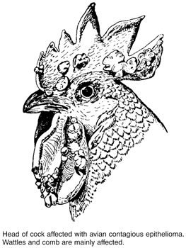
The virus infects the skin through abrasions, wounds and punctures and may be transmitted by insect vectors (especially mosquitoes). Various secondary organisms are usually responsible for deaths.
The period of incubation is usually between three and 12 days, and bad housing conditions, severe weather and poor feeding serve to lower vitality and render an outbreak much more serious.
Signs There are three types of lesion: (i) nodular eruptive lesions on comb and wattles; (ii) a cheesy, yellowish membrane in the mouth and throat; (iii) oculo-nasal form (possibly due to a different virus).
The mouth lesions consist of patches of a greyish, fairly firm, cheesy-looking material, which is of considerable thickness, and not easy to detach. This is the ‘false membrane’. In many cases the entrance to the trachea is partially blocked with these deposits, and the breathing is consequently obstructed. The smell from the mouth is always foul.
Treatment is economically unsound. The best measures consist of the slaughter of all affected birds and the inoculation of the healthy ones with ‘pigeon-pox vaccine’.
Prevention Newly purchased birds should be isolated for three weeks before being added to the flock, and after returning from shows, laying trials, etc., the same procedure should be adopted.
Vaccination (in regions where vaccine is available) can be given at six weeks of age; or, more usually, between three and five months of age. (See AVIPOX.)
This is an acute infectious disease of fowls (also of ducks, geese, turkeys, game and wild birds) caused by Salmonella gallinarum. The disease (also called KLEIN’S DISEASE) has a worldwide distribution, but has been virtually eradicated from the UK.
Most outbreaks occur in pullets near point of lay, but birds of all ages are susceptible – even chicks. The disease is usually introduced into a flock by the purchase of ‘carrier’ fowls, and thereafter spreads by contamination of food and water with the droppings of such birds. The incubation period is from four to six days.
Signs are not always characteristic. There is generally marked drowsiness, loss of appetite, and great weakness. The fowls prefer to sit about in dark corners. The comb and wattles are sometimes pale and anaemic; they may in other cases be markedly congested. Diarrhoea is usually present. Death, following progressive weakness, occurs in from four to 14 days after the onset of the signs. The percentage mortality varies from about 20 to 30 per cent, and many or most of the recovered birds become ‘carriers’, which serve to spread the disease to other birds.
Diagnosis If fowl typhoid is suspected, samples of blood from the surviving and apparently healthy birds should be submitted to the agglutination test, and all reactors should be isolated and destroyed – the carcases being burned or buried in quicklime. The remaining birds should be treated with antibiotics, moved to fresh premises, and retreated.
After removal of the reacting birds, the houses, utensils, etc. should be disinfected.
There are about 37 species of fox with the red fox (Vulpes vulpes) being most common. They are OMNIVOROUS and not a pack animal. In Europe, North America and other parts of the world, wild foxes often become victims of RABIES, and spread this disease to farm livestock which they may attack. A history of aggressiveness and atypical behaviour does not, however, point conclusively to rabies; DISTEMPER or partial domestication by well-meaning, but misguided, people, may be the reason. (See also FOX ENCEPHALITIS; ‘CHASTEK PARALYSIS’.)
The fox acts as host of the roundworm Toxocara canis and of the Toxascaris leonina, and if silver fox cubs are reared by a cat, they may become infected with Toxocara mystax of the cat. The fox harbours the dog tapeworms Taenia serialis and T. multiceps, and Echinococcus granulosus. Leptospirosis occurs in foxes in the UK and may be spread to farm livestock (five strains have been isolated). Flukes may infest foxes, and sarcoptic mange is quite widespread in urban foxes.
Fox encephalitis is of commercial importance on the fox ranches of North America, where these animals are bred for their fur. The disease is considered identical to RUBARTH’S DISEASE of dogs.
Signs Young foxes in good condition are most frequently affected. A violent convulsion is followed by a lethargic or ‘sleep-walking’ state. This may be followed by excitability and more convulsions – during which the slamming of a door or any loud noise may prove fatal. The illness runs a very rapid course, from one hour to three days, 24 hours being the average duration.
Control By means of serum and preventive inoculation.
This disease, or one caused by a similar virus, may have accounted for the deaths of (wild) foxes in Britain, but the deaths may have occurred as the result of eating birds poisoned by dieldrin. Signs are similar.
(See DIGITALIS - DIGITALIS POISONING.)
Small, lively dogs classed as smooth-haired or wire-haired, according to the coat; usually with a short tail. In both breeds deafness and lens luxation may be inherited but by unknown mechanisms. Pulmonary STENOSIS and aortic stenosis are probably polygenic traits but ACHALASIA is possibly an autosomal dominant trait. Atopic dermatitis occurs only in the wire-haired terrier and is inherited by an unknown mechanism; while cerebellar ataxia in the smooth is an autosomal recessive trait. (See also SMOOTH-HAIRED FOX TERRIERS; WIRE-HAIRED FOX TERRIERS).
Simple fractures are the commonest variety, and consist of those in which the bone is broken clean across, with or without tearing and laceration of the soft parts surrounding it, but without any wound leading from the fracture through to the skin. They are spoken of as being transverse, longitudinal, or oblique, according to the direction of the break.
Compound fractures are those in which the skin is injured, so that a direct or indirect communication between the fracture and the outside air exists. The broken end of the bone very often penetrates through the skin and is found exposed. Bleeding is apt to be severe; infection of the ends of the bones with pathogenic organisms may occur.
Closed fractures are covered by skin and/or other tissues.
Open fractures are seen when the bone protrudes via a wound. These fractures can be contaminated or infected, which may affect healing.
Incomplete or greenstick fractures are those in which the bone is broken only partly across, or in which the tough periosteum (the tissue covering the bone, which is thicker in juvenile animals than adults) is not torn. They occur in the shin-bones (tibiae) of horses which have been kicked, and in the bones of young animals. The bone cracks like a twig half-way across, and then splits for some distance along its length, just as does a branch which has been cut halfway through and then bent.
Fissured fractures are mere cracks in the bone which are found in the skull and face bones after blows or falls. They are usually not serious unless haemorrhage accompanies them and the blood clot presses upon a nerve or on the brain itself.
Deferred fractures occur when the bone has actually been fractured, but the fractions do not separate until or unless some extra severe strain is put upon the part.
Distracted fractures are those in which muscular contraction causes the detached fragment to be drawn away from the main body of the bone.
Depressed fractures occur in the skull bones as a rule, and consist of fractures in which a fragment of bone is forced in below the level of the surrounding surface. They may give rise to very serious clinical signs when the depressed portion presses upon the brain substance.
Complicated fractures are those in which there is some other serious injury produced in addition to the fracture, e.g. dislocation of the dog’s hip along with fracture of the shaft of the femur; tearing of a large nerve, etc.
Comminuted fractures are those in which there is much splintering, the term ‘sequestra’ being applied to those splinters of bone which are separated and eventually die.
Impacted fractures are those in which, after the break has occurred, one fragment is jammed inside another, usually at an angle.
Ununited fractures are those in which, after the usual time has elapsed for the fracture to heal, it is found that union has not taken place. The failure to unite may be simply due to ‘delayed union’, on account of debility or illness or due to the fact that the limb or other member is not kept at rest sufficiently for the process of healing to occur. In other cases of ununited fracture, a piece of muscle or other tissue becomes displaced between the broken ends of the bones and effectively prevents their union.

Classification of fractures. A. Transverse fracture with excellent stability after reduction. B. Oblique fracture with no stability after reduction. C. Slightly oblique fracture which, by virtue of the irregularity of the fracture lines, provides a useable degree of stability after reduction. D. A typical distracted fracture. (With acknowledgements to the British Veterinary Association.)
Causes of fractures Disease, such as osteomalacia, in which there is a reduction in the density of bone and of its tensile strength, is one cause. However, the common cause is external violence. (See also ELECTRIC SHOCK, ‘STRAY VOLTAGE’ AND ELECTROCUTION.)
Horses Fractures result from kicks, falls or blows; errors in judgement during jumping; accidentally placing a foot into rabbit-holes when galloping; and accidents when the animal collides with some stationary object, or is struck by a vehicle.
Fractures incurred by 53 race horses at a New York track were found to be due to three lesions: OSTEOCHONDROSIS, chondro-osteo necrosis, and degeneration of tendons and ligaments. (See also SHOEING OF CATTLE.)
Cattle Fractures result from injuries during fighting, slipping, and falling when struggling; from running, BULLING and mounting or during service; from jumping fences, hedges and ditches; from crowding accidents at markets, etc.; and from crushes in cattle-trucks.
Fracture of the third phalanx in a medial front claw is commonly associated with fluorine poisoning, and causes cattle to stand with their legs crossed. (See also SHOEING OF CATTLE.)
Pigs and sheep The causes are usually similar, but legs are broken more easily. Careless use of the shepherd’s crook is responsible for many. Falling over precipices and getting a limb stuck in a gate, fence, or hurdle may also result in a broken bone.
Dogs and cats In dogs and cats, road accidents account for the majority of fractures, especially of the femur, tibia, pelvis, radius and ulna, spine and jaw.
Signs The chief signs of a fracture are uselessness of the part, CREPITUS of the fragments, and sometimes unnatural mobility and deformity. If a limb is affected there is usually an unnatural mobility, inability to sustain weight, distortion or deformity, shortening of the length of the limb (due to overlapping of the fragments), a thickness or swelling at the seat of the fracture, and a variable amount of pain. (See also ‘Fractures of special parts’, below.)
Healing of fractures When the bone breaks, many blood vessels are torn, and accordingly a large clot of blood forms around the ends, between them, and for some distance up the inside of the bone. Later, great numbers of white blood cells find their way into this clot, which becomes ‘organised’ – blood vessels and, later, fibrous tissue being formed in it (soft callus). Next, lime salts are gradually deposited in this fibrous tissue, which thus develops into bone (hard callus). In this process a thick ring of new bone forms round the broken ends, filling up all crevices; and when union is complete, this thickening is again gradually absorbed, restoring the bone as it was before the injury.
Treatment There are many ways of stabilising a fracture after reduction and apposition are brought about by manipulation of the fractured bones under anaesthesia. External methods include bandages and splints and specially moulded casts of plaster of Paris, and various proprietary mixtures impregnated into bandages. (See SPLINTING MATERIALS.) Whenever splints, plaster, or other bandages are being applied to fractured limbs it is essential to ensure that the surface of the skin is well padded with cotton-wool, and that the pressure is evenly distributed. Failure in this respect may result in parts of the skin developing pressure sores or becoming gangrenous through obstruction to the blood-flow. Thus other methods are now often replaced by sophisticated surgical techniques. (See SPLINTING MATERIALS.)
Special types of extension splints, having transverse pins which transfix the bone, have been used with success in appropriate cases. Medullary pins, driven down the marrow cavity of long bones; wiring; and plating have all been used with success. (See BONE-PINNING.)
Bone Grafts These are used to a limited extent in veterinary surgery to repair fractures of the femur, humerus, tibia, radius and ulna; or to replace comminuted fragments, to lengthen bones or to correct delayed or faulty union.
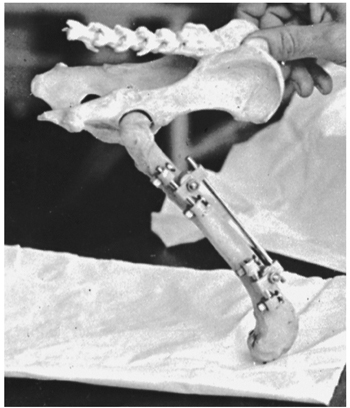
Metal splints which have transverse pins to penetrate and fix the bone are used in treating fractures in small animals, and have succeeded in cases where other methods would be ineffective.
The allografts are harvested aseptically from the patient or from an outside source such as a conspecific animal or from synthetic sources. Ordinary orthopedic techniques are used to secure the implant.
1. The cranium Cases of traumatic fracture of the skull result in concussion; if not severe they may recover with conservative treatment and nursing. Depressed fractures involving the cranial cavity or the cranial nerves are difficult to treat and usually carry a poor prognosis. (See also CONCUSSION.) Surgical treatment may be successful in cases in which fractures involve bones of the jaws and face.
2. The face bones Fractures may be simple or serious according to bones involved. Nasal bones, often fractured from accidents, may be accompanied by swelling, pain, haemorrhage, difficulty in breathing, and much watering of the eyes. Jaw-bones broken from falls, kicks, etc. usually interfere with feeding. Lower-jaw fractures usually result in an open hanging mouth, escape of saliva, and altered expression; frequently, loose teeth, torn lips, and haemorrhage are seen. Bones of orbit fractured by falls on to side of head, collisions, etc. interference with vision and with movements of the lower jaw, in most cases serious. Treatment usually necessitates operation – removal of broken pieces, elevation of depressed portions, removal of loose teeth, wiring or plating broken parts together. Feeding must be carefully undertaken when jaws are injured – sloppy food, mashes, etc., for horses; hand-feeding for dogs. Sometimes in small animals a feeding tube may be needed.
3. The vertebrae Commonest in horse and dog through accidents (e.g. in horses being cast in stall or for surgery, road-traffic accidents in dogs, falls from heights). If a vertebra is fractured, paralysis results. There is often a fatal termination, or a need for euthanasia. Tail-bones are often broken in dogs, cats and cattle through getting caught in doors, gates, fences, etc.
4. The ribs These are usually due to trauma. Most rib fractures heal spontaneously without any surgery or treatment. Flail chest describes multiple adjacent rib fractures, which can then severely affect breathing. First rib fracture sometimes happens through muscular action in a side-slip and violent recovery in horses, when it often results in RADIAL PARALYSIS. Otherwise, broken ribs show little or nothing characteristic except local pain and deformity, unless many are involved, when breathing may be short and/or difficult. (See ‘FLAIL CHEST’; PNEUMOTHORAX.)
5. The pelvis In dogs and cats, loss of conformity of the pelvis may lead to DYSTOKIA or intractable chronic CONSTIPATION and so surgery is recommended when a pelvic fracture is comminuted.
6. The scapula Fractures are uncommon. Mostly, they occur through the neck of the bone, or on the projecting spine. The musculature covering the bone may impede diagnosis but assists recovery, and it may act as natural bandage.
7. The humerus Lameness, intense in all animals, follows fracture; the limb is usually quite useless. Horses and cattle do not make good recoveries except when young, but healing in small animals is more satisfactory. The majority of fractures affect the middle and distal third and almost all require surgery for repair. Fractures of the distal humerus are common in young dogs and may also affect the elbow. (See BONE-PINNING.)
8. The radius and ulna One or both bones may be broken; fracture of the ulna is less serious unless the elbow-joint is involved. In dogs and cats, if one is broken the other acts as a natural splint. Lameness is always marked, and there is pain on pressure. Local swelling and deformity are usually noticed. Young horses should be placed in slings. Surgical fixation has been carried out successfully in the dog and the horse.
9. Coronoid process Young dogs with mild lameness due to fragmented medial coronoid processes probably do not benefit from surgery, but dogs with chronic, moderate or severe lameness do.
10. Bones of knee These are seldom fractured, but if they are it is usually impossible to bring about recovery without stiffening of the joint (ankylosis).
11. The metacarpals In the horse, good recoveries are made in cases of clean transverse fractures without complications or splinters. Prognosis is best in fractures occurring in the middle of the cannon. The limb is bandaged with a plaster or proprietary resin-impregnated bandage and the horse slung; the plaster is left in position for at least 6 weeks. In the dog, the treatment depends on which bones are involved. Metacarpals three and four are the main weight-bearing bones and if both of these are affected surgery is normally indicated. Fractures where only one or two bones are affected usually respond well after setting and a supporting dressing of the affected limb, as the other unaffected bones act as natural splints.
12. The pastern bone Fractures may be transverse, oblique, or longitudinal (‘split pastern’), often comminuted. Severe lameness always results. In the horse, simple transverse fractures can be treated satisfactorily if the temperament of the horse will allow rest and slinging; oblique, longitudinal, and all comminuted cases are unsatisfactory and if recovery occurs, usually some deformity or blemish is left.
13. The second phalanx, coffin and navicular bones Fractures in these bones are rare; they may the caused by direct violence, and sometimes follow an operation of neurectomy (un-nerving); may be seen in cattle as a result of weakening of bone through FLUOROSIS. Fracture of the coffin-bone, if simple and joint surfaces are not involved, makes good recovery as a rule, since the hoof acts as splint and bandage. Fracture of the second phalanx (short pastern bone) is usually difficult to resolve.
Most fractures of the navicular bone are sagittal and minimally displaced, but the prognosis is usually poor because the fibrous callus causes permanent lameness. Such fractures have been repaired by inserting a 50 mm screw which exerts compression between the two fragments. The pilot hole is drilled and the screw is inserted precisely along the transverse axis of the navicular bone by means of a mechanical guide, the process being monitored by image-intensifying fluoroscopy. The fractures are said to heal without superfluous callus formation.
14. The femur Very commonly fractured in dogs after road traffic accidents. Shaft, neck, or one of the trochanters may be involved. Frequently in dogs, dislocation of the hip-joint accompanies fracture, but dislocation may occur without damage to the bone. Extreme lameness, shortening of the limb, local swelling, and great pain on movement are usually seen. There may or may not be crepitus. In horses, fracture of pelvis very often accompanies fractured femur and makes diagnosis difficult. A fractured femur usually necessitates euthanasia in large animals, but in small animals the chances of recovery are usually improved by surgery. (See BONE-PINNING.)

Two Rush-type intramedullary pins used to repair a supracondyloid fracture of the femur.
15. The patella Fracture is a very serious condition, resulting in a lowering of the affected stifle and inability to advance the limb. There is great pain. Treatment is union of the fragments by wire sutures; this may be difficult to perform satisfactorily, and complete recovery may not occur.
16. The tibia Many fractures of tibia become compound from sharp points of broken bones penetrating through the skin. (See BONE-PINNING which has been used successfully in the dog, cat and horse.) The poor blood supply of the tibia means that it takes longer to heal than other long bones.
17. Bones of hocks In fracture of the os calcis (point of hock) – the proximal epiphysis becomes displaced from the rest of the bone by an undue pull of the Achilles’ tendon (gastocnemius or hamstring). Fractures of other bones of hock are less common (with the exception of the SCAPHOID in the racing greyhound).
An aminoglycoside antibiotic with activity against some Gram-positive and some Gram-negative bacteria. It is mainly used in intramammary cerates, topical applications and to treat intestinal infections. It is little absorbed. An injection is available for treatment of acute bacterial MASTITIS in cows caused by E. Coli, KLEBSIELLA, STAPHYLOCOCCUS AUREUS or Trueperella pyogenes. It is not widely used in human medicine.
(See TULARAEMIA.)
(See FARM ANIMAL WELFARE COMMITTEE; ‘FIVE FREEDOMS’ FOR FARM ANIMALS.)
Usually defined as a sterile heifer born twin to a normal bull calf; the most widely accepted explanation being that sex hormones from the earlier developing male twin pass across to the female twin, with the result that sexual differentiation of both male and female proceeds under control of male hormones.
However, as long ago as 1917 it had been suggested that hermaphrodites might occur in female single births, as a consequence of early fetal death and resorption of the male twin in the uterus. During the 1970s chromosome analysis had revealed the presence of both male and female cells in single-born bull calves. Dr W. V. S. Wijeratne and colleagues were the first to demonstrate this condition – technically known as secondary chimerism – in single-born freemartins. (Primary chimerism can occur where two sperms fertilise the same ovum.)
Not every female fetus having a male twin sharing the uterus will become a freemartin, because in some instances death of the male twin fetus occurs before about day 39 of pregnancy – when a common blood supply may become established. Moreover, in between 5 and 10 per cent of heterosexual twin pregnancies a common blood supply is not established.
About 90 per cent of heifer calves born twin to a bull calf are freemartins. Many freemartins can be detected on clinical examination, since the vagina is often only one-third of the normal length and, in addition, there is often an enlarged clitoris and a vulval tuft of hair.
The condition is associated with anastomosis of the placental blood vessels (see diagram).
Pig freemartins may also occur. (See also H-Y ANTIGEN.)
Highly reactive molecules, formed in the presence of oxygen and capable of damaging living tissue. They have been implicated in human heart disease and arthritis. They may also be a cause of sudden death of pigs – those being transported for long distances or subject to other forms of stress. However, it has been suggested that protection may be given by feeding vitamin E, which ‘scavenges’ radicals.
A system where birds have daily access to the outside, but will also have access to housing. The maximum number of birds outside per square metre is nine. There must be two metres (6 ft 6 in) of opening per 1000 birds. In 1975 three per cent of whole eggs sold were free range; in 2011 50 per cent of eggs consumed were free range. IMPACTION of the GIZZARD is common due to the excessive ingestion of coarse fibrous material. They are more likely to be exposed to wild birds and animals and develop AVIAN TUBERCULOSIS and SALMONELLOSIS. Poultry eating EARTHWORMS of Eisinea foetida produce garlic flavoured eggs. Tree-covered pens reduce FEATHER PICKING and second-quality eggs.
(See BRANDING.)
Fremitus is a sensation which is communicated to the hand of an observer when it is laid across the chest in certain diseases of the lungs and heart. Friction fremitus is a grating feeling communicated to the hand by the pleura or pericardium (see HEART DISEASES) when it is roughened as in pleurisy or pericarditis. On rectal examination of cows over five months pregnant, fremitus can be felt in the middle uterine artery.
A small dog resembling the English bulldog of the 19th century. BRACHYCEPHALIC airway obstruction syndrome is inherited by an unknown mechanism, as is SPONDYLOSIS DEFORMANS, intervertebral disc disease and HEMIVERTEBRAE. Haemophilia, both A (Factor VIII deficiency) and B (Factor IX deficiency). may be found as sex-linked recessive traits. Ununited anconeal process (elbow) is a dominant trait and patellar luxation may be a recessive trait. Inherited CATARACT occurs (DNA test available). NICTITATING MEMBRANE GLAND PROLAPSE is seen.
(See DOGUE DE BORDEAUX.)
(See BUDGERIGAR FLEDGLING DISEASE.)
A black and white breed of milking cow which was predominant in the UK from the 1960s to late 1980s. It produces good volume of milk and is well fleshed so that the male calves can be used for beef production. It became superseded by crossing with the HOLSTEIN, producing the HOLSTEIN-FRIESIAN CATTLE. Most UK milking herds became pure Holstein; however, the Friesian is more robust than the Holstein and is able to survive longer with more lactations. It also appears to have fewer lameness and fertility problems. Many organic herds favour its use.
These horses originate from the Netherlands, where they were bred in Medieval times as war horses. They have to be black without any white present and with a long mane and tail. The limbs are FEATHERED and they have a high-stepping gait.
Also known as the British Friesland. A white sheep, naturally polled, with a thin white face; fine boned and a long bald tail. They are used for milk. The rams weigh about 75 kg (165 lb) but can be 120 kg (265 lb) and the ewes 50 kg (110 lb), with about 5 kg (11 lb) fleece.
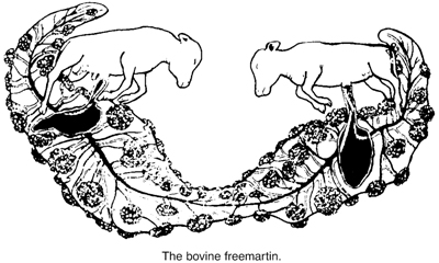
(See FOOT OF THE HORSE.)
They are amphibians from the Order Anura, usually with short bodies, webbed feet, absence of tail, protruding eyes and a forked tongue; some produce mild toxins. Their hind legs are developed for jumping. They are mainly semiaquatic and can live in humid climates outside water because they have a permeable skin. They occur in most parts of the world but more species are found in the tropics, and some of these in particular are kept as pets. In recent years they have suffered a fatal fungal dermatitis due to BATRACHOCHYTRIUM DENDROBATIDIS. A dead vaccine has been developed producing IgG and IgY antibodies.
A roughly quadrilateral plate-like bone which forms part of the roof of the cranium and passes forward between the eyes to meet the nasal bones. In the horned breeds of cattle and sheep it is extended laterally to form the horn cores. (See BONES; also SINUSES OF THE SKULL.)
(See SINUSES OF THE SKULL.)
Frost-bite may affect any animal exposed for long periods to severe cold, resulting in freezing of the skin, particularly of the extremities, with precipitation of blood proteins, occluding blood vessels. It is less common in animals than humans because of the protection given by the winter hair coat. Hairless species and parts of the body are, however, susceptible. For example, in poultry on the wattles, comb and face; and around the CORONARY BAND of horses.
TRITRICHOMONIASIS in hawks and falcons. (See FALCONS, DISEASES OF.)
(See EMBRYO TRANSFER.)
These are used in nutrition of animals and man. They are POLYMERS of FRUCTOSE molecules. Those that are short chains are called FRUCTO-OLIGOSACCHARIDES and long chain ones are called INULIN.
Used to stabilise the intestinal microflora in many species including horses, calves, chickens and fish (rainbow trout, tilapia, shrimp). They can be used after enteritis.
A plasma protein which helps to indicate the average blood glucose measurement in the previous one to two weeks. It is a combination of fructose with ammonia or an amine.
It is a sugar (monosaccharide) and ISOMER of GLUCOSE also known as ‘fruit sugar’, ‘grape sugar’ or laevulose. It is found in honey and fruits and is absorbed.
Fruit-eating.
A potent diuretic suitable for the treatment in dogs and cats of some cases of OEDEMA associated with cardiac insufficiency, renal dysfunction or trauma. Frusemide acts about one hour after oral administration and is effective for about four hours.
Overheated fat gives off acrolein, which can be highly poisonous and was, indeed, used in chemical warfare in 1914–18. A dog died after being shut up in a kitchen for half an hour with a smoking chip pan. Ante-mortem signs were distressed breathing and cyanosis.
Cage birds are susceptible to fumes from an overheated non-stick frying pan coated with plastic polytetrafluoroethylene (TEFLON). It is quite a common accident in birds kept in kitchens. Death occurs rapidly following exposure. (See ‘KITCHEN DEATHS’.)
(See FOOD STANDARDS AGENCY.)
(See FOLLICLE STIMULATING HORMONE.)
A lysosomal storage disease caused by the absence of an enzyme – alpha-L-fucosidase.
It is an inherited disease in the English springer spaniel, affecting mainly those between 18 months and four years old. The signs of this ultimately fatal disease include ataxia, change in temperament, depression, apparent deafness and impaired sight. Swallowing may be difficult. Loss of weight occurs. A DNA test exists to identity affected dogs and carriers of the disorder. (See LYSOSOMAL STORAGE DISEASE.)
An extract from seaweed found in some herbal remedies for dogs and cats, which claims to assist in the treatment of obesity, constipation and iodine deficiency.
The destruction of a superficial lesion with electric or radiation energy. (See RADIOSURGERY.)
(See CARBON MONOXIDE; ‘FRYING PAN’ DEATHS; SLURRY; ANAESTHESIA, GENERAL; AEROSOLS.)
(See DISINFECTION.)
Fundus is the base or innermost part of a hollow organ distant from its opening.
Broadly speaking, these include both the invasion of tissues by fungi, and the effects on organs of fungal poisons (see MYCOTOXICOSIS).
Ringworm offers a good example of the invasion of tissues by pathogenic fungi; one should, perhaps, say potentially pathogenic fungi, for many are present in the alimentary canal of healthy animals, and cause lesions only when circumstances favour invasion or multiplication. (See MASTITIS IN COWS, MYCOSIS, for an example of the latter; also ASPERGILLOSIS; BLASTOMYCOSIS OF DOGS; HISTOPLASMOSIS; MUCORMYCOSIS; MONILIASIS; STREPTOTHRICOSIS; CRYPTOCOCCOSIS; Fusarium; MORTIERELLA; COCCIDIOMYCOSIS; RHINOSPORIDIOSIS; SPOROTRICHOSIS.)
(See MYCOTOXICOSIS.)
(See FUR RING; FUR SLIP.)
(See HAIR-BALL.)
A nitrofuran compound used against antibacterial and antiprotozoal infections. Its use in food animals is no longer permitted in the EU.
This is seen in chinchillas and is caused by boredom, nutritional deficiencies or stress.
Commonly called the ‘wishbone’, the furcula comprises a fusion of the clavicles or ‘collarbones’ of birds. Together with the coracoids, scapulae and sternum it forms the pectoral girdle and acts to maintain the proper spacing of the shoulder joints.
Furfuraceous is a term applied to skin diseases which produce a bran-like scaliness.
(See MITES, PARASITIC.)
A diuretic (removing fluid) acting on the ascending loop of Henle in the kidney. It is used to treat OEDEMA due to CONGESTIVE HEART FAILURE or RENAL INSUFFICIENCY and HYPERTENSION. It can result in excessive potassium loss, increased retention of uric acid and glucose.
A condition in which fur is found encircling the penis of chinchillas. The usual clinical presentation is PARAPHIMOSIS, the fur ring being found on closer inspection of the penis. Clinical signs include excessive grooming, discomfort, POLLAKURIA, or dysuria. Often, the animal injures its own penis in its attempts to remove the fur.
Seen in chinchillas as hair loss and localised alopecia following rough handling. Regrowth occurs in four to six months.
This is the dermatological term for a boil (or furuncle). It follows the rupture of an infected follicle into the dermis and subcutaneous tissues, where it may spread extensively. The cause of the original infection may be bacterial, fungal or parasitic. Often there is associated disease. The condition occurs in all species. (See INTERDIGITAL CYSTS.) Perianal furunculosis also occurs in dogs.
Furunculosis in fish is caused by Aeromonas salmonicida. Raised furuncles can be seen all over the body and they may be complicated by secondary fungal infection. A sudden increase in water temperature can trigger the appearance of the disease.
Furunculosis in salmon is caused by Aeromonas salmonicida but may be triggered by a sudden rise in water temperature. Young fish stop feeding and may die soon afterwards. Older fish are more resistant; they develop large, boil-like swellings on the shoulder and back. If these burst, a reddish fluid rich in bacteria is released. The bacterium persists in some fish between outbreaks and is present in wild fish. Treatment is by medicating the feed with sulfonamides or tetracyclines but the fish may be reluctant to take medicated food. It is a NOTIFIABLE DISEASE under the Diseases of Fish Act 1937.
(Ulex europaeus.) A very common and plentiful shrub in waste lands in Britain, it was formerly often cut and used as fodder after chaffing or bruising. The plant contains a very small proportion of a poisonous alkaloid which is called ulexine, and is practically identical with cystine from broom. It is a nerve and muscle poison, but it is seldom present in dangerous amounts.
(FELINE UROLOGICAL SYNDROME; see FELINE LOWER URINARY TRACT DISEASE)
A mould that can produce toxins including T2 TOXIN. Mouldy shelled maize containing F. moniliforme has caused diarrhoea and ataxia in cattle; and in BROILERS the same species, contaminating maize and wheat, has with F. culmorum, F. Tricinctum and F. nivale been implicated in poor growth rate, poor feathering, and abnormal behaviour. Fusarium species may also cause keratoconjunctivitis. In pigs, it causes refusal of food. (See under EYE, DISEASES; also MYCOTOXICOSIS; ZEARALENONE.)
(See FUROSEMIDE.)
An antibiotic used in topical applications for otitis externa (See EAR, DISEASES OF), PYODERMA, and conjunctivitis.
An anaerobic bacterium, formerly known as Fusiformis necrophorus, which causes FOUL-IN-THE-FOOT of cattle, CALF DIPHTHERIA, ABSCESSES in the liver and other organs. Also involved in foot-rot in sheep. (See table under BACTERIA, and FOOT-ROT OF SHEEP.)
An area of the large colon of LAGOMORPHS, more muscular and more highly innervated than the rest of the organ. During digestion, it is responsible for control of the passage of food to and from the CAECUM.
(See FEDERATION OF VETERINARIANS OF EUROPE.)