
Histology is the study of the structure and function of cells and tissues. Cells can be seen as the basic unit in the body—like one brick of Lego in a giant Lego castle.
These cells are grouped together to make tissues, of which there are four basic types. You could think of these as being like the foundations, woodwork, bricks, wallpaper, and electric circuitry used in building a house. The tissue types are connective tissue, which supports, protects, and connects; epithelial tissue, which lines and covers; muscular tissue, which provides movement; and nervous tissue, which is excitable and conductive, allowing for control and rapid communication of information and commands throughout the organism.
The cell is the basic unit of activity in the body. You can think of each cell as being like a factory—each one takes in raw materials, processes them, and creates products and waste. Cells are amazing, and should be thought of as being individuals in their own right. An emerging model in biology is that each cell has a consciousness pervading and orchestrating it—the consciousness of the whole organism.

FIGURE 3.1. Basic cell
Our body is made of about 50 trillion cells. The largest human cells are about the diameter of a human hair, but most are smaller, about one-tenth of the diameter of a human hair. Look at a single strand of your hair. It is not thick, being about 100 microns in diameter. (A micron is a millionth of a meter, so 100 microns is a tenth of a millimeter, which is about 0.004 inches.) Look down at your little toe—it contains two to three billion cells, depending on how big you are.
Bacteria are about the simplest cells that exist today. Interestingly, it seems that looking closely at cells may tell us of our evolution. The organelles, or functional parts of each cell, look very like certain bacteria. The theory is that millions of years ago some bacteria, hanging out in the primordial soup, got together with good effect—in other words, found that survival went well in cooperation with each other. Eventually, the first single-celled organism—an amoeba—was formed. Over time, amoebae grouped together successfully to make multicellular organisms, of which we are a wonderfully complicated example. Of course, this can only ever be a theory; it is not possible to really prove by scientific methods what happened all that time ago.
A bacteria is a single, self-contained, living cell. An Escherichia coli bacteria (or E. coli) is typical. It is about one-hundredth the size of a human cell. Bacteria are also a lot simpler than human cells. They consist of an outer wrapper called the cell membrane, and a watery fluid called the cytoplasm on the inside. Cytoplasm is about seventy percent water. The other thirty percent is filled with proteins called enzymes that the cell has manufactured, along with smaller molecules like amino acids, glucose molecules, and ATP. At the center of the cell is a ball of DNA (similar to a wadded-up ball of string). If you were to stretch out this DNA into a single long strand, it would be incredibly long compared to the bacteria—about a thousand times longer! Very similar to our cells, in fact. On the surface of our skin alone there are ten times more bacteria than our bodies contain cells—our bodies contain about 2 kg (about 4 and a half pounds) of bacteria in normal circumstances.
Every one of the billions of cells in our body has its own kind of independent life; it has its skin or cell membrane, its own need for food, it excretes, makes energy, communicates with other cells, and (in many cases) can reproduce itself. There is an old maxim used in many traditional healing systems: the microcosm in the macrocosm, and the macrocosm in the microcosm. This philosophy, first recorded in ancient Greece, means that patterns found in the largest scale—the cosmos or the universe—are repeated in the smallest—the single organism, the atom, even the subatomic level. We can see reflections of what is in the very small and the very large.
For example, imbalances in a society are the imbalances of the society’s individuals writ large, and imbalances in an individual are reflections of the imbalances in society. A single leaf reveals the condition of the whole tree. Holistically thinking, every part of the whole affects every other part. We can benefit from considering the health of our cells. If the cells are healthy and getting their needs met, the whole organism will be well. This concept is gaining more and more ground in the emerging “new biology” of such great thinkers as Bruce Lipton, who says:
You may consider yourself an individual, but as a cell biologist I can tell you that you are in truth a cooperative community of approximately 50 trillion single-celled citizens.… As a nation reflects the traits of its citizens, our human-ness must reflect the basic nature of our cellular communities.1
Each cell is enclosed in an amazing membrane made of phospholipids, cleverly designed to be semipermeable, to allow some things in and keep some out.
Phospholipids are molecules that have an electrically charged phosphate-nitrogen head, and a neutral fatty acid tail. Two layers of these molecules are arranged so that the electrically oriented phosphate ends face outward in contact with the extracellular fluid, and inward in contact with the intracellular fluid. The lipid fatty acid tails are in the middle of the two layers.
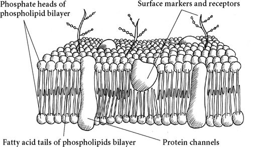
FIGURE 3.2. Cell membrane
Traditionally, the nucleus is seen as the brain of the cell, orchestrating things with its DNA, but in fact, a cell can survive for months without its nucleus, but dies instantly without its membrane. Bruce Lipton eloquently describes this in The Biology of Belief, a must-read book for anyone interested in cellular biology. He says that it looks rather like the membrane is the brain of the cell, controlling what goes on, and actually switching genes on and off as needed in response to the environment. The nucleus, with its DNA, while seen by conventional biology as the control center of the cell, is mainly needed for reproduction and so seems to be more like the gonads!
A significant portion of the lipid part of the cell membrane is made of essential fatty acids. You will remember that these are called essential because they cannot be made by the body but must be eaten in the diet. For our cell membranes to stay healthy and function at optimum level, we especially need the Omega-3 fatty acids.*
Because of this bipolar attribute of the membrane, it is selectively permeable—some things freely cross it, while others do not. Like dissolves like: Because of the layers of fats in the membrane, which are nonpolar, charged or polar particles such as sodium and potassium ions cannot freely cross. An exception to this is water—a pretty exceptional substance. Although water is polar it can still diffuse through the bilayer.
Water enters and leaves a cell according to the osmotic pressure of the fluids inside and outside. If there is more salt in a cell, water will be drawn in and the cell will expand; if there are more salts in the extracellular fluid, water will leave and the cell will shrink. The business of what can get in and out of a cell is very important, as you might think. The membrane also contains structural proteins, as well as special proteins for transporting substances. Protein receptors, which are also in the cell, are being created and reabsorbed all the time, so the membrane is not static in structure but always being adapted and mended.
There are various ways for substances to cross over the cell membrane and enter or leave the cell. Most of these involve the movement of water as well.
Simple diffusion involves the random scattering of very small particles from a high to a low concentration, down the concentration gradient—oxygen and carbon dioxide do this easily, dissolving through the phospholipid layer as described above. Imagine someone farts in a room full of people—at first there is a high concentration of the smelly gas around that person, but gradually it diffuses through the air in the room until eventually it is spread so far and so thinly that no one can detect it any more.
Facilitated diffusion is a process that honors the concentration gradient, but allows bigger substances that cannot pass through the lipid membrane to cross. Carriers or channels in the membrane are used, creating a kind of gate or turnstile. Substances that cross the membrane in this way include glucose, amino acids, and some ions. Each substance has its own selective channel or carrier to allow it to enter the cell. They may either be always open, or may open and close according to chemical or electrical signals.
Osmosis is the diffusion of water through a selectively permeable membrane, moving from where there are low levels of solutes to where there are more of those solutes—in other words, there is this tendency for equalization, seen in diffusion, when a substance will move from a high concentration to a low concentration. If the substance cannot cross the membrane, it will pull water to cross in its direction to dilute it. This pull is known as osmotic pressure. As well as moving through the water-filled channel or pore that runs through the middle of some transport proteins, water can move by thrusting through the lipid layer of the membrane—surprising, since water and fat don’t usually mix. The amount of water in and around the cell is controlled by osmotic pressure versus hydrostatic pressure.
Osmotic pressure is the pressure exerted by the presence of a high concentration of particles, such as proteins. The tendency is for equalization, so the strong solution will attract water into it if water can get in.
Hydrostatic pressure is like the water pressure in a hose pipe—if you squeeze the end, you make the tube narrower and so the hydrostatic pressure increases, which pushes the water out more strongly.
Active transport is like facilitated diffusion in that a carrier is used, but to work the carrier must use energy (from ATP). Then it can move substances against their concentration gradients. This is like a turnstile that takes money (or a token of ATP) to allow entrance or exit. Sodium ions (Na+) and potassium ions (K+) pass, like water, through channels in membrane proteins. Sodium ions are present at a higher concentration outside the cell, so they have a net simple diffusion into the cell through special sodium channels. Potassium ions are present at a higher concentration inside the cell, so have a net simple diffusion out of the cell through special potassium channels. A diffusion equilibrium of Na+ and K+ (where there is a balanced number of each inside and outside the cell) is avoided by an active transport system: the sodium-potassium exchange pump. For every three sodium ions pumped out of the cell, two potassium ions are pumped in. So it is that movement of sodium and potassium is closely linked in the body.
Diuretics, which cause the body to lose fluid via the kidneys, cause loss of potassium and sodium ions too. This was discovered when the first drug diuretics killed people by upsetting their potassium balance. Interestingly, the strong herbal diuretic dandelion leaves are extremely high in potassium.
Vesicular transport moves very large particles, macromolecules, and fluids. The cell kind of extrudes stuff out of itself, or engulfs things that are outside and kind of swallows them. Phagocytosis is the word for cells swallowing things. As you might imagine, it takes energy (in the form of ATP as usual) for these processes to work.
An electrical charge or membrane potential is found across the membrane, and this makes nerve conduction and muscle contraction possible. The membrane potential is caused by a slight difference in electrical charge inside and outside of the cell. It involves sodium (Na+) and potassium (K+) ions. The effect is that the inside of the membrane is normally at about −70 mV compared to the outside. This is the resting membrane potential. It generates our body’s electrical field, and is utilized in nerve conduction and muscle contraction.
The cell membrane is covered with proteins called membrane receptors, which particular chemicals recognize and attach to, e.g., hormones, neurotransmitters, enzymes, and even drugs. This binding affects cellular activity in a particular way. Only substances that a cell has a receptor for can affect that particular cell. When a substance binds with its receptor, the receptor shimmers and dances and changes shape, transmitting some kind of change or information to the cell.*
The health of a cell and the condition of its receptors are of vital importance. You can have a situation where a person has all the clinical symptoms and signs of a hormone deficiency, but the blood levels are normal when tested. For example, someone can have normal thyroid hormone (thyroxine) levels but have all the symptoms of an underactive thyroid. New thinking is that this could well be due to a problem with the cells’ receptors for thyroxine. It is very difficult to study receptors, as there are many thousands at any one time on a cell’s membrane, and the cell reabsorbs them and makes new ones in seconds.
Drug addiction and withdrawal can be connected with cell receptors; sometimes the more there is of a chemical that affects a cell, the more receptors the cell makes for it. Sometimes a cell reduces the number of receptors when more of the chemical is present so the cell doesn’t get overstimulated. This means that more of a drug is needed to get a similar response. Heroin or morphine, from the opium poppy, is identical to our body’s own painkillers (endorphins), so many cells in the body have receptors for this drug. If a person takes the drug, a tolerance builds up, hence needing more and more of it to get the same effect. Then when the drug is withdrawn, the cells are crying out for it, and this is experienced as withdrawal symptoms. The good news is that in time when the drug is no longer present, the cell readjusts its receptors to a normal level, and the withdrawal period is over.
The cells are filled with a fluid called cytoplasm. (When found in a word, cyte always refers to cells.) Outside the cells is a similar fluid called interstitial or tissue fluid. The cell exchanges nutrients and waste products with the tissue fluid. This tissue fluid and cytoplasm is what is meant by the internal environment of the body, which is kept in balance by homeostatic mechanisms. What’s interesting is that each cell has its own independent life—taking what it needs from the tissue fluid, and putting out waste products as well as anything it makes for exportation—but the cell’s primary work is to keep itself going. At the same time, the body’s cells are connected and react to things together. There is more and more scientific evidence to back up what seems obvious to anyone with a body and a trust in nature—the fact that there is an innate, overall, underlying intelligence that creates a so-called field of coherence throughout the body.*
Holistic thinking acknowledges that we are also part of a greater whole—just as our cells can have the illusion of separateness, doing their own thing, but in fact being completely affected by the health of the overall organism. So we are part of our family, community, society, the Earth, and the entire universe, completely dependent on the health of the whole for our own best functioning. James Lovelock’s Gaia hypothesis describes us as an integral part of the body of the Earth, subject to homeostatic mechanisms just as our own bodies are.2 It may not be possible for us to remain completely in optimum health while we are part of an unbalanced and unhealthy society—but at the same time, as we become more balanced we will have a healthful effect on the whole.
Within the cytoplasm are found the small components of the cell, called organelles, or little organs. These include the nucleus, mitochondria, endoplasmic reticulum, Golgi apparatus, lysosomes, centrioles, and packages of cellular chemical products for secretion.
The cytoplasm itself is made of proteins in water; there are about ten thousand water molecules for every protein molecule in a cell.
The nucleus, surrounded by a nuclear membrane and full of fluid called protoplasm, is where our genetic material is found. This consists of DNA (deoxyribonucleic acid). Arranged in a double helix shape, this beautiful and complex molecule contains the plans used to make all the cells and tissues of the body. Yet it is made of only four varieties of molecule, arranged in countless different ways. When cell division takes place, the DNA unravels, copies itself, and is replicated into two cells. When a particular protein is needed to be made, the DNA plan for it is copied by a sister substance, messenger RNA (ribonucleic acid), which goes off and creates the new protein. We share DNA with all other animals—mammals, reptiles, insects—and with plants. In fact, human beings, animals, bananas, and oak trees are at least about forty-two percent the same in terms of DNA. There are only so many basic designs, and all the incredible variety of this beautiful Earth of ours comes from similar genetic roots—everything is our kin, our ancestor. All the other beings really are our relations, as many Native Americans say. Human beings are actually only five percent different from our closest relatives, bonobos monkeys and chimpanzees, and the difference between two humans is a mere 0.01 percent. Time to wake up to our connectedness!

FIGURE 3.3. Contents of a cell
DNA tends to be seen as fixed and immovable; you’re born with it and stuck with it. This has been the prevailing trend in mainstream science and is reflected in public perception. However, it really is not true that our genes are responsible for everything. The environment is vital. It is also now known that DNA does not just work from a fixed standpoint. It has the ability to adapt to environment and create new chemicals as needed, for example, antibodies for a new cold virus just encountered. The DNA does not switch itself on and off; it is the cell that seems to do this, in a yet unknown way. Interestingly, only three percent of the uses of genetic material in our genes have so far been analyzed. Who knows what the other ninety-seven percent might be capable of? Also, although there are about 120,000 different proteins in our body, we have only 25,000 genes—not one gene to make each protein, as was hypothesized and would make sense if genes are really in control.3
The mitochondria are the power stations of the cell. Within them, cellular respiration happens—glucose is oxidized and ATP produced for use in energy-requiring processes.
Mitochondria have an interesting genetic twist: The DNA that makes them is separate from that of the rest of the cell. It seems that the sperm cell from our father uses up all its mitochondria powering itself up to meet the egg of our mother. The egg, on the other hand, is full of mitochondria at conception, and it is these that are passed, mother to child, through the generations.
Through this genetic material it is possible to trace our mother’s mother’s mother—our female line, back for countless generations. The remains of an ancient female human were discovered in Africa. Although she lived and died an estimated 140,000 to 200,000 years ago, through looking at the DNA in her mitochondria and comparing to that of all known races of people alive today, it is possible to see that we all come from her—way back then. She is the ancestor of us all. She is known as mitochondrial Eve. Of course, we don’t necessarily literally come from her (and there may well be people who haven’t been genetically analyzed who have different mitochondrial genes), but it is sure that the other women alive at the same time as she was either had the same mitochondrial genes (that is, had a common female ancestor to her) or have no living female descendants today. There is some controversy (of course!) about what it all means. If you are interested, do an Internet search for “mitochondrial Eve.”
The endoplasmic reticulum is a series of tubes that carries out the day-to-day business of the cell. It is made of a phospholipid membrane that encloses spaces to create sacs. It is here that nutrients are processed, and any products of the cell made. It can have a wide variety of functions depending on the particular cell. Attached to it is an area called the Golgi body.
The Golgi body or Golgi apparatus processes the waste or products of the cell, packaging it in parcels and sending it off out of the cell.
The cell is full of microtubules that form a kind of skeleton within it. These are miniscule hollow tubes networking throughout the cell. Quantum events in the cytoskeleton seem likely to be involved in information arising everywhere in the body at the same time.4 Small vesicles of powerful enzymes capable of digesting the cell, called lysosomes, are also present in most cells. These can destroy a damaged or diseased cell.
There are two ways for a cell to reproduce. One, mitosis, is how a living cell reproduces or clones itself. It is a continuous process throughout life. Millions of cells are doing it as you read this. Basically, the cell copies itself, becoming a kind of double cell, then splits into two.
The chromosomes that make up the DNA unravel, copy themselves, and line up on the centrosomes, which have separated onto either side of the cell. Then the cell splits into two parts, each having a full complement of genetic material.
Then there is meiosis—this is the very special kind of cell reproduction resulting in a new organism. It requires special gametes or sex cells—the egg and the sperm. Two gametes will fuse to form a zygote, from which the new human grows. We will discuss this more with reproduction.
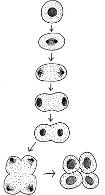
FIGURE 3.4. Cell division
A Visualization Treat for Your Cells
Find a quiet place to relax where nothing will disturb you for fifteen to thirty minutes. Sit comfortably or lie down and breathe gently for a few minutes, saying to yourself as you breathe in, “I breathe in healing, relaxing energy.” and as you breathe out, “I breathe out all tension, anxiety, negativity.”
Also try “every time I breathe out, I become twice as relaxed.”
After a few minutes like this, imagine you are floating along on a cloud of golden light, warm and safely enveloped. Begin to breathe in this healing light. As you breathe, feel the warm, loving, golden energy filling your lungs. It begins to spread through your body, up into your head, neck, and shoulders, down into your arms and hands. It spreads down your back and into your belly. The warm golden healing light fills your pelvis and moves down your legs into your feet. Your whole body is full of warm, healing golden light.
Imagine the cells of your body, billion upon billion, each one filled with this healing golden glow. Imagine one of your cells, anywhere you like. See it, feel it, think it filled with a warm, healing light energy. The cell is expanding, relaxing, happy and joyful as it bathes in the healing light. Every cell in your body is celebrating, enjoying the warm and golden healing light.
Your cells know what to do; your body knows what to do. We are completely as we are supposed to be, and our bodies, minds, and spirits are equipped with wonderful healing mechanisms. Allow yourself to enjoy this knowledge, allow the warm golden light to spread its glow throughout your body, and throughout your mind and spirit. All is well.
Afterwards, gently bring your attention back into the place you are in, and resume your daily activities, knowing you are filled with light and your cells are zinging with joy!
Some cells in the body normally replicate themselves, while others never do. This affects their capacity for regeneration if they have been damaged. Cells that are continually replicating themselves include epithelium, bone marrow, blood, spleen, and lymphoid tissue. Cells that can replicate, but rarely do under normal circumstances, include the liver,* kidney, pancreas, smooth muscle, bone cells, and fibroblasts (which make fibers in connective tissue).
There are cells that were considered to be permanent—unable to replicate after normal growth is complete: nerve cells, and skeletal and cardiac muscle. However, science has now found that skeletal muscles have limited powers to regrow due to satellite cells that can grow new cells, and cardiac muscle has modest ability to divide, although injuries to cardiac muscle are usually replaced by scar tissue.
Research is being done into gene therapy and stem cell therapy to enhance this process; natural healers will know that there are many ways to encourage the body’s own healing mechanisms. There is a great story in Deepak Chopra’s book Unconditional Life: Mastering the Forces That Shape Personal Reality of a miraculous healing of a skeletal muscle, back in the days when this was considered more or less impossible by scientific thinking of the day—not that long ago, really.
It was also believed until recently that we do not make new nerve cells. However, a study on rats undertaken in the year 2000 showed that brains continue to grow well after puberty; the adult brain is capable of growth and regeneration. It is not all the downhill tumble to senility we were led to believe!5
The implications of this knowledge for practitioners of medicine or healing are interesting: If cells are damaged, how easy is it for the body to repair or replace them? What can we do to encourage this process? Also, do we accept that certain things are impossible, or can we hold a belief in the possibility of miraculous healing for our patients? Bearing in mind that accepted physiological facts do turn out to be wrong now and then, it seems reasonable to hold out belief for optimum healing. “Be realistic—plan for a miracle,” as the bumper sticker says!
Studies have shown that our beliefs about other people are important—the power of our mind to heal applies not only to our thoughts about ourselves, but to what kind of thoughts we send to others.* Our thoughts are shaped by our beliefs.
The best thing is to try it for yourself. There are many interesting books you might use to get started. As well as Lynne McTaggart’s The Intention Experiment, take a look at Creative Visualization by Shakti Gawain, and You Can Heal Your Life by Louise Hay.
As we have said, cells are arranged into tissues, which in turn get together to make the organs and systems of the body. There are four types: epithelial, connective, muscular, and nerve.
Epithelial Tissue
Epithelial tissue covers the surface of the body, lines hollow organs and tubes inside the body, and forms glands. It consists of tightly packed cells arranged in continuous sheets on a basement membrane. They can be either single- or multilayered (that is, simple or compound). Epithelium adheres firmly to the underlying connective tissue via its basement membrane. It continually renews itself; the lower levels divide by mitosis and the older cells slough off. We all have firsthand experience of this with our skin, which now and then we see coming off—at least the dead top layers. Did you know that most of the dirt in the subway (and in your house) consists of human skin cells? Every day a new layer is made, and an old one sloughed off. The multilayered skin takes about thirty days to be completely renewed, while the single-layered epithelial lining of the gut is renewed every day.
There are various types of simple and compound epithelium, named for its appearance. Examples of simple are columnar, squamous, and cuboidal.
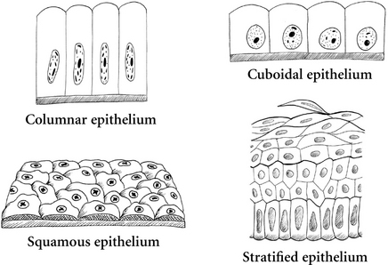
FIGURE 3.5. Epithelial tissue
Examples of compound epithelial tissue include stratified (the skin) and transitional (the bladder).
Sometimes epithelium is ciliated. Cilia are small hair-like projections of the cell membrane, which have the ability to move. They are found in the air pipes of the lungs, and in the fallopian tubes.
The epithelium of the lungs, gut, and urinary and reproductive systems forms what are called mucous membranes. Interspersed with the epithelial cells are special mucus-producing cells called goblet cells.
The ciliated mucous membrane of the lungs is ingenious: Inhaled dust and other particles stick to the mucus, which forms a lining over the surface of the epithelial cells. The cilia constantly move in one direction, shifting the mucus up toward the throat, and when it reaches the throat it can be drawn into the mouth and spat out. This is known as the ciliary escalator. One of the injurious effects of smoking tobacco is that nicotine depresses the ciliary escalator, thus preventing the lungs from clearing themselves just when they need it most. Luckily, this effect wears off as soon as smoking is stopped—as the epithelial lining does its amazing job of renewing itself, the cilia work again. This is the reason many smokers cough in the morning—overnight while asleep and without the influence of tobacco, the membrane repairs itself and the lungs begin to free themselves from accumulated toxins and debris. Many a hapless smoker, on noticing that the first cigarette of the day “cures” their cough, is able to fool himself or herself that smoking is healthful. (Actually, the cough is not a symptom—it is the cure, the body’s attempt to remedy a harmful situation. This illustrates the very important fundamental holistic principle that symptoms are not diseases. Much more on this later!)
Connective tissue is the most abundant tissue in the body. A binding and supporting tissue, it often has a very rich blood supply. (This does not apply to cartilage and ligaments; they have no direct blood supply at all, which is why they are white in color.)
The cells of connective tissue are widely scattered in a matrix of extra cellular material. There are many types: areolar, adipose, fibrous, elastic, cartilage, bone, blood, lymphoid, and reticular. Some of these seem very different from each other—blood and bone, for example—but if you look at the composition and formation of these tissues, they have a lot in common.
Areolar tissue is found all over the body, like a kind of packaging tissue. Sometimes it is called loose connective tissue. It consists of a ground substance protein and water background, with scattered cells making collagen and elastic fibers. The dermis, found in the skin under the stratified epithelial layer known as the epidermis, is made of this type of connective tissue. What is particularly interesting about it is that it can move from a more liquid to more solid gel state and back—which affects how well substances can diffuse through it.
A technique called skin rolling aims to loosen up the areolar tissue to get fresh fluid into it. Skin rolling lifts and squeezes the superficial layers of fascia underneath the skin, breaking down any adhesions, to release the sticking. It can often be very painful at first, but eases with each skin roll. Lift the skin with the thumb and push underneath and against the forefinger that is anchoring skin and fascia, moving along methodically until you reach the end of the available skin (e.g., from the inferior edge of the trapezius to the superior, or shoulder, edge). It needs to be a continual roll. Then work deeper with massage techniques.6
Adipose tissue is basically fat or a collection of cells that fill up with fat. It is useful for protection—padding under the skin (the subcutaneous fat) and around vital organs—and as a very concentrated energy store. And, of course, it gives us women our lovely curves. One of the reasons too much of it can harm us is because it tends to collect around our organs—an excess of fat around the heart makes life a lot harder for this organ. Have you come across the apple-or-pear-shaped thing? Pear-shaped people tend to put most of their fat around their bottoms and thighs, while apple-shaped people tend to deposit fat around their chest and middle. It seems when it comes to carrying excess fat, the pears have the advantage, as the apples will be more prone to gathering fat around the heart, leading to a greater workload for the heart and thus increasing the risk of heart disease. Adipose tissue makes the hormone leptin and is involved in regulating our sensation of hunger.
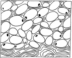
FIGURE 3.7. Adipose or fatty tissue
Fibrous tissue consists of collagen fibers made by cells called fibroblasts. Collagen forms thick ropes with tensile strength. The fascia around and within muscles, which comes together to form tendons, is fibrous connective tissue, as is periosteum, the tough fibrous covering of bone. Ligaments are primarily made of fibrous tissue, though they also contain some elastic fibers, as they need to be stretchy. The connective tissue outer coverings of organs are rich in fibrous tissue for protection and strength. Our blood vessel walls are full of it. We’ll be hearing more about this important substance.
Elastic tissue does what you might think: It gives stretch and recoil where needed—in skin, lungs, and arteries, for example. The elastic fibers in the dermis of the skin give our skin its ability to snap back when stretched. There is a tendency for this to diminish with age. Having said this, our bodies are of course renewable, with elastic fibers that can be repaired and made new.
Cartilage is amazing stuff—incredibly strong, a little stretchy, and totally flexible. Cartilage is found at most joints, where two bones meet each other. It allows for movement and protects the bones from grating on each other as the joint moves. We have some in our earlobes and nose, and an intricate assortment makes up the voice box, or larynx, which along with the vocal cords enables us to speak. Cartilage is a blue-white color; it has no blood supply of its own, and relies on surrounding tissues to get its nutrients. Because of this, it is slow to heal when damaged. Our first skeleton, formed when we are in the womb, is made of cartilage. As developing embryos, we first make a cartilaginous blueprint of our bones, then we begin to lay down calcium salts to form our bones. At birth our skeleton is mostly bone, with some cartilage left from which the bones grow. We grow throughout childhood, the bones growing from special cartilaginous growing plates until they fuse in the late teens or early twenties, after which we will grow no more in height.

The body makes the background substance of cartilage, loose and fibrous connective tissue from glucosamine (amino acid and sugar mixed). This is made in the body by an enzyme called glucosamine synthetase. As we age, this enzyme becomes less effective, which is why healing is slower in the elderly. Studies have shown that taking a supplement of glucosamine daily can help arthritis, aging or slowly healing skin, ligament and tendon injuries, and possibly heart disease and IBS (irritable bowel syndrome).7
Bone is what gives structure, shape, and support to our bodies, and protects our vital organs. It is made from cartilage with a beautiful and intricate pattern of calcium phosphate salts laid down within it. Bone is a vital and living tissue; it has a very rich blood supply. It can heal itself well from injuries and breaks by a process of calcification—repairing the breaks by laying down lots of calcium.
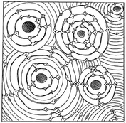
FIGURE 3.9. Bone
If you take a bone from a newly dead body and dissolve all the salts out of it, you are left with a completely flexible cartilage tissue that you could tie in a knot. Throughout life, our bones are reabsorbed and made anew, as we exchange the calcium salts between them and the blood. It’s thought that over about seven years the whole skeleton is replaced.
Blood is considered to be a connective tissue. It contains a background substance—the plasma—with cells loosely interspersed in it. Actually, blood cells are made in the bone marrow, so there is one obvious connection.

FIGURE 3.10. Blood
Lymphoid tissue, or reticular tissue, is specialized connective tissue found in the lymphatic system, in lymph nodes and vessels. In the lymph nodes it forms a mesh that is filled with the white blood cells of the lymphatic system. Here, debris from the lymph fluid is trapped and filtered out.
A membrane is a special covering that includes connective and epithelial tissues. Our bodies have four main types of membrane: cutaneous, mucous, serous, and synovial. The first three are continuous sheets of covering material made of an epithelial layer closely bound to an underlying bed of connective tissue, and synovial is formed from connective tissue.
Cutaneous membrane refers simply to the skin: a thick layer of compound epithelium over a thicker layer of loose connective tissue containing interesting structures like sweat glands, hair follicles, and so on. The following chapter is dedicated to skin.
Mucous membranes are lovely, wet, slippery membranes made of either compound or simple epithelial cells interspersed with goblet cells over a layer of loose connective tissue. Mucous membranes are adapted for absorption and secretion. Some secrete a lot of mucus (the lung and the gut); some do not (the urinary tract).
Serous membranes contain a layer of epithelial cells resting on a loose connective tissue base. The epithelial cells secrete a watery fluid. They are found in the heart (the pericardium), the lungs (the pleura), and the gut (the peritoneum).
Synovial membranes are found in synovial joints. (These are known as diarthroidal or movable joints in America.) They are made of loose connective tissue, and secrete a special lubricating fluid into the joint capsule.
Muscle tissue has the special property of contractibility, and so is responsible for almost all the movement in the body. There are three types: skeletal, smooth, and cardiac.
Skeletal muscle is what you will already be accustomed to thinking of as muscle—biceps, lats, abs, and the other gym favorites. As the name suggests, this type of muscle moves the skeleton.
Muscle cells contain tiny microfilaments or myofibrils made of proteins called actin and myosin, which lie together in such a way as to be able to move over each other in a ratchet mechanism. In skeletal muscle these are arranged in lines, which makes the muscle look striped under the microscope—hence its other name, striated muscle. Each movement of the fibers uses energy as ATP, and much of the heat we generate comes from muscle contraction. It is also called voluntary because we control it consciously and voluntarily, unlike the other two types.
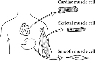
FIGURE 3.11. Muscle tissue
Smooth muscle is under involuntary control. Its cells are arranged in sheets, which wrap around tubes and hollow organs in the body. It moves food through the gut, and is found in all the body’s tubes and all its hollow organs except for the heart.
Cardiac muscle is only found in the heart. It looks striped, like skeletal muscle, but the cells are a special shape unique to it. It can never rest for long—it must keep on beating all of our lives. It is under involuntary control.
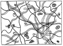
Nerve tissue is very specialized. The cells, called neurons or nerve cells, are excitable and conductive. This allows for sending messages to do with control throughout the body. This tissue also contains special supporting cells called neuroglia cells, or glial cells. These are important in that they nourish and protect the neurons and help in regeneration. They form a scaffold all around the neurons.
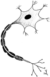
FIGURE 3.12. Neuron, or nerve cell
*Omega-3 is very long and highly flexible. When it is incorporated into the cell membrane, it helps make the membrane itself elastic and fluid so that signals pass through it efficiently. But if the wrong fatty acids are incorporated into the membrane, the receptors can’t react as well to their substances.
*Candace Pert describes this beautifully in her fascinating book Molecules of Emotion: Why You Feel the Way You Feel.
*Lynne McTaggart’s The Field: The Quest for the Secret Force of the Universe lays out the evidence coherently.
*The now well-known plant milk thistle (Psylibum marianus) has an effect of stimulating regeneration of liver cells.
*One study looked at the power of curses—what happens when we send negative thoughts to another? In the experiment, 195 separate cultures of a fungus were “cursed.” Seventy-seven percent showed retarded growth compared to the control group. (J. Barry, “General and Comparative Study of the Psychokinetic Effect on a Fungus Culture,” Journal of Parapsychology, 1968, 32(94):237–243, cited in The Intention Experiment by Lynne McTaggart.)