
After Chapter 1.3, you will be able to:
Throughout this section, refer to Figure 1.4, which identifies various anatomical structures inside the human brain. As we discuss different parts of the brain, it’s important to remember the functions of these brain structures. Different parts of the brain perform remarkably different functions. For instance, one part of the brain processes sensory information while an entirely different part of the brain maintains activities of the internal organs. For complex functions such as playing a musical instrument, several brain regions work together. For the MCAT, you will need to know some of the basics about how the brain integrates input from different regions.
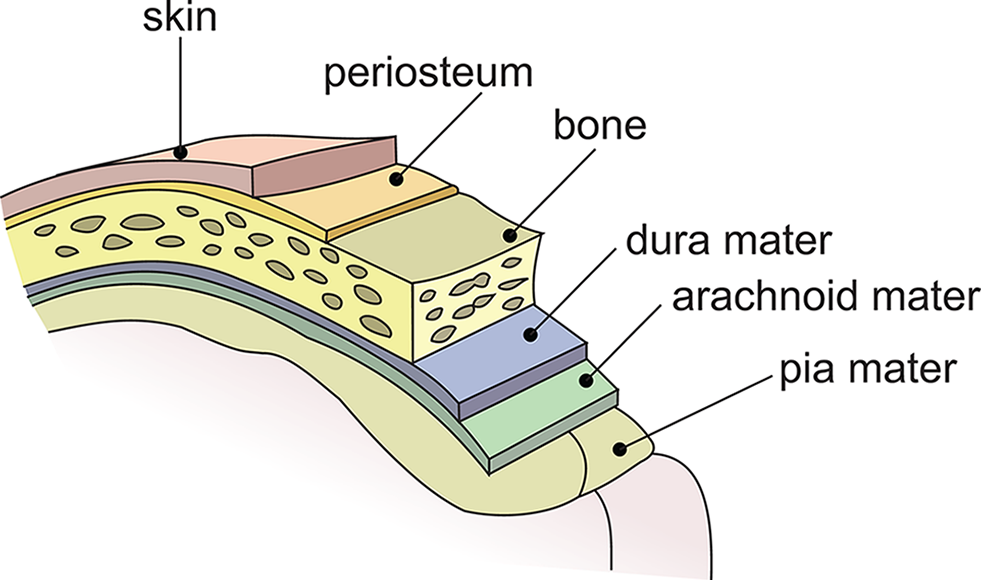
The brain is covered with a thick sheath of connective tissue called the meninges. The meninges help protect the brain, keep it anchored within the skull, and resorb cerebrospinal fluid. They are composed of three layers: the dura mater, the arachnoid mater, and the pia mater, as shown in Figure 1.5. Cerebrospinal fluid is the aqueous solution in which the brain and spinal cord rest; it is produced by specialized cells that line the ventricles (internal cavities) of the brain.
The human brain can be divided into three basic subdivisions: the hindbrain, the midbrain, and the forebrain. Notice that brain structures associated with basic survival are located at the base of the brain and brain structures with more complex functions are located higher up. The meaningful connection between brain location and functional complexity is no accident. In evolutionary terms, the hindbrain and midbrain were brain structures that developed earlier. Together they form the brainstem, which is the most primitive region of the brain. The forebrain developed later, including the limbic system, a group of neural structures primarily associated with emotion and memory. Aggression, fear, pleasure, and pain are all related to the limbic system. The most recent evolutionary development of the human brain is the cerebral cortex, which is the outer covering of the cerebral hemispheres. In humans, the cerebral cortex is associated with everything from language processing to problem solving, and from impulse control to long-term planning. Most of the key brain regions described in the following sections are summarized in Table 1.1.
| Major Divisions and Principal Structures | Functions |
|
|
|
|
|
|
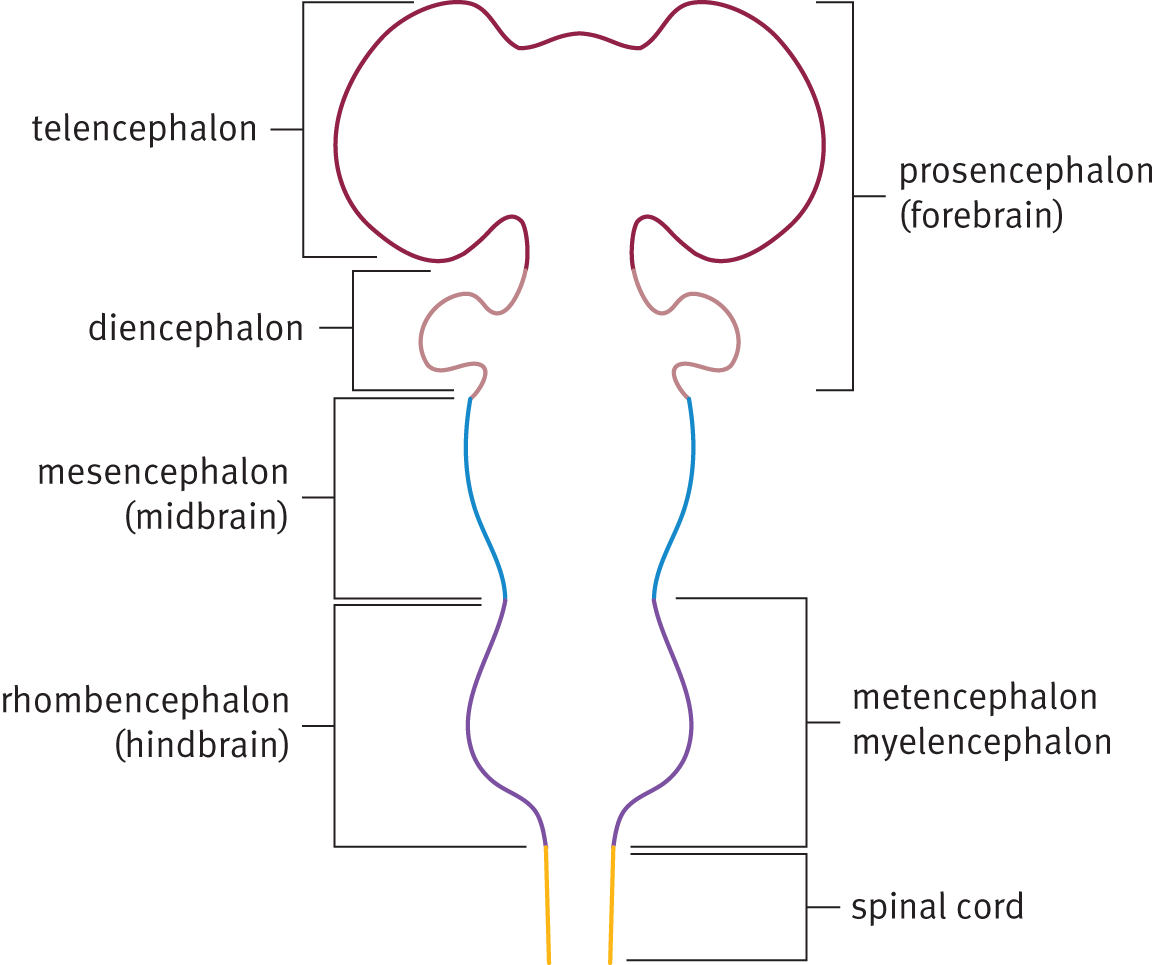
In prenatal life, the brain develops from the neural tube. At first, the tube is composed of three swellings, which correspond to the hindbrain, midbrain, and forebrain. Both the hindbrain and forebrain later divide into two swellings, creating five total swellings in the mature neural tube. The embryonic brain is diagrammed in Figure 1.6, and its subdivisions are described further in the following sections.
Located where the brain meets the spinal cord, the hindbrain (rhombencephalon) controls balance, motor coordination, breathing, digestion, and general arousal processes such as sleeping and waking. In short, the hindbrain manages vital functioning necessary for survival. During embryonic development, the rhombencephalon divides to form the myelencephalon (which becomes the medulla oblongata) and the metencephalon (which becomes the pons and cerebellum). The medulla oblongata is a lower brain structure that is responsible for regulating vital functions such as breathing, heart rate, and blood pressure. The pons lies above the medulla and contains sensory and motor pathways between the cortex and the medulla. At the top of the hindbrain, mushrooming out of the back of the pons, is the cerebellum, a structure that helps maintain posture and balance and coordinates body movements. Damage to the cerebellum causes clumsiness, slurred speech, and loss of balance. Notably, alcohol impairs the functioning of the cerebellum, and consequently affects speech and balance.
Just above the hindbrain is the midbrain (mesencephalon), which receives sensory and motor information from the rest of the body. The midbrain is associated with involuntary reflex responses triggered by visual or auditory stimuli. There are several prominent nuclei in the midbrain, two of which are collectively called colliculi. The superior colliculus receives visual sensory input, and the inferior colliculus receives sensory information from the auditory system. The inferior colliculus has a role in reflexive reactions to sudden loud noises.
Above the midbrain is the forebrain (prosencephalon), which is associated with complex perceptual, cognitive, and behavioral processes. Among its other functions, the forebrain is associated with emotion and memory; it is the forebrain that has the greatest influence on human behavior. Its functions are not absolutely necessary for survival, but are associated instead with the intellectual and emotional capacities most characteristic of humans. During prenatal development, the prosencephalon divides to form the telencephalon (which forms the cerebral cortex, basal ganglia, and limbic system) and the diencephalon (which forms the thalamus, hypothalamus, posterior pituitary gland, and pineal gland).
Neuropsychology refers to the study of functions and behaviors associated with specific regions of the brain. It is most often applied in research settings, where researchers attempt to associate very specific areas in the brain to behavior, and in clinical settings when patients are treated for brain lesions. Neuropsychology has its own experimental methodology and technology.
Studying human patients with brain lesions is one way that researchers have determined the functions of the brain. One problem in studying human brain lesions is that they are rarely isolated to specific brain structures. When several brain structures are damaged, it becomes difficult for researchers to attribute a specific functional impairment to any single brain region; the impairment could just as easily be attributed to any other region that suffered damage.
One method for studying the relationship of brain regions and behaviors is to study brain lesions in lab animals. The advantage of this approach is that precisely defined brain lesions can be created in animals by extirpation. Researchers can also produce lesions by inserting tiny electrodes inside the brain and then selectively applying intense heat, cold, or electricity to specific brain regions. Such electrodes can be placed with great precision by using stereotactic instruments, which provide high-resolution, three-coordinate images of the brain. Notwithstanding the ethical or cruelty concerns such studies have raised, they have greatly increased our understanding of comparable neural structures in humans.
Another method involves electrically stimulating and recording brain activity. Before operating on the brain, one can stimulate a patient’s cortex with a small electrode. This causes individual neurons to fire, thereby activating the behavioral or perceptual processes associated with those neurons. For instance, if the electrode stimulates neurons in the motor cortex, it leads to specific muscle movements. If the electrode stimulates the visual cortex, the patient “sees” flashes of light that are not really there. By using electrical stimulation, neurosurgeons can thus create cortical maps. This method relies on the assistance of the patient, who is awake and alert. Because there are no pain receptors in the brain, only local anesthesia is required. Electrodes have also been used in lab animals to study deeper regions of the brain. Depending on where they are implanted, the electrodes can elicit sleep, sexual arousal, rage, or terror. Once the electrode is turned off, these behaviors cease.
Electrodes can also be used to record electrical activity produced by the brain itself. In some studies, individual neurons are recorded by inserting ultrasensitive microelectrodes into individual brain cells, recording their electrical activity. Electrical activity generated by larger groups of neurons can be studied using an electroencephalogram (EEG), which involves placing several electrodes on the scalp. Broad patterns of electrical activity can thus be detected and recorded. Because this procedure is noninvasive (it does not cause any damage), it is commonly used with human subjects. In fact, research on sleep, seizures, and brain lesions relies heavily on EEGs, as shown in Figure 1.7.

Another noninvasive mapping procedure is regional cerebral blood flow (rCBF), which detects broad patterns of neural activity based on increased blood flow to different parts of the brain. rCBF relies on the assumption that when a specific cognitive function activates certain regions of the brain, the blood flow to those regions increases. For example, listening to music may increase blood flow to the right auditory cortex because that is where music is processed in most individuals’ brains. To measure blood flow, the patient inhales a harmless radioactive gas; a special device that can detect radioactivity in the bloodstream can then correlate radioactivity levels with regional cerebral blood flow. This research method uses noninvasive computerized scanning devices.
Some of the other common scanning devices and methods of visualization used for brain imaging include: