 Chapter 4
 Aligned 2016 CACREP Standards
Standard 2.F.3.b. Theories of learning
Standard 2.F.3.c. Theories of normal and abnormal personality development
Standard 2.F.3.e. Biological, neurological, and physiological factors that affect human development, functioning, and behavior
Standard 2.F.3.f. Systemic and environmental factors that affect human development, functioning, and behavior
• • •
In prior chapters, we reviewed the nervous system and cellular function. A next step in learning foundational information about child hood and adolescence is examining the developmental process with regard to neurophysiological and social development. In this chapter, we build on prior information from Chapters 2 and 3 to understand this developmental process, starting at conception (i.e., fertilization) and extending through the elementary years. We thus adhere to our definition of childhood stated in the Preface as encompassing 0 to 11 years of age, although we also want to recognize that some children mature to adolescence earlier than age 11.
Human growth and development is a large topic area, and we were selective in the information we decided to cover in this chapter. Because this text is focused on applied and practical information pertinent to counseling, we chose to focus on neurodevelopmental information that would be useful to the counseling process and inform subsequent chapters. We do not cover typical theories of human development (e.g., Erik Erikson, Jean Piaget) in this chapter. We also note that although identity development begins in childhood, we cover ethnic, gender, and sexual identity development in Chapter 8, on adolescent development.
As with prior chapters, we recommend that you consult the glossary to clarify the meaning of unfamiliar terminology. In writing this chapter, we aquired information from the National Institute of Child Health and Human Development (2017) and the Centers for Disease Control and Prevention (CDC; 2018a, 2018b, 2019). We also consulted the following texts when writing this chapter: Developmental Neuroscience by Fahrbach (2013), Developmental Cognitive Neuroscience: An Introduction (4th ed.) by Johnson and de Haan (2015), and Fundamentals of Human Neuropsychology (7th ed.) by Kolb and Whishaw (2015). We recommend these texts if you want to learn more about child neural development.
During the embryonic period, a special organ forms in the uterus called the placenta. The placenta not only provides the fetus with oxygen and nutrients via the umbilical cord and attempts to filter out harmful substances but also secretes estrogens, progesterone, and human chorionic gonadotropin hormone. This latter hormone reduces the mother’s immune function so that the fetus is not rejected as a foreign organism. In addition, human chorionic gonadotropin secretes human placental lactogen, which prepares the mother’s breasts for lactation.
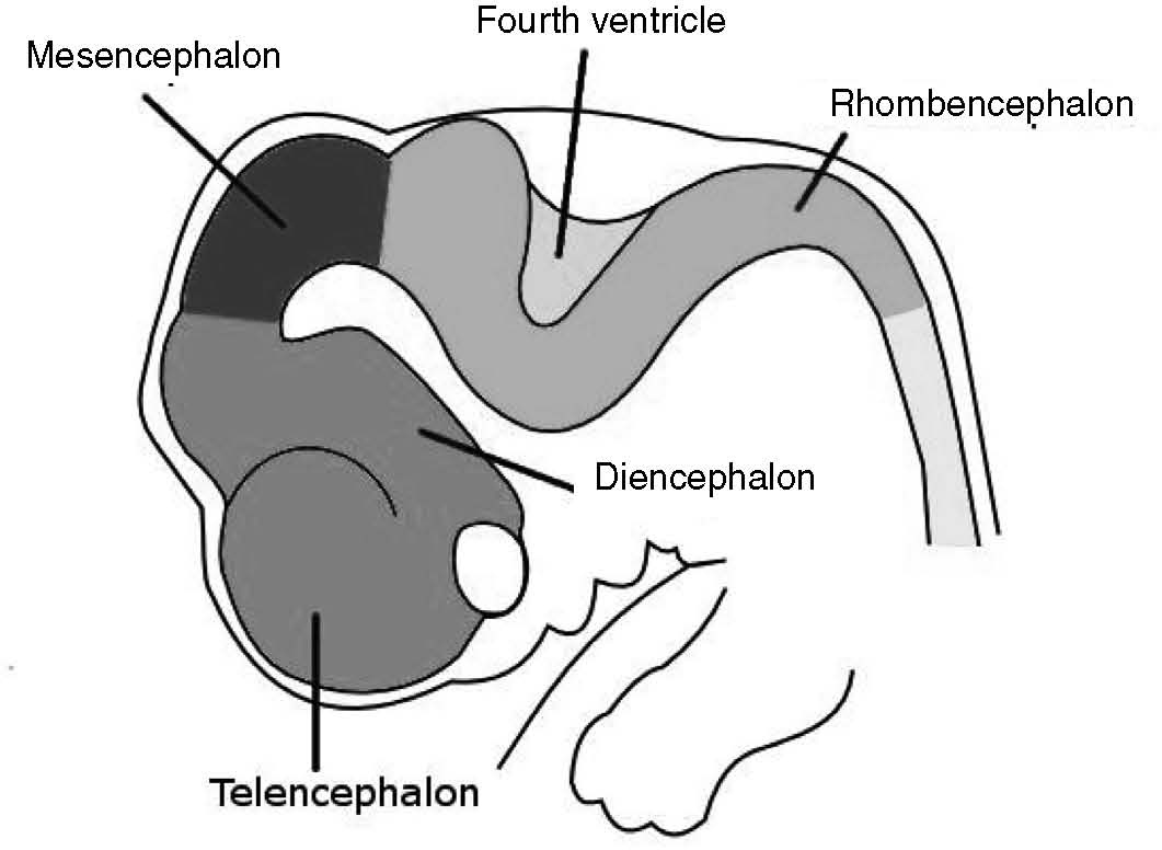
The development of the nervous system is one of the first processes to begin upon fertilization and one of the last to be completed following delivery. By the third week of gestation, the neural plate folds to form a neural groove before transitioning into the neural tube. Within the neural tube, neurons and glia begin to form. The neural tube will eventually become the brain and spinal cord (i.e., the central nervous system). By Week 5, several important neural tube structures have developed that will eventually become important structures in the adult brain. The telencephalon will become major structures of the cerebrum, including the amygdala, hippocampus, frontal cortex, hypothalamus, and pituitary gland; the diencephalon will become the thalamus and pineal gland; the mesencephalon will become the cerebellum; and the myelencephalon will become the medulla oblongata. These neural tube structures and others are depicted in Figure 4.1.
During the second trimester, a few significant developments occur. By Week 20, the hypothalamic-pituitary-adrenal axis is established. The thyroid hormonal system soon follows, which assists with the development of the fetus’s metabolism. The baby’s muscular system develops to the extent that the mother can feel kicks from the fetus. In the third trimester, the brain increases fourfold in size, and brain tissue volume likewise increases in a steady linear fashion as new neurons and glia (i.e., gray matter) are generated (Glass et al., 2015).
Fetal, newborn, and infant development are critical periods because events during these stages of development can have profound and lasting impacts. Multiple factors can result in congenital problems, such as the age of the parents, nutritional and vitamin deficiencies, infections, the presence of toxins, and side effects of medication.

FIGURE 4.1 Neural Tube at Six Weeks' Gestation
Note. From Kurzon, 2007. In Wikimedia Commons.
Parental age has been associated with risk for congenital problems. Parents in the United States are conceiving children at older ages. The birth rate for women 40 to 44 years of age has more than doubled since 1990 (Matthews & Hamilton, 2014). This increase in age is associated with a greater risk for congenital problems in the fetus. In an older study from 2002, a mother’s risk of conceiving a child with Down syndrome increased fourfold between 35 and 40 years of age and tenfold between 35 and 45 years of age (Morris, Mutton, & Alberman, 2002). In a large study of 5.8 million children from five countries that examined the relationship between parental age and a child’s development of autism, fathers older than 50 were 66% more likely to conceive a child with autism compared to fathers ages 20 to 29 (Sandin et al., 2016). A 15% increase in risk was observed for mothers older than 50, and an 18% increase in risk was found for mothers younger than 20 years old. These risk ratios were found when the researchers controlled for confounding factors and even the age of the other parent. In the study, it appeared that risk did not increase significantly until parents were older than 40 years of age. Note that other hypothesized factors (e.g., vaccinations) show no evidence of a causal link to these developmental disabilities (Hviid, Hansen, Frisch, & Melbye, 2019).
The exact mechanism of the effect of parental age on congenital problems is unknown. Several hypotheses have been proposed. First, the effect of parental age could be caused by DNA copy errors. Sperm cells will have copied 600 times by age 40 compared to 200 times at age 20 (Malaspina, Gilman, & Kranz, 2015). Similar copy errors have been proposed in ovarian cells. Second, DNA mutations occur with increasing age. For example, sperm telomeres increase in length with age rather than decrease in length as is true of other cells (Sharma et al., 2015). Longer sperm telomere length appears to be a risk factor for schizophrenia (Malaspina et al., 2014). Third, epigenetic errors could account for this age effect (Perrin, Brown, & Malaspina, 2007).
In utero, fetal growth and development is influenced by the mother’s diet and nutrition. Folic acid is essential to the development of the brain and spinal cord, and deficits in the mother’s folate intake can result in significant neural tube defects that result in paralysis, vision and hearing loss, intellectual disability, and even death in the baby (CDC, 2018b). The fetus also needs other vitamins to develop, such as iron, vitamin B12, and so on. For these reasons, a prenatal vitamin supplement is often recommended (National Institute of Child Health and Human Development, 2017).
Viral infections such as rubella, toxoplasmosis, and Zika virus can cause a reduction in head size known as microcephaly, which is associated with severe neurodevelopmental abnormalities and intellectual disability (CDC, 2018a). Zika virus gained attention because no vaccine exists, and it can be transmitted through mosquito bites and through sexual intercourse.
Lead is a known neurotoxin. Exposure to lead in utero can increase the risk of miscarriage, low birth weight, stillbirth, and premature birth. It can also cause intellectual impairment, developmental disorders, and impulse control problems. At the cellular level, lead is able to cross the blood-brain barrier rapidly and has neurotoxic actions that include apoptosis (programmed cell death) and interference with myelination during early childhood (Sanders, Liu, Buchner, & Tchounwou, 2009). Pregnant mothers and children may be exposed to lead through corroded water pipes or lead paint in their environment.
Exposure of the fetus or newborn to alcohol via breastfeeding can lead to fetal alcohol spectrum issues, such as intellectual and developmental disabilities, abnormal facial features, and problems with organ function. Current research indicates that no amount of exposure to alcohol during pregnancy is safe for the fetus. Although brain damage is dose dependent (i.e., it worsens as alcohol consumption increases), even a small amount of exposure to alcohol can cause changes to brain structure (Eckstrand et al., 2012). Research has found that exposure to alcohol during pregnancy reduces gray matter volume in the cingulate cortex, frontal lobe, temporal lobe, and caudate nucleus (Eckstrand et al., 2012). As we learned in Chapter 2, these brain regions are associated with executive functioning, attention, and memory.
Smoking or consuming tobacco and cannabis can also have significant negative effects on the fetus, such as doubling the risk of stillbirth. Even passive exposure to tobacco (i.e., exposure to secondhand smoke) doubles the risk of stillbirth and increases the risk of premature birth and low birth weight (Khader, Al-Akour, Alzubi, & Lataifeh, 2011). Nicotine readily crosses the placenta, and concentrations of nicotine can be 15% higher in the placenta than in the mother (Wickström, 2007). Nicotine is also excreted through breastmilk, so the baby will be exposed even if the mother smokes or consumes tobacco away from the newborn. Cannabis use has been associated with low birth weight alongside lasting deficits in attention, impulse control, and memory (Grant, Petroff, Isoherranen, Stella, & Burbacher, 2018). Inhalation of tobacco and cannabis smoke is problematic given the other toxins involved, such as carbon monoxide.
Several complications can occur during pregnancy, labor, and delivery. Detailing the full extent of these is beyond the scope of this chapter, but a few are worth mentioning. Preterm birth is fairly common in the United States, affecting 11.5% of all births (Glass et al., 2015). Expected birth terms are typically 37–42 weeks’ gestation. Births before 37 weeks’ gestation are considered premature. Extremely premature birth is defined as birth before 28 weeks’ gestation. The estimated age of viability, defined as an approximately 50% likelihood of survival, is considered to be birth at 23 to 24 weeks (Glass et al., 2015). Neonatal care is continually improving, and the age of viability has reduced over time.
Although newborns are better able to survive and thrive at earlier ages of premature delivery, extremely premature birth is associated with increased risk of persisting cognitive deficits. In a large study of nearly 900 children, approximately one quarter of children who were born at or before 28 weeks had significant cognitive deficits at 10 years of age (Kuban et al., 2016). Males appear to be at greatest risk (Kuban et al., 2016). Extremely premature infants are more likely to have impairments in executive function with regard to impulse control, cognitive flexibility, planning and organizational ability, and working memory (Luu, Ment, Allan, Schneider, & Vohr, 2011). These impairments resemble core symptoms of attention-deficit/hyperactivity disorder (ADHD), and the incidence of ADHD is 3 times higher in extremely premature infants than children born at term (Franz et al., 2018). These neurological impairments are believed to be caused by missed opportunities for brain development in the third trimester (remember, the brain increases fourfold in size in the third trimester).
During the first 3 months after delivery, a series of developmental changes occur as the newborn adjusts to the external world. The infant period is proposed to last from birth to 24 months after delivery, with the first month called the neonate stage. During this time, the immune system develops and the cerebellum triples in size, associated with rapid growth in motor abilities. The hippocampus also develops during the first 4 months, resulting in improvements to facial recognition (Farroni, Massaccesi, Menon, & Johnson, 2007). At approximately 3 years of age, gut microbiota stabilize. At this stage of development, the bacterial composition of the child’s gut resembles that of the adult gut (Bokulich et al., 2016).
During the first years of life, infants largely learn via their relationships with their caregivers. For example, infants learn to mimic vocal sounds and tones from their parents and guardians. Because their environment is new and fraught with potentially threatening stimuli, infants also learn to navigate and explore their environment while relying on their caregivers to provide protection and support. Attachment to caregivers is thus an essential survival mechanism during early childhood.
The neuropeptide oxytocin has an important role in the bonding of parents or caregivers and children during pregnancy and the first year of life. It is broadly thought of as a bonding chemical that facilitates close relationships and associated feelings of trust, intimacy, and empathy (Feldman, Gordon, & Zagoory-Sharon, 2011; Hurlemann et al., 2010). Oxytocin is also involved in parent-infant synchrony (or attuned emotional behaviors), such as gaze, facial expression, and touch (Feldman et al., 2011). It seems that physical touch, such as being held and breastfeeding, are particularly important antecedents to oxytocin release (Feldman et al., 2011).
Oxytocin is produced by the hypothalamus and projects to (i.e., has directional neural pathways with) the nucleus accumbens and the pituitary gland (Ross & Young, 2009). Oxytocin receptors are also located in the amygdala, which is important given the calming and soothing effect of oxytocin in response to anxiety and fear (Kirsch et al., 2005; Knobloch et al., 2012). Oxytocin inhibits the release of adrenocorticotropic hormone by the pituitary gland and thus can downregulate the stress response in the hypothalamic-pituitary-adrenal axis. When infants are upset or frightened, it is fairly common for them to seek the comfort of their parents. A child’s bonding with their caregiver results in the release of oxytocin, which can help assuage fear activation and stress response. It is interesting that the secretion of oxytocin seems to be influenced by novel interactions. Oxytocin levels are higher in mothers who interact with other unfamiliar children than in mothers who interact with their own children (Bick & Dozier, 2010).
Sex differences exist in response to the secretion of oxytocin. Females seem to have greater activation of the amygdala on receiving oxytocin compared to males, which is believed to be caused by oxytocin’s interaction with estrogen (Lischke et al., 2012). Estrogen enhances the secretion of oxytocin and binding to receptors in the amygdala (Lischke et al., 2012). In males, testosterone suppresses oxytocin (Okabe, Kitano, Nagasawa, Mogi, & Kikusui, 2013). Okabe et al. (2013) proposed that testosterone’s suppression of oxytocin is a function of evolution, as oxytocin is associated with empathy responses that may compromise activities such as hunting and attacking threats in the environment.
Animal studies have found that low secretion of oxytocin has profound effects on social behavior. Studies that have replaced genes associated with oxytocin receptors with nongenetic DNA in mice (sometimes referred to as knockout studies) have found impaired recognition of others, increased aggression, and impaired interaction between rat offspring and their mothers (Nishimori et al., 2008). Although it is unethical to replace human genetic sequences, these behaviors are also observed in humans who have oxytocin deficits. For example, a meta-analysis of 16 studies found significant changes and mutations to oxytocin receptor genes in children with autism compared to controls (LoParo & Waldman, 2015). Social impairments are a central symptom cluster of autism, and thus oxytocin receptor dysfunction might be a potential biomarker of the disorder (LoParo & Waldman, 2015). Oxytocin also appears to have antidepressant effects in animal studies, and some researchers have proposed that a lack of parental bonding and oxytocin might be a pathway to clinical depression later in life (McQuaid, McInnis, Abizaid, & Anisman, 2014). See Reflection Question 4.1.
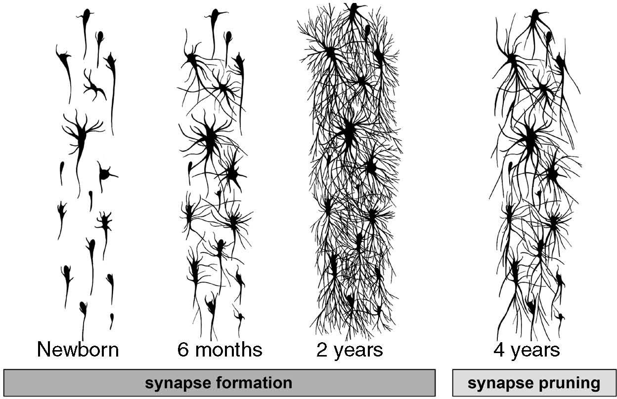
The brain develops at a rapid pace during the first years of life. By approximately 2 years of age, all of the major brain structures (e.g., structures of the subcortex and cerebral cortex) resemble those of the adult brain (Johnson & de Haan, 2015). Neuronal growth (i.e., neurogenesis) is fastest during the fetal stage (Johnson & de Haan, 2015). After birth, the brain experiences tremendous growth in synaptic connections (synaptogenesis). In addition, there is substantial growth in the myelination of these neurons. You may recall that the fatty myelin sheath assists with electrical conduction, or the speed with which electrical messages can be passed within cells. As a result of synaptogenesis and myelination, the connections between neurons become more numerous and messages are passed more quickly. Dendrites also begin to branch out and develop more receptors. Figure 4.2 depicts this neural developmental process.
Neural development during early childhood helps us understand why infants become more adept at a number of tasks over time. In regard to motor neurons and motor movement, infants become able to grab objects, move their facial muscles to make sounds and words, crawl, and walk on two feet. In regard to sensory development, infants become better able to see longer distances as they develop and differentiate sensory input, such as identifying different smells. Children also begin to develop more complicated higher order cognitive functions, such as learning sequences (e.g., bedtime routines), developing and expressing language, and using declarative memory retrieval (e.g., requesting objects they played with several days before).

FIGURE 4.2 Synapse Formation and Pruning
Note. From Rachel Chaney, illustrator. Used by permission. All rights reserved.
Not all areas of the central nervous system myelinate at the same rate. Myelination in the brain tends to develop in a back-to-front direction, with neurons associated with sensory and motor functions myelinating more quickly than areas of the brain associated with prefrontal function (Casey, Tottenham, Liston, & Durston, 2005; Johnson & de Haan, 2015). Myelination of the sensorimotor cortex is largely complete by the second year, whereas myelination of the prefrontal cortex lasts through early adulthood (i.e., until a person is in their 20s). This slow development of the prefrontal cortex and associated executive functioning helps us understand why young children have difficulty with frustration tolerance and impulse control.
The sensorimotor cortex develops before the prefrontal cortex because infants must learn to communicate when they are hungry and need to feed, among other functions. The infant’s cry for nourishment is an essential survival function and does not require primary activation of the prefrontal cortex. In contrast, imagine an infant who has an adult-like cognitive ability for rational thought at birth but is unable to see their caregiver, unable to call out when hungry, and so on. The infant’s survival would be under threat! Thus, it is important that the parts of the nervous system that are associated with basic sensorimotor function develop first.
By approximately the end of the second year, the child has the most synaptic connections they will ever have during their lifetime (Johnson & de Haan, 2015). The high number of synaptic connections enable the child to learn tasks at a rapid pace. Children at this age have more synaptic connections than they functionally need. Synaptic connections are therefore eliminated (pruned). The number of neurotransmitters that are produced and excreted also reduces during this period (Johnson & de Haan, 2015). By the time a child reaches 10 years of age, 50% of their synapses will have been eliminated (Kolb & Whishaw, 2015). Synaptic pruning is experience dependent, which means that synaptic connections that are not activated through a child’s interactions with the environment are pruned (Kolb & Whishaw, 2015). Thus, the synapses that are eliminated will differ between individual children based on their experiences. Microglia have an important role in synaptic pruning, as they engulf synapses via phagocytosis (Shafer et al., 2012). Figure 4.2 visualizes the developmental trajectory of synaptic pruning.
Autism has been associated with reductions in synaptic pruning and an increase in the density of dendritic spines (i.e., more receptors; Tang et al., 2014). In a study by Tang and colleagues (2014), typically developing adolescents had 41% fewer synapses than typically developing infants, whereas adolescents with autism had only 16% fewer synapses than infants with autism. It appears that synaptic pruning is essential to healthy functioning of the central nervous system, and a preponderance of synapses can result in overcommunication between brain structures, which creates confusion and noise within the brain (Tang et al., 2014). In the study, children with autism also appeared to have higher than average numbers of degraded and older neurons, a marker of problems with glia function (Tang et al., 2014).
After infancy, children continue to seek nurturance, protection, and support from their parents and guardians as they further explore the world around them and yearn for some degree of independence. Children lean on early attachment bonds that they forged with their caregivers during infancy. At this stage, children develop cooperative play skills and enjoy playing with caregivers and peers. Even during independent activity, children will often seek attention from their caregivers. Caregivers at this stage often balance attentively supporting their child’s independent activities with intervening to address problems that the child cannot resolve without assistance. Because children continue to need guidance and direction at times, parents and guardians take a fairly active role during early childhood. For example, a child might be unable to clean up a spill without assistance or might need guidance in resolving a peer conflict.
The needs of the child will change with age, as the child matures and develops. In this section, we review key factors in early child development, such as the role of play, sibling relationships, and early school experiences.
During early childhood, play behavior has a central role in child development (Ginsberg & Committee on Communications, 2007). In rats, play behavior activates the prefrontal cortex and striatum, which are associated with cognition, motivation, and conditioned reward responses (Van Kerkhof, Damsteegt, Trezza, Voorn, & Vanderschuren, 2013). Play also facilitates transcription of brain-derived neurotrophic factor in the amygdala and dorsolateral prefrontal cortex (Gordon, Burke, Akil, Watson, & Panksepp, 2003). Accordingly, young children often process their emotional experiences through play. Children benefit most from unstructured and self-directed play, which facilitates creativity and mental processing (Ginsberg & Committee on Communications, 2007). In school settings, play helps children transition into a more structured environment and develop academic readiness skills (Blanco, Ray, & Holliman, 2012). Play with other children also facilitates social skills such as sharing, negotiation, conflict resolution, and self-advocacy (Blanco et al., 2012). Play with caregivers also grants an opportunity for the adult caregiver to gain insights into the child’s inner world (Ginsberg & Committee on Communications, 2007). Playing together is a healthy method of communicating to children that their caregivers are attentive to them and care about their perspectives. Play is so crucial to child development that Ginsberg and Committee on Communications (2007) cautioned parents and guardians to limit access to technology (e.g., screen time) and to structure the daily household routine so that adult caregivers have ample time for unstructured play with their children.
Impairments in play skills have important effects on the overall development of social skills and communication (Lang et al., 2010). For example, children with autism often experience deficits in symbolic or imaginative play while also engaging in repetitive and self-stimulatory behavior that can interrupt their ability to fully engage in play (Lang et al., 2010). Children with autism may need assistance developing play skills, which may serve as a building block for scaffolding subsequent social skills, such as attending to and learning social cues. Early intervention seems critical, as play behavior seems to be associated with the quality of the interaction between the caregiver and the child with autism (Naber et al., 2008). Play skills are best taught through behavioral techniques (Wong et al., 2015), which are examined in a subsequent chapter.
More than 80% of children live in households with other siblings, slightly higher than the percentage of children who live in a household with a father figure (McHale, Updegraff, Whiteman, 2012). Younger siblings often learn from their older siblings, who serve as social role models (McHale et al., 2012). A child’s interaction with their siblings can have an important role in their development of self-regulation. Padilla-Walker, Harper, and Jensen (2010) found that positive sibling perceptions were associated with longitudinal self-regulation and prosocial behavior and were a protective factor against externalizing behavior problems (e.g., aggression). In contrast, sibling hostility has been associated with internalizing problems, such as anxiety and depression (Campione-Barr, Greer, & Kruse, 2013; Padilla-Walker et al., 2010). Siblings of children with developmental disabilities such as autism appear to be at heightened risk for social and behavioral problems (Orsmond & Seltzer, 2007). Counselors will at times work with child adjustment issues related to sibling relationships. It can sometimes be helpful to include siblings in the counseling process to work through relational conflict. In-home counseling can be especially facilitative in this regard.
School serves a variety of functions for children. Not only does school assist children in developing linguistic, mathematics, and science knowledge and skills, it can also serve as a facilitator of peer interaction and social skills development. In school, children learn to work cooperatively, develop patience, and navigate difficult relationships (Ginsberg & Committee on Communications, 2007).
Elementary school can also bring attention to problems that are less pronounced in the home environment, such as difficulty attending to tasks for extended periods and trouble sitting still. Elementary school is often the time when neurodevelopmental issues such as ADHD are first assessed. In the next chapter, we explore controversies related to the diagnosis of ADHD in children. Some children may need accommodations to reach their academic potential, and we recommend that counselors have sound knowledge of the special education system and eligibility process in the state where they are practicing. In our experience, families often need guidance in accessing school-based services and advocating for their child. We recommend that counselors in clinical settings develop relationships with school counselors to better understand the services provided in schools and the eligibility process. See Reflection Question 4.2.
Counselors can benefit from a working knowledge of neurophysiological and social development to comprehensively conceptualize early childhood experiences and problems that arise. In this chapter, we learned that children will likely benefit from play-based activities, need an approach that affirms and strengthens caregiver bonds and attachments, and require the counselor’s understanding that they will not have developed the prefrontal functioning to benefit from traditional insight-oriented talk therapy that requires substantial frustration tolerance. We also learned that preventing congenital problems (to the extent possible) is important, as many congenital issues persist throughout childhood into adulthood if acquired. In subsequent chapters, we introduce several counseling approaches for working with children, including play-based creative approaches and behavioral interventions.