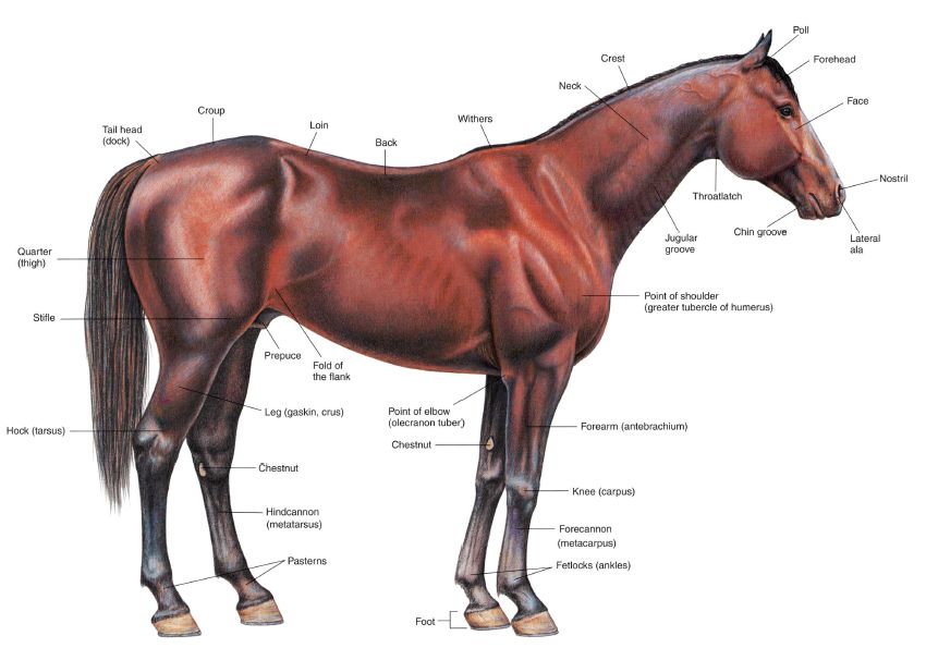
PLATE 1.1 Right lateral view of a stallion.
PLATE 1.2 Left lateral view of a mare.
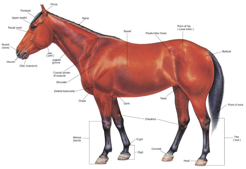
PLATE 1.3 Body regions of the horse. Right lateral view.

PLATE 1.4 Skeleton of the horse. Left lateral view. C = cervical vertebra, T = thoracic vertebra, L = lumbar vertebra, b = bone
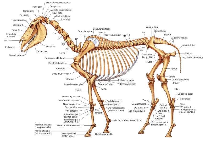
PLATE 1.5 Cutaneous muscles and major fasciae of the stallion. Right lateral view, m = muscle
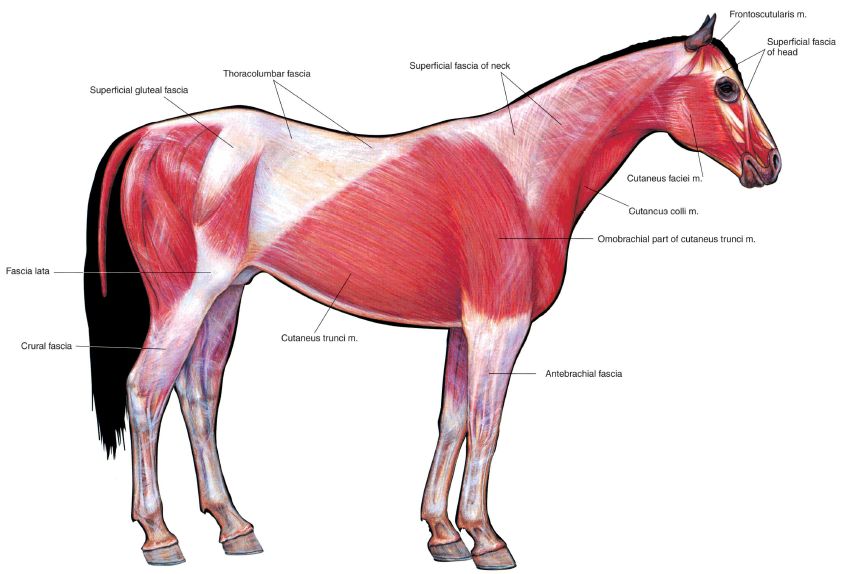
PLATE 1.6 Superficial muscles and veins of the mare. Left lateral view. m = muscle, n = nerve, v = vein

PLATE 1.7 A. Parasagittal section of the equine digit. B. Palmar (plantar) view of major structures of the equine digit. Navicular bursa obscures joining of collateral sesamoidean ligaments on the navicular bone, b = bone
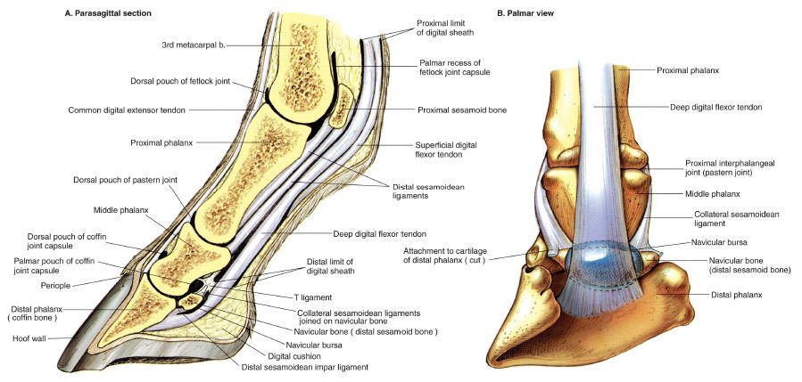
PLATE 1.8 Relations of the hoof. A. Separation of the hoof to show its relations to regions of the coriuni. B. Three-dimensional dissection to show relations of the hoof wall, coronary and laminar corium, and distal phalanx. C. Solar surface of the hoof.

PLATE 1.9 Stay apparatus of the equine forelimb. The continuum of tendons and ligaments with minimal muscular activity stabilizes joints of the forelimb in the standing position, m = muscle

PLATE 1.10 Slay apparatus and reciprocal apparatus of the hindlimb. A. One hindlimb partly flexed with its toe on the ground, and the foot of the opposite limb fixed with minimal muscular activity by the stay apparatus. B. Stay apparatus of the hindlimb. C. The reciprocal apparatus, m = muscle
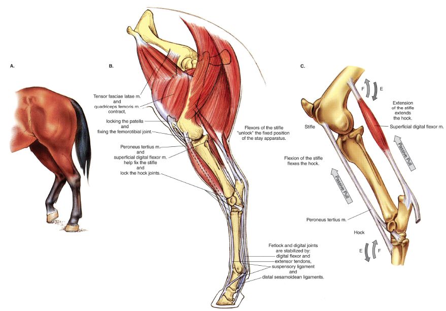
PLATE 1.11 Deep muscles and in situ viscera of the stallion Right lateral view, m = muscle, b = bone
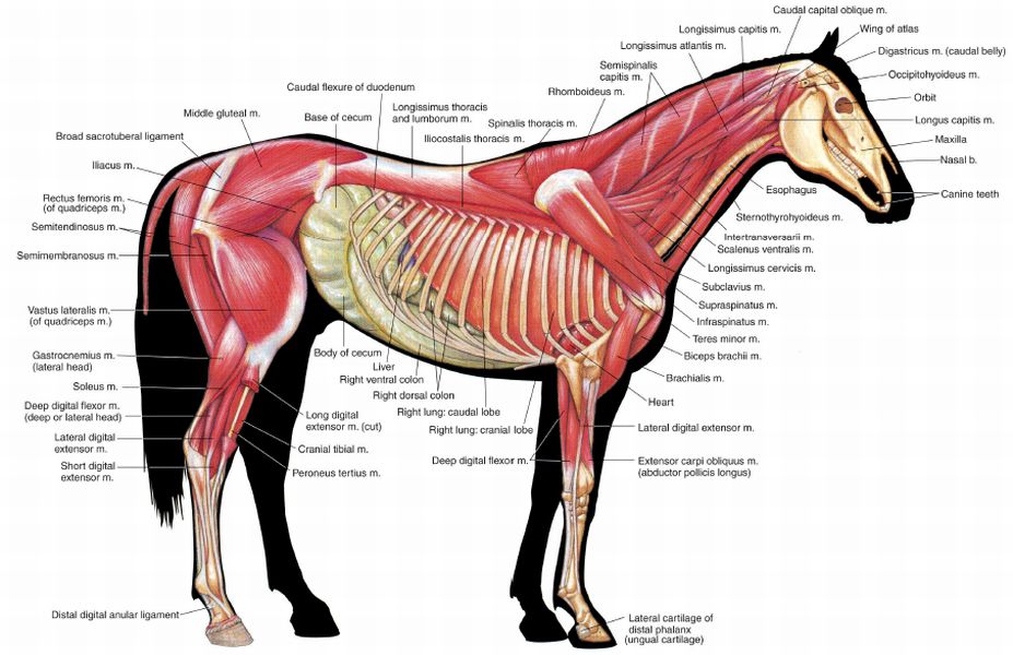
PLATE 1.12 Deep cervical muscles, major joints, and in situ viscera of the mare. Left lateral view, n = nerve, v = vein, m = muscle, a = artery, j = joint, lig = ligament

PLATE 1.13 Median section of the horse's head. Nasal septum mostly removed, b = bone, m = muscle

PLATE 1.14 A. Occlusal (grinding) surfaces of an equine lower first incisor tooth related to continuous eruption and wear. Approximate levels at advancing ages indicated on a longitudinal section. B. Complete dentition of the male horse circa 5 years of age.

PLATE 1.15 Isolated stomach and intestines of the horse.

PLATE 1.16 Equine cecum, large (ascending) colon, and transverse colon in situ. A. Right lateral view. B. Left lateral view.

PLATE 1.17 Clinical condition: Right dorsal displacement of the large colon. A. Right lateral view. B. Dorsal view. This displacement is a common cause of colic in adult horses. Most commonly, the large colon moves from the left side of the abdomen, courses caudad between the right body wall and the cecum, and comes to lie again in the left portion of the abdomen with the pelvic flexure facing toward the diaphragm. In many cases, the pelvic flexure will not migrate that far craniad and will instead be located in the caudal aspect of the abdomen on either side of the body or the median plane.
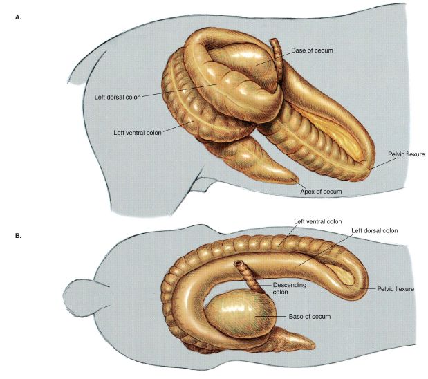
PLATE 1.18 Clinical condition: Left dorsal displacement of the large colon. A. Left lateral view. B. Cross-section of the left side of the abdomen, Caudocranial view. C. Dorsal view. In this displacement, the left colon moves dorsad and becomes entrapped over the nephrosplenic ligament The abnormal position of the left colon can often be confirmed by rectal examination, and, many times, left dorsal displacement can be corrected by anesthetizing and rolling the horse to free the entrapment.

PLATE 1.19 Reproductive organs, urinary organs, liver, heart, and adjacent major vessels related to the skeleton of the stallion. Intestines and lungs are removed. Right lateral view, v = vein, a = artery, m = muscle
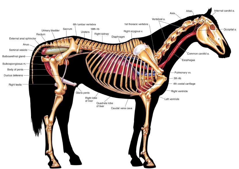
PLATE 1.20 Heart and some adjacent major vessels, abdominal and pelvic viscera, and udder (mammary glands) of the mare. Intestines and lungs arc removed. Left lateral view, a = artery, v = vein, m = muscle

PLATE 1.21 Relations of the reproductive organs of the stallion. Median section, right lateral view, m = muscle
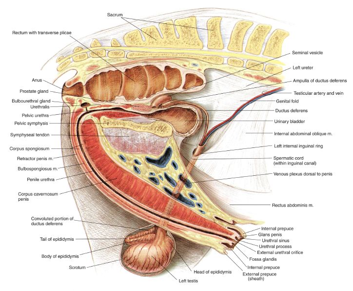
PLATE 1.22 Relations of the reproductive organs of the mare. Partial median section. l.eft lateral view, m = muscle
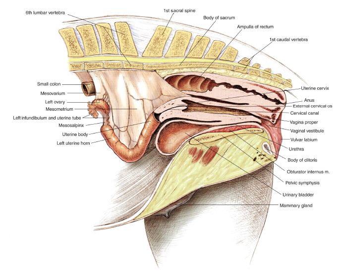
PLATE 1.23 Neonatal organs of the foal. Left lateral view.

PLATE 1.24 Major arteries of the marc. Left lateral view, a = artery

PLATE 1.25 Major veins of Ihe stallion. Portal system excluded. Right lateral view.
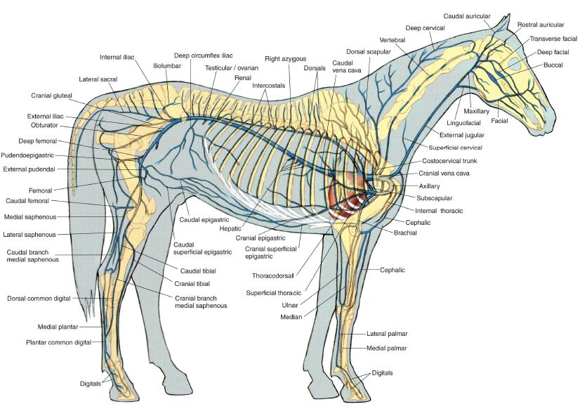
PLATE 1.26 Lymph nodes and vessels of the horse. Right lateral view. Arrows indicate the flow of lymph. Lymph node groups in the horse consist of up to dozens of lymph nodes ranging in size from a few millimeters to 2 centimeters in diameter. In = lymph node
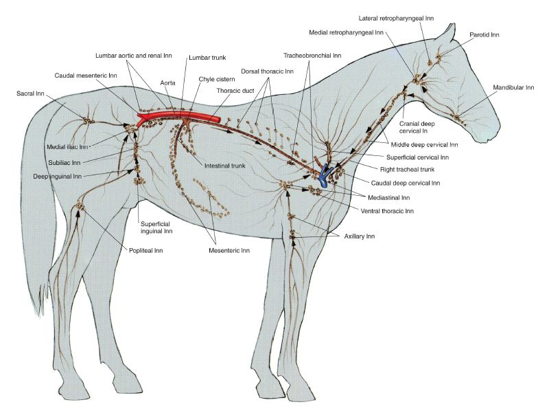
PLATE 1.27 Central and somatic nervous system of the stallion. Right lateral view.
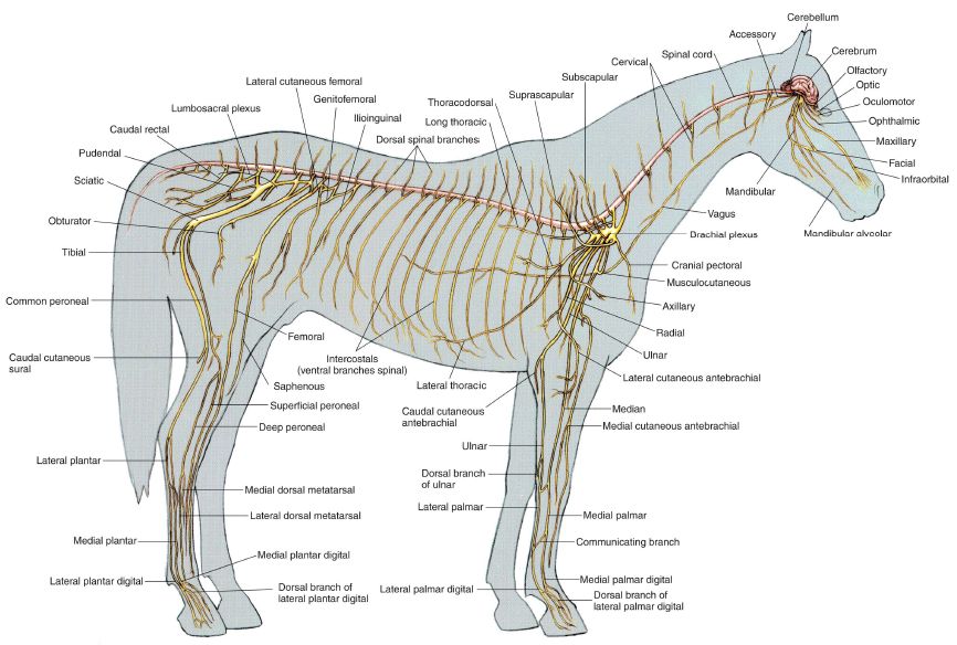
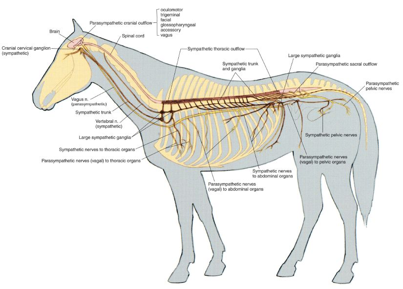
PLATE 1.28 Autonomic nervous system of the mare. Left lateral view, n = nerve