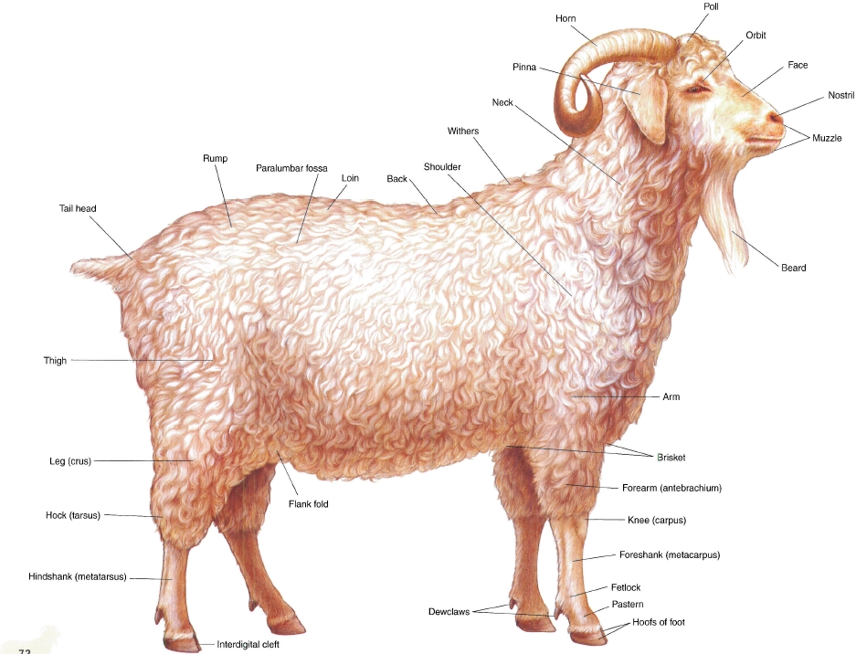
PLATE 4.1 Right lateral view of an Angora buck (billy).
PLATE 4.2 Left lateral view of a Toggenberg doe (nanny). Dorsal vertebral regions are indicated, v = vein
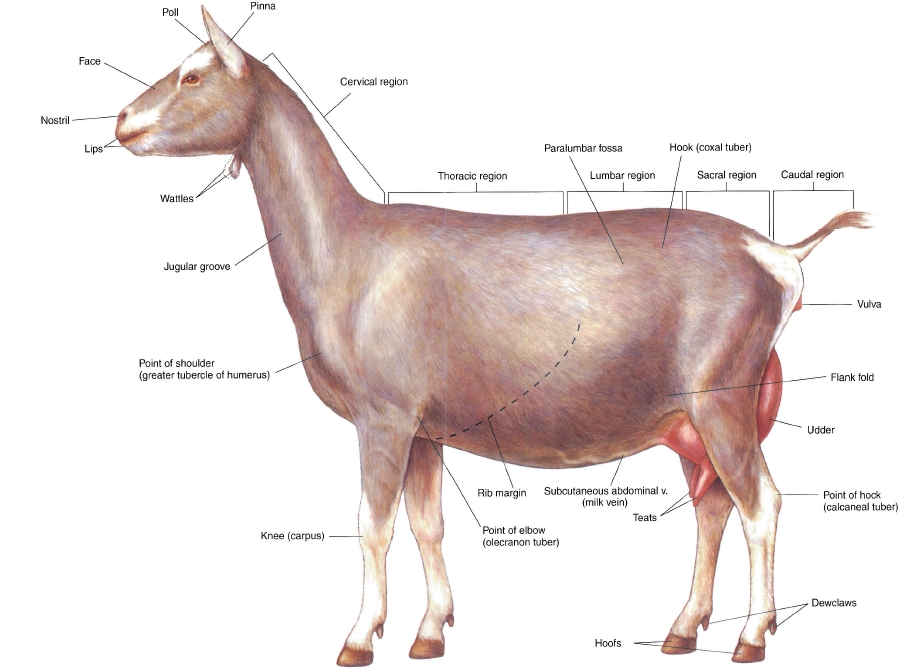
PLATE 4.3 Body regions of the goat. Right lateral view.
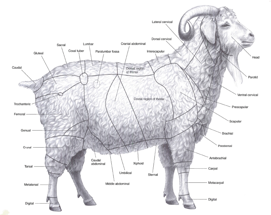
PLATE 4.4 Skeleton of the goat. Left lateral view, b = bone, C = cervical vertebra, T = thoracic vertebra, L = lumbar vertebra
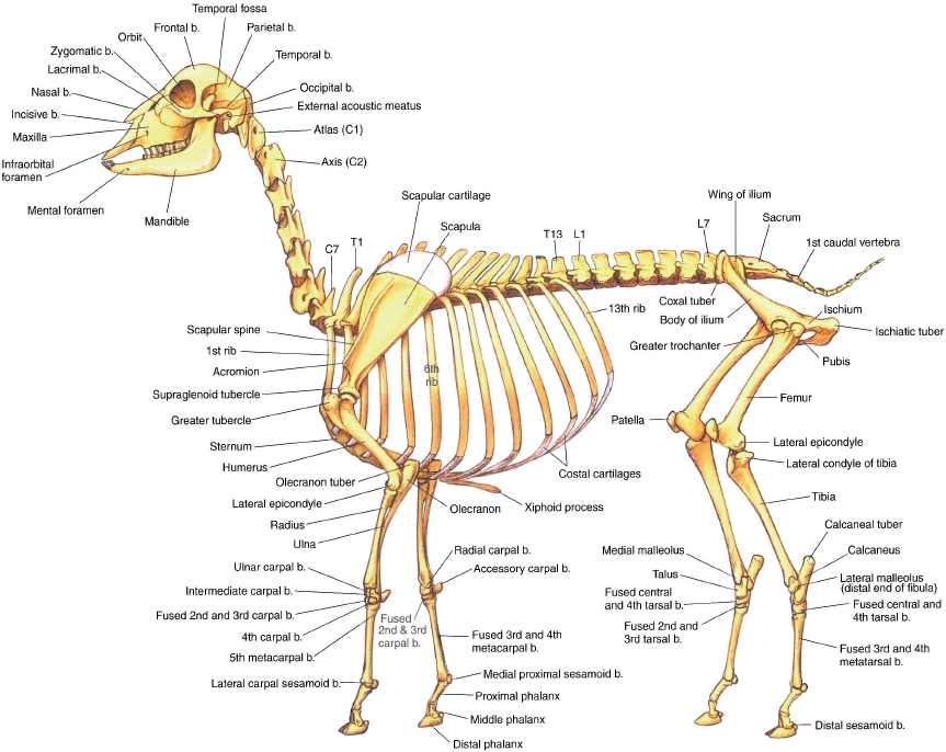
PLATE 4.5 Cutaneous muscles and major fasciae of the buck. Right lateral view, m = muscle
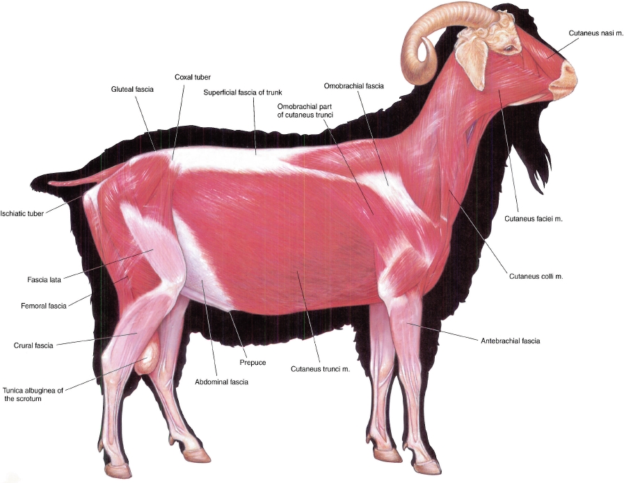
PLATE 4.6 Superficial muscles and veins of the doe. Left lateral view, m = muscle, v = vein
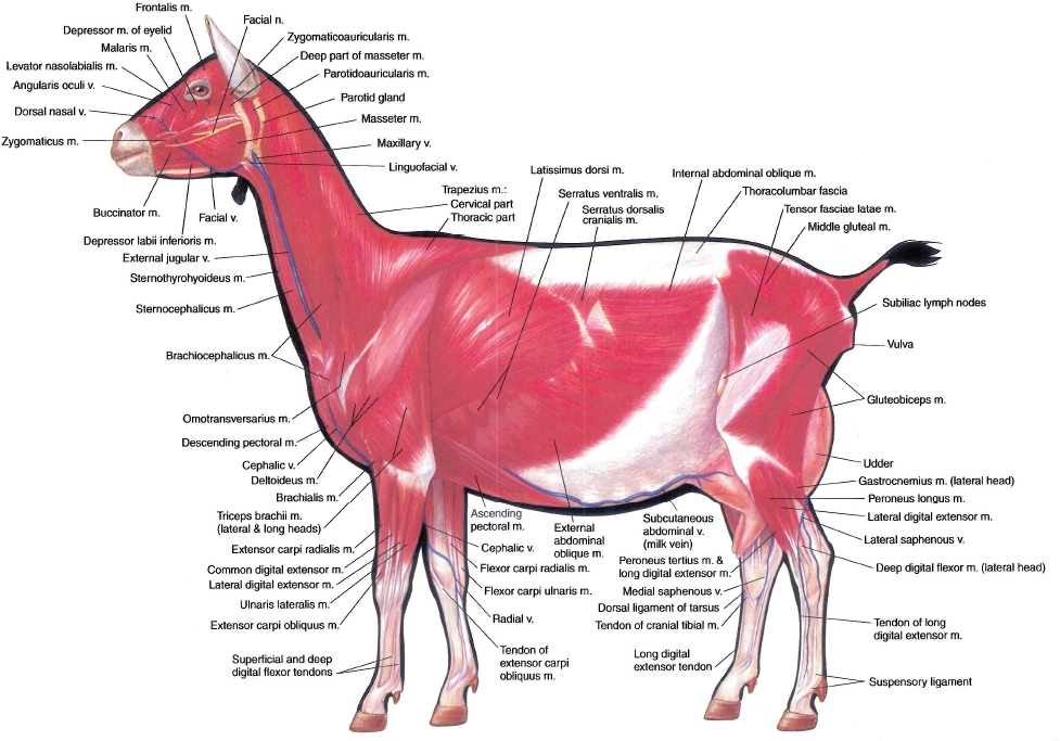
PLATE 4.7 Major structures of the caprine left distal metacarpus and digits. A. Dorsal view, arteries excluded. B. Palmar view, veins excluded, n = nerve, m = muscle, a = artery
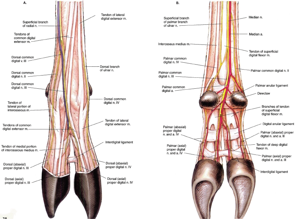
PLATE 4.8 A. Untrimmed hoofs of the goat. B. Trimmed hoofs of the goat. C. Parasagittal section through the fetlock and digit. For artiodactyls, claw is synonymous with hoof. When kept on soft ground, a mature goat's hoofs should be trimmed every 4-5 months, b = bone
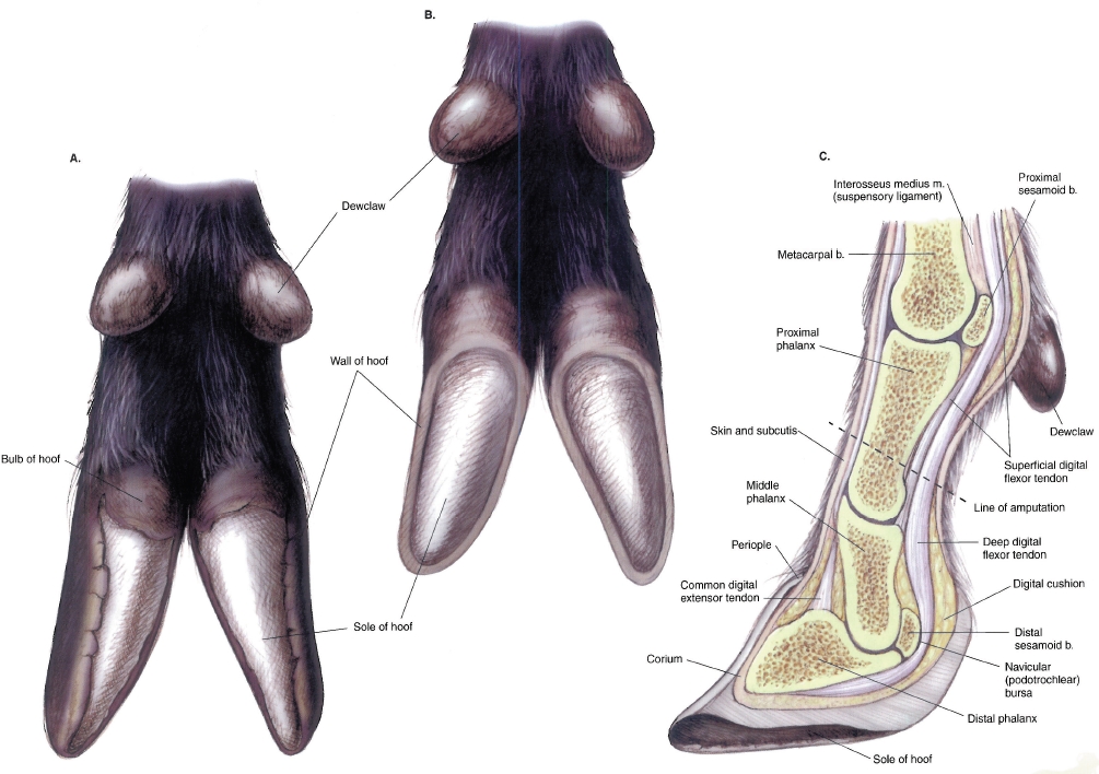
PLATE 4.9Deep muscles and in in situ viscera of the buck. Greter omentum is removed. Right lateral view. m = muscle, n = nerve, a = artery, b = bone
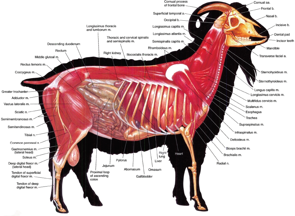
PLATE 4.10 Major structures of the caprine left distal metacarpus and digits. A. Dorsal view, arteries excluded. B. Palmar view, veins excluded, n = nerve, m = muscle, a = artery
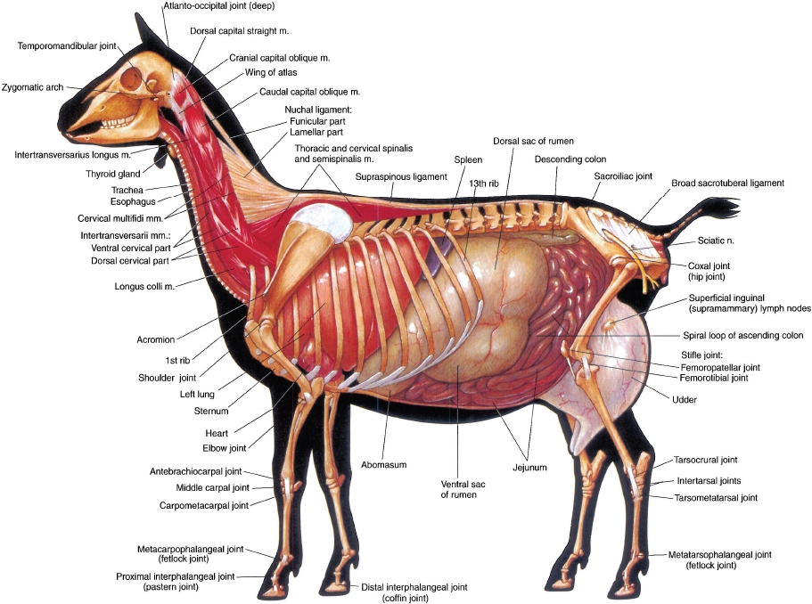
PLATE 4.11 Superficial structures of the goat's head. Dashed line indicates the site of a dehorning incision, a = artery, b = bone, n = nerve, M = molar tooth, P = premolar tooth
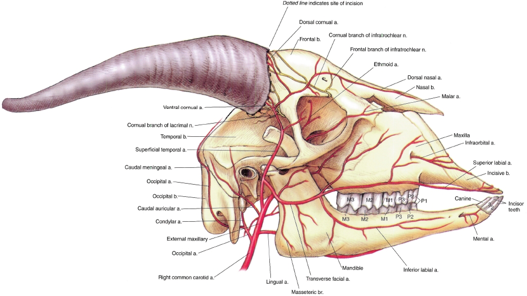
PLATE 4.12 Median section of the caprine head. Most of the nasal septum is removed, m = muscle, b = bone
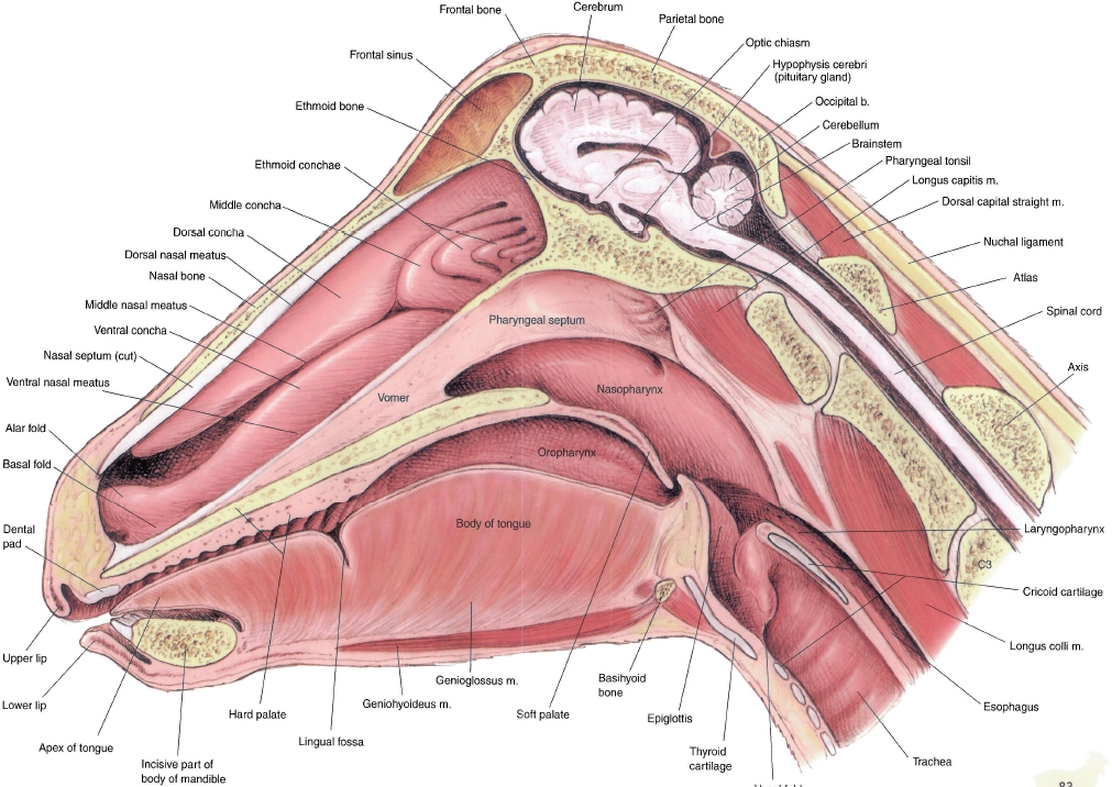
PLATE 4.13 Reproductive organs, abdominal viscera, heart, and adjacent major vessels related to the skeleton of the buck. Intestines and lungs removed. Right lateral view, m = muscle, v = vein, a = artery, b = bone
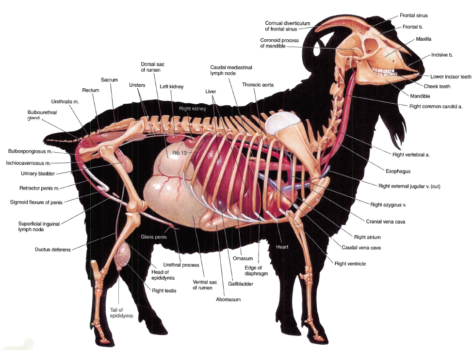
PLATE 4.14 Reproductive organs, abdominal viscera, heart, and adjacent major vessels of the doe. Ribs 2 and 12 and the lungs and intestines are removed. Left lateral view, a = artery, b = bone, v = vein
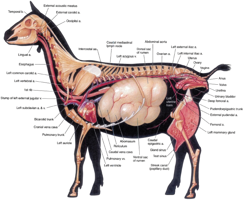
PLATE 4.15 Relations of the reproductive organs of the buck. Right pelvic limb and body wall are removed. Right lateral view, a = artery, m = muscle, v = vein
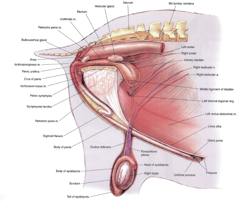
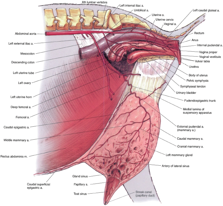
PLATE 4.16 Relations of the reproductive organs of the doe. Median section, a = artery, m = muscle