Chapter Five
STRUCTURE
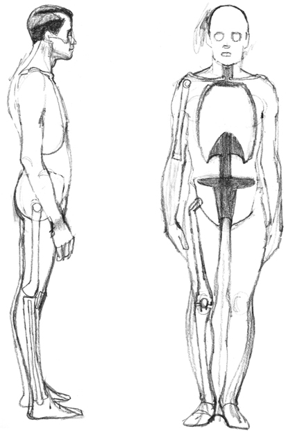
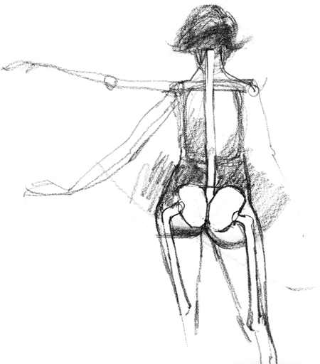
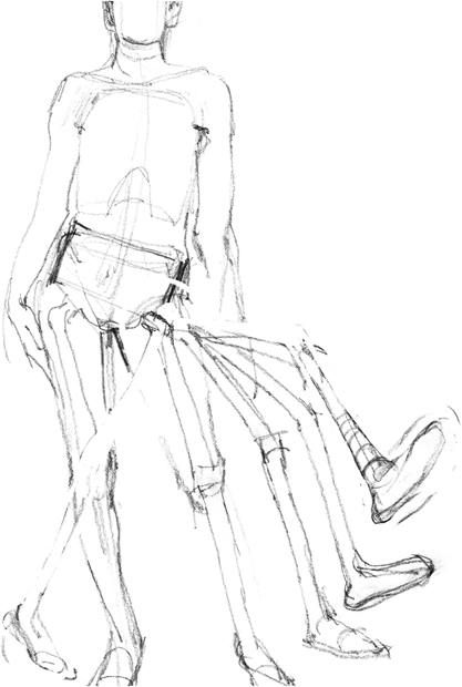
What gives the human figure its balance, flexibility and mobility is its design on an S-curve. The S-curve is constantly visible in the human figure. While a straight line is a rigid and fragile form, a curved shape is capable of flexibility and can manage the crucial task of self-balancing. The drawings here illustrate the profile, front and back views of bodies shifting weight and the way the S-curve controls and allows such actions. It begins at the top of the spine (base of the skull), follows the spine to the pelvis, then emerges at the front of the upper leg to the arch of the foot. The S-shape is also visible when people walk, sit, kneel, lie down, swim, dance, etc.
Muscles
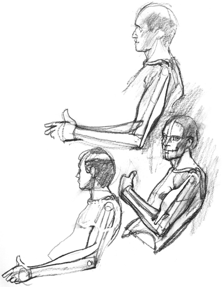
The skeleton supports and is supported by the muscles. This interrelation creates a system of “tension” that is an arrangement of rigid and flexible parts. It is as if the skeleton is bound in a suit of rubber bands that work with and against each other, operating our bones, pulling them back and forth and up and down as we walk, run, move and breathe. The drawings in this chapter include illustrations of muscles joining bones at the elbow and shoulder, pulling them from their origin and causing operation of the bones where they insert. The “origin,” in this terminology, is the static root of the muscle. It is the point from which the muscle pulls. The “insertion” is where action results. For example, when an arm is flexed, the biceps pulls the insertion, located on the forearm just above the inner elbow, towards its origin at the shoulder and causes movement of the forearm. When an arm is extended, the triceps pulls its insertion, located on the forearm just beyond the outer elbow, towards its origin, located behind and below the shoulder. When the triceps is flexed, the biceps is extended. When the biceps is flexed, the triceps is extended. These muscles resemble rubber bands operating opposite each other to give the arm its power and movement.
There are approximately thirty-one points where the bones are visible under the skin. Examples are the elbows, ankles, wrists, knees, pelvis, collar-bones, shoulder blades, knuckles, upper tip of the spine, bridge of the nose and the cheekbones. An artist must be aware of and capable of correctly indicating these points to give a feeling of the boniness of the human form. No matter the degree of fleshiness of a particular human figure, these points are almost always visible. This is particularly true at the joints.
The skeleton is operated by a system of muscles in varying degrees of tension. When opposing muscles are in equal tension, the body is still, as when standing erect. When the tensions are unequal, the body moves.
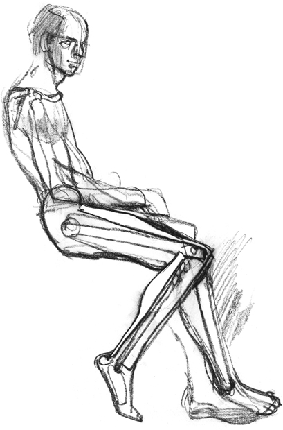
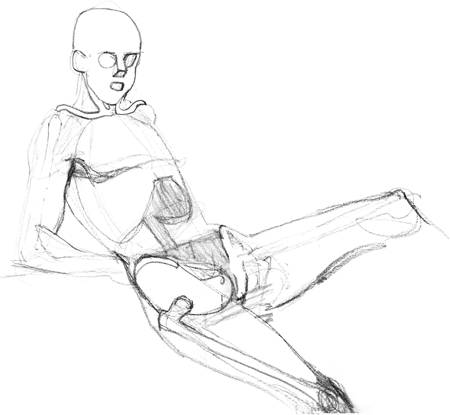
The skeleton forms the inner structure of the body. It is the framework or inner architecture of the human form. The bones that provide protection for the vital organs form container-like masses; the bones that enable bodily motion are long, rigid rods.
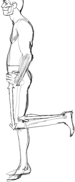
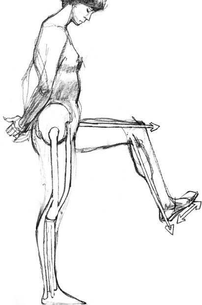
A muscle connects two different bones by passing over one or more joints so that when the muscle contracts it can pull the bones together. The muscle, shown here highlighted on the thigh, has its “origin” on the bony flange of the pelvis and its “insertion” in the ligaments of the kneecap. These ligaments are in turn attached to the lower leg bone. When it is contracted, this muscle will straighten the lower leg.
The muscle highlighted on the lower leg, the calf muscle, has its “origin” on the upper leg bone and its “insertion” on the heel bone. When it contracts it pulls the heel up and toes down, providing the push-off that propels the body forward when waking.
Muscles form opposing pairs. When one contracts, the other relaxes and stretches. The biceps and triceps form such a pair. The biceps is in the front of the upper arm bone and pulls the lower arm toward the shoulder when it contracts. The triceps opposes this motion; when it contracts it pivots the lower arm at the elbow so the lower arm moves away from the shoulder.
Muscles are connected to bones and other muscles in such a way that they seem to flow into each other to form units within larger units.
The body is moved by muscles pulling in opposite directions. These visible lines of thrust and counter-thrust give a healthy body a sense of energy or action even when motionless.
Muscles are arranged not only in opposing pairs, but also often in alternating masses. For example, the large muscles of the thigh are massed in front, while the large muscles of the lower leg are massed behind, creating a graceful S-curve.
The body is incredibly complex; it's the artist's job to understand that complexity in simpler shapes and mechanisms. In these drawings, you can see a simplified view of the body's inner structure.
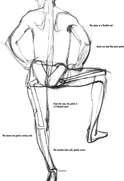
The rib cage is a protective and flexible container for the vital organs such as the lungs and heart. It supports the chest muscles, the shoulder muscles and the back muscles. The figure is unified by the spine, which is a flexible connecting tube made up of moving parts called vertebrae.
The pelvis, as illustrated, is a simple bowl-shaped, rigid container that gives the body a unifying, supportive foundation. In the pelvis is the intersection of the shifting upper and lower fields of the body. The figure cannot be drawn convincingly if the pelvis has been located or rendered inaccurately.
Joints
As discussed in the previous chapter, there are, for the artist's purposes, three categories of skeletal joints: ball-and-socket, hinge and irregular.
The ball-and-socket joints permit roughly 95 percent freedom of action. They provide adjacent limbs the ability to extend in a nearly complete 360-degree circle. The disadvantage of this range of motion is that these joints would be relatively unstable if they weren't surrounded by the largest muscles in the body.
The collarbone and the shoulder blade meet to form a cup-shaped socket at the top of the shoulder. Into this inserts the rounded end of the upper arm bone, completing a ball-and-socket joint. Covering this joint and keeping it in place is the deltoid muscle. In a similar manner, the rounded end of the upper leg bone inserts into the cup-shaped socket of the pelvis. It is supported in this position by the buttocks and a variety of lesser muscles.
The hinge joints such as the knee or elbow bend only in one direction and are less flexible but more rigid and stronger than the ball-and-socket joints. They lock into a rigid position and provide exceptional leverage. The arm is a lever with its power (insertion of the biceps) located between the fulcrum (the elbow) and the point of resistance (the hand). This provides us with an exceptional ability to pull objects in one direction — towards the body. The elbows allow movement only in two directions. They are highly specialized for activities such as pulling roots out of the ground and tearing fruit from branches. The legs, which also possess this highly specific ability to lock into position because of their hinge-type knee joints, guarantee balance and support as the body moves about. The need for stable joints explains why these limbs are hinged in this way at these locations.
Irregular joints are found in the wrist and ankle, where a number of small bones are located, allowing for the rather complex movements of the hand and foot.
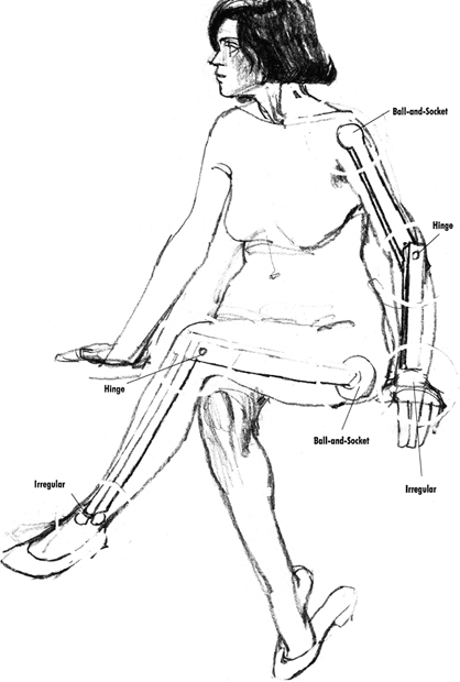
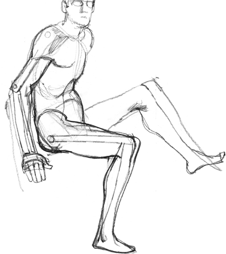
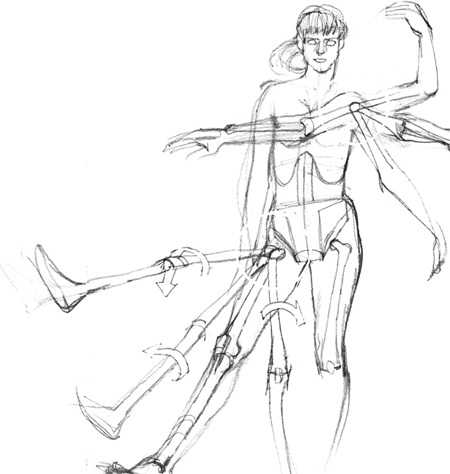
The hip joint and the shoulder are both ball-and-socket joints. These joints have a very wide range of movement. Notice that both the arm and the leg are connected to the trunk by ball-and-socket joints.
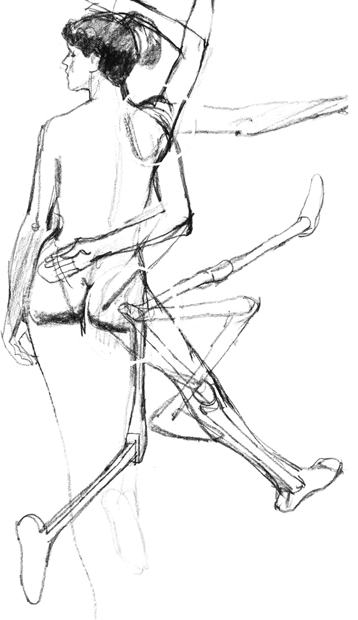
Because the knee and elbow allow movement only along one axis, any rotation of the limbs must occur at their ball-and-socket linkage with the trunk of the body.
The wide range of movement at the hip gives the figure great mobility as well as grace. Because a ball-and-socket joint is not a strong joint, the hip is surrounded by the largest muscles of the body, the thigh and buttock muscles.
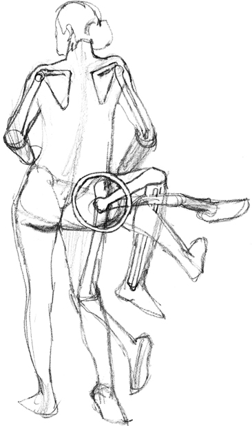
Drawing a figure with limbs shown in multiple positions is excellent practice. The hip joint allows the foot and leg to describe a kind of cone of motion.