A Brief Introduction to Your Brain and Brain SPECT Imaging
ADD is a brain-based disorder. As such, it is important to learn about your brain and how we look at it. The human brain typically weighs about three pounds and it has the consistency of soft butter. It is housed in a very hard skull that has many sharp boney ridges and was never meant to hit a soccer ball or be in the ring with a 300-pound mixed martial arts fighter who wants to literally smash your head repeatedly against the canvas.
The most noticeable structure in human brains is the cerebral cortex, the wrinkly mass that sits atop and covers the rest of the brain. The cortex has four main areas or lobes: frontal, temporal, parietal, and occipital.
The frontal lobes consist of the motor cortex, which is in charge of movement; the premotor cortex, which plans movement; and the prefrontal cortex (PFC), which is considered the executive part of the brain. The PFC is the most evolved part of the human brain: the center of focus, forethought, judgment, organization, planning, impulse control, empathy, and learning from the mistakes you make. It makes up 30 percent of the human brain. Compare that to our closest cousin, the chimpanzee, whose PFC is only 11 percent; a dog, whose PFC is just 7 percent; or a cat, whose PFC is only 3.5 percent. It’s a good thing a cat has nine lives, because their PFC isn’t going to do much to keep them out of trouble.
Outside View of the Brain
left side view of brain

Inside View of the Brain

The temporal lobes, underneath your temples and behind your eyes, are the seat of auditory processing, naming things, getting memories into long-term storage, and emotional reactions. They are called the “What Pathway” in the brain as they name what things are. The parietal lobes, to the top side and back of the brain, are the centers for sensory processing and direction sense. They are called the “Where Pathway” because they help us know where things are. And the occipital lobes, at the back of the cortex, are concerned primarily with vision. Information from the world enters the back part of the brain (temporal and parietal lobes), is processed, and then passes to the front part of the brain for decision making. The cerebellum at the back bottom part of the brain is involved with motor and thought coordination. It is essential for processing complex information.
Sitting beneath the cortex is the deep limbic or emotional system. This is the part of the brain that colors our emotions and is involved with bonding, nesting, and emotions. Also, beneath the cortex are two large structures called the basal ganglia, involved with motivation, pleasure, and smoothing motor movements. Deep in your frontal lobes is the anterior cingulate gyrus, involved with error detection and sifting attention.
The cortex is divided into two hemispheres, left and right. While the two sides overlap in function, the left side in right-handed people is generally the seat of language, and tends to be the analytical, logical, detail-oriented part of the brain; while the right hemisphere sees the big picture and is responsible for hunches and intuition. The opposite is often, but not always, true in left-handed people.
BRAIN SPECT IMAGING
Over the last twenty-three years the Amen Clinics have used a variety of tools to look at and evaluate brain function. The most common of which is a study called brain SPECT imaging. SPECT (single photon emission computed tomography) is a nuclear medicine study that evaluates blood flow and activity patterns in the brain. It looks at how the brain works. It is different than CAT scans and MRIs, which are anatomy studies that look at the structure of the brain. SPECT looks at function and, in my opinion, is much more helpful in psychiatric illnesses like ADD.
SPECT scans basically show us three pieces of information about activity and blood flow in the brain:
- healthy activity
- too little activity
- too much activity
It also helps us see if the brain has been hurt from a physical trauma, or if it has had some sort of toxic exposure. At the clinics we read each scan with a high level of detail. For illustration purposes in this book I will show you two types of scans: surface and active. Surface scans look at the outside surface of the brain, and show areas of low activity. Active scans show areas of high or increased activity.
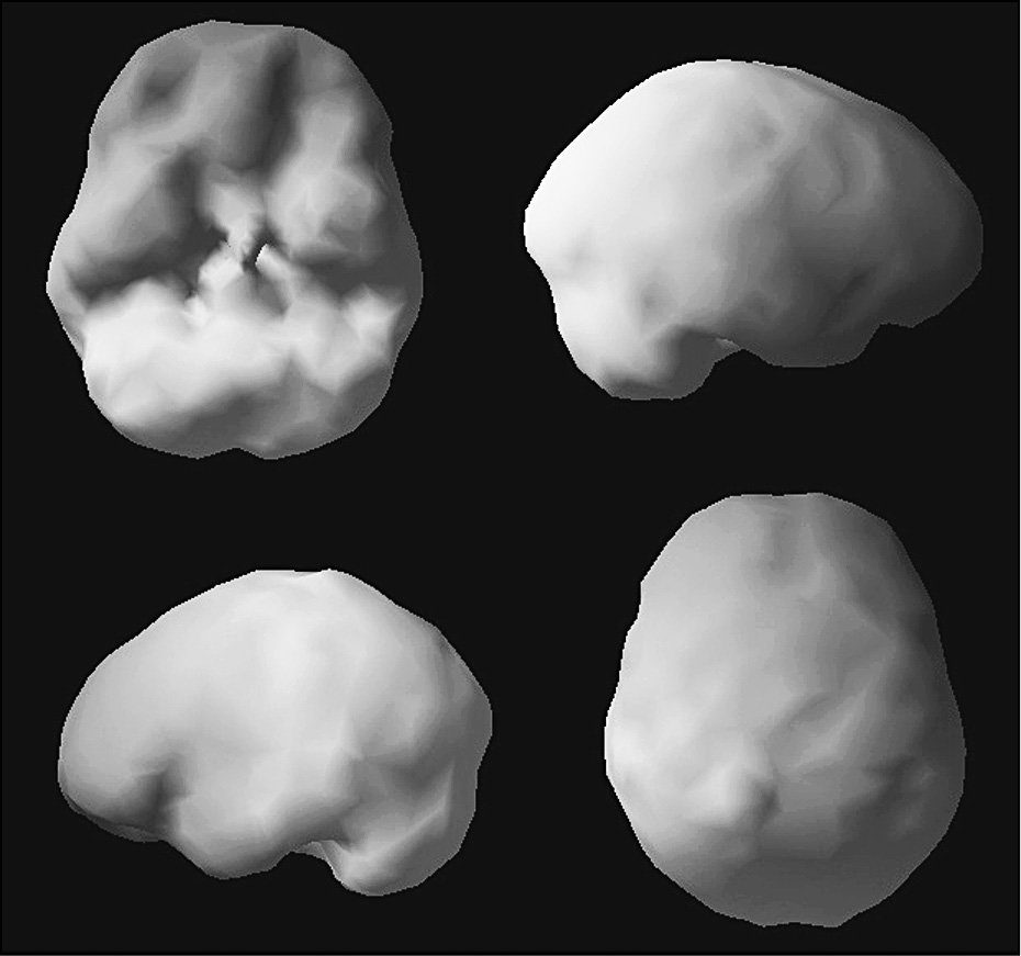
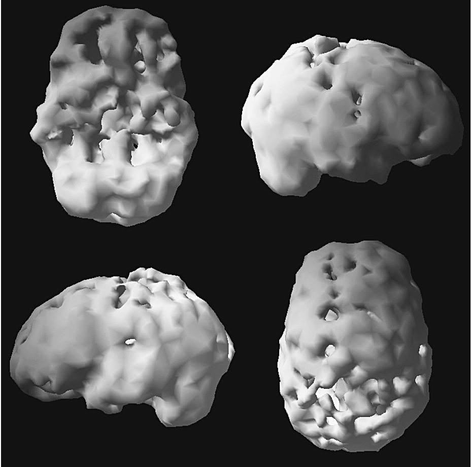
Throughout the book you will see many brain SPECT images. Here is an example of two “surface” SPECT scans. A healthy scan shows full, even, symmetrical activity. The top left image is looking underneath the brain. The bottom right image is looking down from the top, the other images look at the brain from the side. The surface scans look at the top 45 percent of brain activity. Anything below that shows up as a hole or a dent. So, the scan images with “holes” throughout the book are typically not missing activity, they are low in activity. The image next to the healthy one is of a person who was affected by toxicity from drug and alcohol exposure.
Healthy vs Toxic Outside Surface SPECT Studies

Healthy

Drug affected
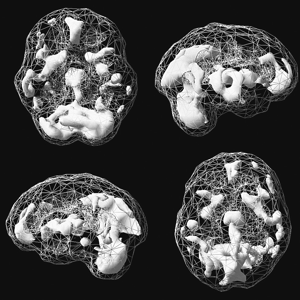
Here is an example of a set of “Active” SPECT scans. The grey background is average activity. The white areas represent the top 15 percent of brain activity, which in adults is mostly in the back, bottom part of the brain, the cerebellum. Overall, children have much more activity than adults. Again, the top left image is looking underneath the brain. The bottom right image is looking down from the top, the other images look at the brain from the side.
Healthy vs Hyperactive Active SPECT Studies

Healthy

Anxiety with obsessive features, note increased activity deep in the brain
For more information on brain SPECT imaging and the research behind it, visit amenclinics.com/the-science/brain-spect-abstracts.