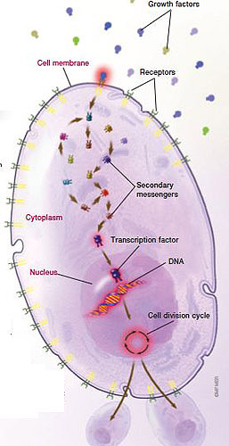
The normal functioning of a cell is controlled at many levels, including its surface (cell membrane), interior (cytoplasm) and growth control center (nucleus). Changes that may lead to cancer development and growth can occur at any of these levels.
Cell membrane
At the cell surface, chemical messengers that signal a cell to divide (growth factors) and nutrients bind to receptors on the cell surface. Cancer cells overexpress receptors that capture growth factors and nutrients.
Cytoplasm
Signals from growth factors are sent to the cell nucleus by way of a cascading series of so-called secondary messengers. Cancer cells can cause numerous growth-promoting changes in these signaling pathways.
Nucleus
Within the tightly coiled DNA of each cell are the genetic instructions to make all of the proteins a cell needs to carry out its work. Which genes are turned on depends on the signals received from the cytoplasm, conveyed by transcription factors. Transcription factors bind to the DNA of targeted genes.
Cell division is controlled within the nucleus, too. Before a cell can divide, it must pass through a tightly governed cycle with several checkpoints. This is to ensure that an injured cell doesn’t divide until damaged DNA has been repaired. Cancer cells lack these checkpoint mechanisms, allowing altered cells to grow and proliferate.
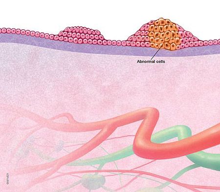
Cancer is characterized by the overgrowth of abnormal cells, a multistep process called carcinogenesis. Over time, the abnormal cells accumulate into a mass, called a growth or tumor, that can invade nearby normal tissue and spread.
Normal cell (left)
Normal cells grow and divide in an orderly fashion.
Hyperplasia (middle)
The intricate system of cell development and growth is disrupted, causing an overproduction of normal-appearing cells.
Atypical hyperplasia (right)
As the excess cells stack upon one another, some start taking on an abnormal appearance.
Noninvasive cancer (left)
The abnormal cells continue to change in appearance and multiply, evolving into cancer. The cancer remains confined within normal borders.
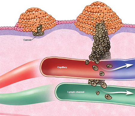
Invasive cancer (right)
As the cancer cells invade deeper into surrounding tissue, eventually they can spread to nearby lymph channels and tiny blood vessels (capillaries), which can carry cancer cells to other parts of the body.
Stage I
Cancer is 2 centimeters (cm) in size or less with no spread to the lymph nodes.*
Stage II
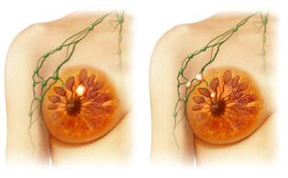
Cancer is 2.1 cm to 5 cm in size, or has spread to up to three lymph nodes under the arm or to lymph nodes under the sternum, or both. Cancer greater than 5 cm with no spread to the lymph nodes is also stage II.
Stage III
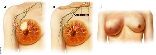
Stage III cancers include a number of criteria that make it a broad category. Here are three examples: tumor greater than 5 cm in size with involvement of at least one lymph node under the arm (A); spread to lymph nodes above the collarbone (B); spread to breast skin causing swelling and redness, known as inflammatory breast cancer (C).
*Lymph nodes containing cells that look like they came from the breast but are less than 0.2 mm are considered negative lymph nodes because there’s no good evidence these are established cancers.
Stage IV
Cancer has spread to distant sites, such as the lungs, liver or bone.
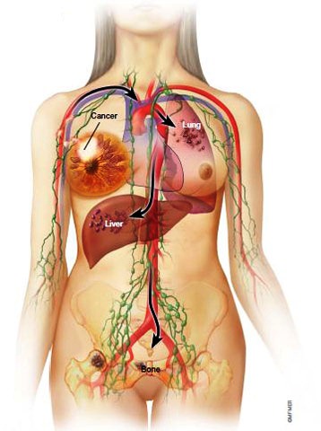
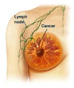
Cancer cells from a breast tumor can spread throughout the body by way of the tiny blood vessels (capillaries), which empty into veins, and by way of lymph channels, which drain into lymph nodes. Fluid and cancer cells from the lymphatic system eventually empty into large veins, which in turn empty into the heart. From the heart, cancer cells can spread to the rest of the body by way of the arteries.
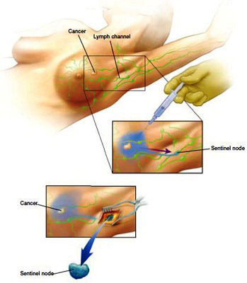
In this procedure, a dye is injected into the area of the tumor. The dye is absorbed into nearby lymph channels. The first lymph nodes to collect the dye, called the sentinel nodes, likely are the first to receive drainage from the breast tumor. The sentinel nodes are removed and examined for cancer cells. A small amount of radioactive material may be used instead of a dye, or a combination of the two.
The two main types of surgery to treat breast cancer are a lumpectomy (left), in which only the tumor is removed, and a mastectomy (right), in which the entire breast is removed.

Radiation therapy uses a high-energy radiation source to kill cancer cells or interfere with their ability to grow and divide.
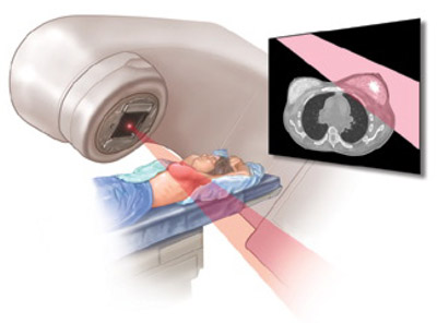
With external beam radiation, the radiation comes from a machine called a linear accelerator located outside of the body. Radiation beams pass through the skin to reach the tumor site.
Medications (therapeutic agents) that kill or slow down cancer cells work at many sites within the cell. They can interfere with processes at the cell membrane. They can block the signaling cascades occurring within the cytoplasm that stimulate cell growth and division. And they can interfere with the normal functioning of DNA so that new proteins can’t be made and new cells can’t be formed.
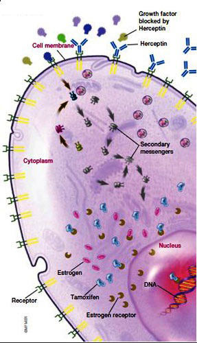
Cell membrane
Medications that act at the outer layer of a cell (cell membrane) block the binding of growth factors to their receptors. Or they bind to the receptors but inhibit their activation. This prevents the transmission of growth signals to the interior of the cancer cell. The drug trastuzumab (Herceptin) is an example. It’s a monoclonal antibody that binds to HER2 (also called erbB2), a growth factor receptor on the cell membrane, shutting down the receptor. Other medications being tested inhibit the erbB1 receptor, or both erbB1 and erbB2.
Cytoplasm
Several new cancer therapies target the secondary messengers within a cell that transmit growth signals from the cell membrane to the nucleus.
Nucleus
Many medications work directly at the nucleus, the control center of the cell. Most chemotherapy medications, for example, interfere with the function of DNA. Some drugs bind directly to DNA, preventing it from unfolding to manufacture proteins or to be duplicated to produce new cancer cells. Other chemotherapy drugs cause breaks in the DNA structure and inhibit the enzymes needed to repair the damage. Still other medications block the protein scaffolding that oversees cell division. The drug tamoxifen interferes with the ability of the receptor for the hormone estrogen to act as a transcription factor and turn on relevant downstream genes. Other agents work within the nucleus to induce cancer cells to undergo cell death.