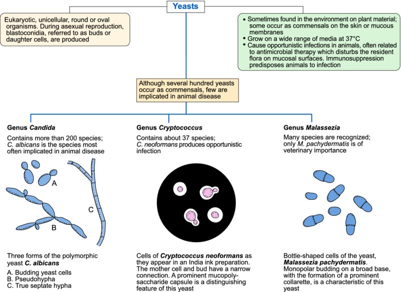
44 Yeasts and disease production

Yeasts are eukaryotic, unicellular, round or oval, single-celled organisms. During asexual reproduction, blastoconidia, also referred to as buds or daughter cells, develop. Yeasts grow aerobically on Sabouraud dextrose agar and species capable of tissue invasion grow well at 37°C. Colonies, which are usually moist and creamy in texture, resemble large bacterial colonies. In addition to their environmental habitat on plants or plant material, yeasts also occur as commensals on the skin or mucous membranes of animals. Immunosuppression or factors such as antimicrobial therapy which disturb the resident flora on mucosal surfaces may facilitate yeast overgrowth leading to tissue invasion. Yeasts of importance in animal disease are Candida species (particularly C. albicans), Cryptococcus neoformans and Malassezia pachydermatis. Macrorhabdus ornithogaster (formerly referred to as ‘megabacteria’) is a yeast found in the proventriculus of several avian species. It is associated with ‘going light’ in budgerigars, a fatal disease characterized by progressive weight loss.
Candida species
Although there are more than 200 species in the genus Candida, the species most often implicated in animal disease is Candida albicans. It grows aerobically at 37°C on a wide range of media, including Sabouraud dextrose agar. Colonies are composed of budding oval cells approximately 5.0 × 8.0 μm. In animal tissues, C. albicans may exhibit polymorphism in the form of pseudohyphae or hyphae. The transition from budding to hyphal forms probably facilitates tissue penetration and increases resistance to phagocytosis due to the larger size of the hyphae.
Opportunistic infections with Candida species occur sporadically. Localized mucocutaneous tissue invasion referred to as thrush can occur in the oral cavity or in the gastrointestinal and urogenital tracts. Predisposing factors include defects in cell-mediated immunity, concurrent disease, disturbance of the normal flora by prolonged use of antimicrobial drugs and damage to the mucosal surface from indwelling catheters. The clinical conditions attributed to C. albicans are presented in Table 44.1. Suitable specimens for culture and histopathology include biopsy or postmortem tissue samples and milk samples. Tissue sections, stained by the PAS or methenamine silver methods, may reveal budding yeast cells or hyphae. Culture is carried out aerobically at 37°C for up to five days on Sabouraud dextrose agar. Criteria used for identification of C. albicans isolates include characteristic colonies yielding budding yeast cells, growth on media containing cycloheximide, biochemical profile, germ tube production when incubated for two hours in serum at 37°C and chlamydospore production in cornmeal agar.
Table 44.1 Clinical conditions associated with Candida albicans.
| Hosts | Clinical conditions |
| Pups, kittens, foals | Mycotic stomatitis |
| Pigs, foals, calves | Gastro-oesophageal ulcers |
| Calves | Rumenitis |
| Dogs | Enteritis, cutaneous lesions |
| Chickens | Thrush of the oesophagus or crop |
| Geese, turkeys | Cloacal and vent infections |
| Cows | Reduced fertility, abortion, mastitis |
| Mares | Pyometra |
| Cats | Urocystitis, pyothorax |
| Cats, horses | Ocular lesions |
| Dogs, cats, pigs, calves | Disseminated disease |
Cryptococcus neoformans
Although the genus Cryptococcus contains more than 30 species, only C. neoformans produces opportunistic infections. The yeast cells are round to oval and 3.5–8 μm in diameter. A daughter cell is formed as a bud from the mother cell on a narrow neck. When recovered directly from affected animals, the yeasts have thick mucopolysaccharide capsules. Cryptococcus species are aerobic and form mucoid colonies on a variety of media, including Sabouraud dextrose agar.
Cryptococcus neoformans is considered to be a species complex consisting of C. neoformans var. grubii (serotype A), C. neoformans var. neoformans (serotype D) and subspecies C. gatii (serotypes B, C). Infection with C. neoformans occurs through inhalation of yeast cells in contaminated dust. Virulence factors of C. neoformans include the capsule, which is antiphagocytic, the ability to grow at mammalian body temperature and the production of phenol oxidase. Infection with C. neoformans is usually associated with defective cell-mediated immunity.
Nasal, cutaneous, neural and ocular forms of cryptococcosis are recognized in cats. The disease in dogs, which is less common than in cats, is often disseminated with neurological and ocular signs. Surgical removal combined with parenteral antifungal drugs is the usual method for treating cutaneous cryptococcosis. Therapy should continue for at least two months.
When first isolated, colonies of Cryptococcus species are mucoid due to the presence of capsular material. They may have a cream, tan or yellowish appearance. Budding yeasts with wide capsules can be demonstrated in India ink preparations. Identification criteria for C. neoformans include the ability to grow at 37°C, brown colonies on birdseed agar, and melanin demonstrable in cell walls using the Fontana–Masson stain on tissue sections.
Malassezia pachydermatis
Malassezia species, commensals on the skin of animals and humans, are aerobic, non-fermentative, urease-positive yeasts which grow at 35–37°C. One species, Malassezia pachydermatis, is of veterinary importance. The cells of M. pachydermatis, which are bottle-shaped, thick-walled and up to 6.5 μm in length, reproduce by monopolar budding on a broad base.
Malassezia pachydermatis is an opportunistic pathogen associated with two clinical conditions, otitis externa and dermatitis in dogs. Colonization and growth of the organism in these locations may be associated with immunosuppression and other predisposing factors such as persistently moist skin folds, poor ear conformation and excessive use of antibiotics. When the yeast cells are present in high numbers on the skin, they apparently induce excessive sebaceous secretion, a feature of seborrhoeic dermatitis. Treatment with miconazole–chlorhexidine shampoo or a combination of topical and oral ketoconazole may be effective. In otitis externa, the production of proteolytic enzymes by M. pachydermatis results in damage to the epithelial lining of the ear canal. The condition is characterized by a dark pungent discharge from the ear canal and intense pruritis.
Numerous, characteristic yeast cells may be demonstrable in exudates or impression smears stained with methylene blue. Malassezia pachydermatis can be cultured aerobically at 37°C for four days on Sabouraud dextrose agar containing chloramphenicol. Identification criteria include colonial appearance, growth without the need for lipid supplementation and characteristic microscopic appearance.