45 Dimorphic fungi

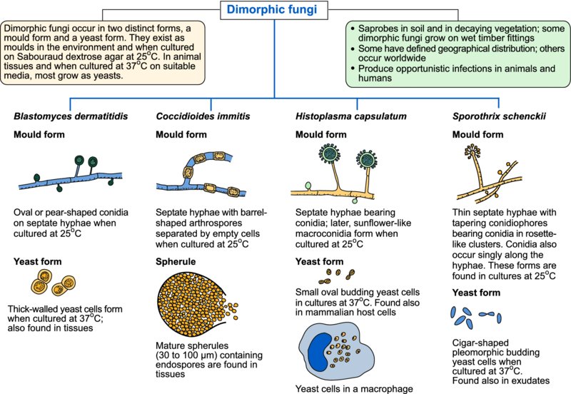
Some fungi, referred to as dimorphic fungi, occur in two distinct forms, a mould form and a yeast form. They exist as moulds in the environment and when cultured on Sabouraud dextrose agar at 25 to 30°C. In animal tissues, and when cultured at 37°C on brain–heart infusion agar with the addition of 5% blood, most grow as yeasts after conversion from the more stable mould form. The dimorphic fungi most often associated with disease in domestic animals are Blastomyces dermatitidis, Histoplasma capsulatum and Coccidioides immitis. The spores of these dimorphic fungi usually enter hosts by the respiratory route. A variant of H. capsulatum, referred to as H. farciminosum, generally enters through skin abrasions and produces lymphocutaneous lesions in horses. Sporothrix schenckii also produces opportunistic lymphocutaneous infections in animals. Table 45.1 summarizes important features of dimorphic fungi associated with disease in animals and humans.
Table 45.1 Dimorphic fungi which are associated with disease in animals and humans.
| Blastomyces dermatitidis | Histoplasma capsulatum | Histoplasma farciminosum | Coccidioides immitis and C. posadasii | Sporothrix schenckii | |
| Disease | Blastomycosis | Histoplasmosis | Epizootic lymphangitis | Coccidioidomycosis | Sporotrichosis |
| Geographical distribution | Eastern regions of North America, sporadic cases in India and the Middle East | Endemic in the Mississippi and Ohio river valleys, sporadic cases in some countries | Africa, Middle East, Asia | Semi-arid regions of south-western USA, Central and South America | Worldwide, most common in subtropical and tropical regions |
| Usual habitat | Acid soil rich in organic matter | Soil enriched with bat or bird faeces | Soil | Desert soils at low elevation | Dead vegetation, rose thorns, wooden posts, sphagnum moss |
| Main hosts | Dogs, humans | Dogs, cats, humans | Horses, other Equidae | Dogs, horses, cats, humans | Horses, cats, dogs, humans |
| Site of lesions | Lungs, dissemination to skin and other tissues | Lungs, dissemination to other organs | Skin, lymphatic vessels, lymph nodes | Lungs, dissemination to bones, skin and other tissues | Skin, lymphatic vessels |
Blastomyces dermatitidis
Blastomycosis, caused by B. dermatitidis, most commonly affects dogs and humans. The teleomorph of B. dermatitidis is a member of the phylum Ascomycota designated Ajellomyces dermatitidis. The disease is encountered in North America, Africa, the Middle East and India. Infection usually occurs by inhalation and pulmonary blastomycosis is the usual form of the disease. Presenting signs include coughing, exercise intolerance and dyspnoea. Amphotericin B, which may be combined with ketoconazole, is effective if administered early in the course of the disease.
When incubated at 25 to 30°C on Sabouraud dextrose agar, mould colonies are white and cottony, usually becoming brown with age. When incubated at 37°C on brain–heart infusion agar with added cysteine and 5% blood, yeast colonies are cream to tan, wrinkled and waxy. Yeast cells may be demonstrated in cytological and histopathological preparations from affected tissues. Serological assays are available.
Histoplasma capsulatum
Although histoplasmosis, caused by H. capsulatum, occurs in many countries, it is endemic in the Mississippi and Ohio river valleys and in other areas of the USA. Disseminated disease in dogs and cats is probably associated with impaired cell-mediated immunity. Granulomatous lesions may be found in the lungs of both dogs and cats. Clinical signs in affected dogs include a chronic cough, persistent diarrhoea and emaciation. Ketoconazole and amphotericin B can be used for treatment.
Epizootic lymphangitis, caused by H. capsulatum var. farciminosum, occurs in Equidae in Africa, the Middle East and Asia. Horses usually acquire infection from environmental sources through minor skin abrasions on the limbs. Characteristic lymphocutaneous lesions consist of ulcerated discharging nodules, usually located along the course of thickened, hard, lymphatic vessels. Yeast cells of H. farciminosum are found in large numbers in lesions, mainly in macrophages. When cultured at 25 to 30°C on Sabouraud dextrose agar, the mould form of H. capsulatum grows as white to buff colonies with aerial hyphae. Septate hyphae bear small conidia and in mature colonies sunflower-like macroconidia may be present. When cultured at 37°C on brain–heart infusion agar with added cysteine and 5% blood, yeast colonies are round, mucoid and cream-coloured. Budding yeast cells are oval to spherical. Histopathological examination of affected tissues reveals pyogranulomatous foci containing yeast forms.
Coccidioides immitis
The geophilic fungus C. immitis can infect many animal species including humans. Although grouped with the dimorphic fungi, C. immitis is biphasic rather than dimorphic because typical yeast forms are not produced. Large spherules containing endospores develop in tissues. Respiratory infections may follow inhalation of arthrospores.
Clinical infections caused by Coccidioides species are limited to defined arid regions of south-western USA, Mexico, and Central and South America. Isolates from outside the endemic region of the San Joaquin Valley in California (formerly referred to as ‘non-California’ C. immitis) have been found to be sufficiently different to be given separate species status with the name C. posadasii. The domestic species most often affected is the dog. Canine coccidioidomycosis may present with non-specific signs including coughing, fever and inappetence. Dissemination from pulmonary lesions often results in osteomyelitis and lameness. Treatment with azole drugs for at least six months may be effective.
Diagnosis is usually based on clinical findings and histopathology. Spherules of C. immitis may be demonstrated in exudates or aspirates cleared with 10% KOH and also in stained tissue sections. Culturing of Coccidioides species is hazardous due to the production of thick-walled, barrel-shaped arthroconidia which are readily aerosolized.
Sporothrix schenckii
This saprobic fungus, which is widely distributed in the environment, grows on dead or senescent vegetation and on wet timber fittings. Infections caused by S. schenckii occur sporadically in horses, cats, dogs and humans.
Sporotrichosis is a chronic cutaneous or lymphocutaneous disease which rarely becomes generalized. Lymphocutaneous sporotrichosis is the most common form of the disease in horses. Fungal spores usually enter through abrasions in the lower limbs, and nodules, which ulcerate and discharge a yellowish exudate, develop along the course of superficial lymphatic vessels. Subcutaneous oedema in the affected limb may result from lymphatic obstruction. In feline sporotrichosis, nodular skin lesions occur most often on limb extremities, head and tail. Nodules ulcerate and discharge a seropurulent exudate. Sporotrichosis in dogs often manifests as multiple, ulcerated and crusted alopecic cutaneous lesions over the head and trunk.
Direct microscopic examination of exudates from feline lesions usually reveals large numbers of cigar-shaped yeast cells. Infected cats carry the organism in their nose and mouth and on their nails, facilitating transmission through biting and scratching. In exudates from other animals, yeast cells are sparse. Sporotrichosis may be treated with itraconazole, fluconazole or voriconazole.