CHAPTER 104
Biology of the Nervous System
The nervous system has two distinct parts: the central nervous system (the brain and spinal cord) and the peripheral nervous system (the nerves outside the brain and spinal cord).
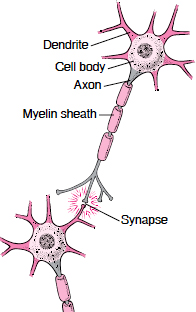
The basic unit of the nervous system is the nerve cell (neuron). Nerve cells consist of a large cell body and two types of nerve fibers:
 Axon: One elongated extension for sending messages as electrical impulses
Dendrites: Usually many branches for receiving impulses
Normally, nerves transmit impulses electrically in one direction—from the impulse-sending axon of one nerve cell to the impulse-receiving dendrites of the next nerve cell. At contact points between nerve cells (synapses), the axon secretes tiny amounts of chemical messengers (neurotransmitters). Neurotransmitters trigger the receptors on the next nerve cell’s dendrites to produce a new electrical current. Different types of nerves use different neurotransmitters to convey impulses across the synapses.
The brain and spinal cord also contain support cells called glial cells. There are several types, including the following:
Astrocytes: These cells help provide nutrients to nerve cells and control the chemical composition of fluids around nerve cells, enabling them to thrive.
Oligodendrocytes: These cells make myelin, a fatty substance that insulates nerve axons and speeds the conduction of impulses along nerve fibers.
Microglia: These cells help protect the brain against infection and help remove debris from dead cells.
Nerve cells routinely increase or decrease the number of connections they have with other nerve cells. This process may partly explain how people learn, adapt, and form memories. But the brain and spinal cord rarely produce new nerve cells. An exception is the hippocampus, an area of the brain involved in memory formation.
The nervous system is an extraordinarily complex communication system that can send and receive voluminous amounts of information simultaneously. However, the system is vulnerable to diseases and injuries. For example, nerve cells can degenerate, causing Alzheimer’s, Huntington’s, or Parkinson’s disease. Oligodendrocytes may become inflamed, causing multiple sclerosis. Bacteria or viruses can infect the brain or spinal cord, causing encephalitis or meningitis. A blockage in the blood supply to the brain can cause a stroke. Injuries or tumors can cause structural damage to the brain or spinal cord.
Brain
The brain’s functions are both mysterious and remarkable. All thoughts, beliefs, memories, behaviors, and moods arise within the brain. The brain is the site of thinking and the control center for the rest of the body. The brain coordinates the abilities to move, touch, smell, taste, hear, and see. It enables people to form words, understand and manipulate numbers, compose and appreciate music, recognize and understand geometric shapes, communicate with others, plan ahead, and even fantasize.
The brain reviews all stimuli—from the internal organs, surface of the body, eyes, ears, nose, and mouth. It then reacts to these stimuli by correcting the position of the body, the movement of limbs, and the rate at which the internal organs function. The brain can also adjust mood and levels of consciousness and alertness.
No computer has yet come close to matching the capabilities of the human brain. However, this sophistication comes with a price. The brain needs constant nourishment. It demands an extremely large amount and continuous flow of blood and oxygen—about 20% of the blood flow from the heart. A loss of blood flow to the brain for more than about 10 seconds can cause loss of consciousness. Lack of oxygen or abnormally low sugar (glucose) levels in the blood can result in less energy for the brain and seriously injure the brain within minutes. However, the brain is defended by several mechanisms that can work to prevent these problems. For example, if blood flow to the brain decreases, the brain immediately signals the heart to beat faster and more forcefully and thus to pump more blood. If the sugar level in the blood becomes too low, the brain signals the adrenal glands to release epinephrine (adrenaline), which stimulates the liver to release stored sugar.
The brain is also protected by a thin barrier that prevents some toxic substances in the blood from reaching the brain. This barrier is called the blood-brain barrier. It exists because in the brain, unlike in most of the body, the cells that form the capillary walls are tightly sealed. (Capillaries, the smallest of the body’s blood vessels, are where the exchange of nutrients and oxygen between the blood and tissues occurs.) The blood-brain barrier limits the types of substances that can pass into the brain. For example, penicillin, many chemotherapy drugs, and most proteins cannot pass into the brain. On the other hand, substances such as alcohol, caffeine, and nicotine can pass into the brain. Certain drugs, such as antidepressants, are designed so that they can pass through the barrier. Some substances needed by the brain, such as sugar and amino acids, do not readily pass through the barrier. However, the blood-brain barrier has transport systems that move substances the brain needs across the barrier to brain tissue.
Viewing the Brain
The brain consists of the cerebrum, brain stem, and cerebellum. Each half (hemisphere) of the cerebrum is divided into lobes. Within the skull, the brain is covered by three layers of tissue called the meninges.
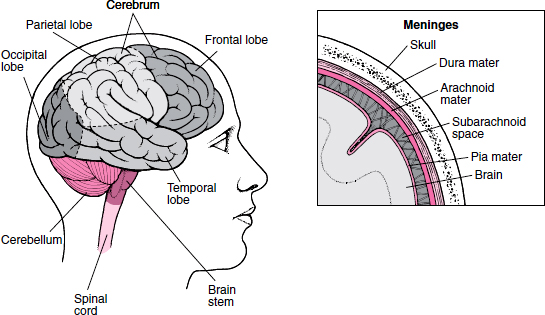
The activity of the brain results from electrical impulses generated by nerve cells (neurons), which process and store information. The impulses pass along the nerve fibers within the brain. How much and what type of brain activity occurs and where in the brain it is initiated depend on a person’s level of consciousness and on the specific activity that the person is doing. The brain has three main parts: the cerebrum, the brain stem, and the cerebellum. Each has a number of smaller areas, each with specific functions.
Cerebrum: The cerebrum, the largest part of the brain, consists of dense, convoluted masses of tissue. The outer layer is the cerebral cortex (gray matter). In adults, the cerebral cortex contains most of the nerve cells in the nervous system. Underneath the cortex is the white matter, which consists mainly of nerve fibers that connect the nerve cells in the cortex with other parts of the nervous system.
The cerebrum is divided into two halves—the left and right cerebral hemispheres. The hemispheres are connected by nerve fibers that form a bridge (called the corpus callosum) through the middle of the brain. Each hemisphere is further divided into a frontal, parietal, occipital, and temporal lobe. Each lobe has specific functions, but for most activities, several areas of different lobes in both hemispheres must work together.
The frontal lobes have the following functions:
Initiating many voluntary actions, ranging from looking toward an object of interest, to crossing a street, to relaxing the bladder to urinate
Controlling learned motor skills, such as writing, playing musical instruments, and tying shoelaces
Controlling complex intellectual processes, such as speech, thought, concentration, problem-solving, and planning for the future
Controlling facial expressions and hand and arm gestures
Coordinating expressions and gestures with mood and feelings
Particular areas of the frontal lobes control specific movements, typically of the opposite side of the body. In most people, the left frontal lobe controls most of the functions involved in using language.
The parietal lobes have the following functions:
Interpreting sensory information from the rest of the body
Controlling body movement
Combining impressions of form, texture, and weight into general perceptions
Influencing mathematical skills and language comprehension, as do adjacent areas of the temporal lobes
Storing spatial memories that enable people to orient themselves in space (know where they are) and to maintain a sense of direction (know where they are going)
Processing information that helps people know the position of their body parts
The occipital lobes have the following functions:
Processing and interpreting vision
Enabling people to form visual memories
Integrating visual perceptions with the spatial information provided by the adjacent parietal lobes
The temporal lobes have the following functions:
Generating memory and emotions
Processing immediate events into recent and long-term memory
Storing and retrieving long-term memories
Comprehending sounds and images, thus enabling people to recognize other people and objects and to integrate hearing and speech
Large collections of nerve cells—the basal ganglia, thalamus, and hypothalamus—are located at the base of the cerebrum. The basal ganglia coordinate and smooth out movements. The thalamus generally organizes sensory messages to and from the highest levels of the brain (cerebral cortex), providing a general awareness of such sensations as pain, touch, and temperature. The hypothalamus coordinates some of the more automatic functions of the body, such as control of sleep and wakefulness, maintenance of body temperature, and regulation of appetite and the balance of water within the body.
A system of nerve fibers—called the limbic system—connects the hypothalamus with other areas of the frontal and temporal lobes, including the hippocampus and amygdala. The limbic system controls the experience and expression of emotions, as well as automatic functions of the body. By producing emotions (such as fear, anger, pleasure, and sadness), the limbic system enables people to behave in ways that help them communicate and survive physical and psychologic upsets. The hippocampus is also involved in the formation and retrieval of memories. Through the limbic system, memories that are emotionally charged are easier to recall than those that are not.
 Did You Know…
The brain rarely produces new nerve cells but can do so in areas of the brain concerned with memory.
Brain Stem: The brain stem connects the cerebrum with the spinal cord. It contains a system of nerve cells and fibers (called the reticular activating system) located deep within the upper part of the brain stem. This system controls levels of consciousness and alertness.
The brain stem also automatically regulates critical body functions, such as breathing, swallowing, blood pressure, and heartbeat, and it helps adjust posture. If the entire brain stem becomes severely damaged, consciousness is lost, and these automatic body functions cease. Death soon follows.
Cerebellum: The cerebellum, which lies below the cerebrum just above the brain stem, coordinates the body’s movements. With information it receives from the cerebral cortex and the basal ganglia about the position of the limbs, the cerebellum helps the limbs move smoothly and accurately. It does so by constantly adjusting muscle tone and posture. The cerebellum interacts with areas in the brain stem called vestibular nuclei, which are connected with the organs of balance (semicircular canals) in the inner ear. Together, these structures provide a sense of balance. The cerebellum also stores memories of practiced movements, enabling highly coordinated movements, such as a ballet dancer’s pirouette, to be done with speed and balance.
Meninges: Both the brain and spinal cord are covered by three layers of tissue (meninges) that protect them. The thin pia mater is the innermost layer, which adheres to the brain and spinal cord. The delicate, spider web-like arachnoid mater is the middle layer. The space between the arachnoid mater and the pia mater (the subarachnoid space) is a channel for cerebrospinal fluid, which helps protect the brain and spinal cord. Cerebrospinal fluid flows over the surface of the brain between the meninges, fills internal spaces within the brain (the four ventricles), and cushions the brain against sudden jarring and minor injury. The leathery dura mater is the outermost and toughest layer. The brain and its meninges are contained in a tough, bony protective structure, the skull.
How the Spine Is Organized
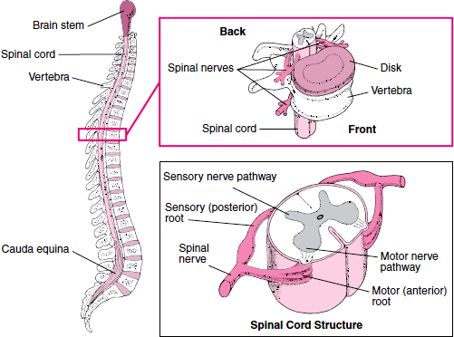
A column of bones called vertebrae make up the spine (spinal column). The vertebrae protect the spinal cord, a long, fragile structure contained in the spinal canal, which runs through the center of the spine. Between the vertebrae are disks composed of cartilage, which help cushion the spine and give it some flexibility. Emerging from the spinal cord between the vertebrae are 31 pairs of spinal nerves. Each nerve emerges in two short branches (roots): one at the front (motor or anterior root) and one at the back (sensory or posterior root) of the spinal cord. The motor roots carry commands from the brain and spinal cord to other parts of the body, particularly to skeletal muscles. The sensory roots carry information to the brain from other parts of the body. The spinal cord ends about three fourths of the way down the spine, but a bundle of nerves extends beyond the cord. This bundle is called the cauda equina because it resembles a horse’s tail. The cauda equina carries nerve impulses to and from the legs.
Spinal Cord
The spinal cord is a long, fragile tubelike structure that begins at the end of the brain stem and continues down almost to the bottom of the spine (spinal column). The spinal cord consists of nerves that carry incoming and outgoing messages between the brain and the rest of the body. It is also the center for reflexes, such as the knee jerk reflex (see art on page 633).
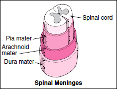
Like the brain, the spinal cord is covered by three layers of tissue (meninges). The spinal cord and meninges are contained in the spinal canal, which runs through the center of the spine. In most adults, the spine is composed of 26 individual back bones (vertebrae). Just as the skull protects the brain, vertebrae protect the spinal cord. The vertebrae are separated by disks made of cartilage, which act as cushions, reducing the forces generated by movements such as walking and jumping.
Like the brain, the spinal cord consists of gray and white matter. The butterfly-shaped center of the cord consists of gray matter. The front “wings” (called horns) contain motor nerve cells, which transmit information from the brain or spinal cord to muscles, stimulating movement. The back horns contain sensory nerve cells, which transmit sensory information from other parts of the body through the spinal cord to the brain. The surrounding white matter contains columns of nerve fibers that carry sensory information to the brain from the rest of the body (ascending tracts) and columns that carry impulses from the brain to the muscles (descending tracts).
Nerves
The peripheral nervous system consists of more than 100 billion nerve cells that run throughout the body like strings, making connections with the brain, other parts of the body, and often with each other. Peripheral nerves consist of bundles of nerve fibers. These fibers are wrapped with many layers of tissue composed of a fatty substance called myelin. These layers form the myelin sheath, which speeds the conduction of nerve impulses along the nerve fiber. Nerves conduct impulses at different speeds depending on their diameter and on the amount of myelin around them.
The peripheral nervous system has two parts: the somatic nervous system and the autonomic nervous system.
Somatic Nervous System: This system consists of nerves that connect the brain and spinal cord with muscles controlled by conscious effort (voluntary or skeletal muscles) and with sensory receptors in the skin. (Sensory receptors are specialized endings of nerve fibers that detect information in and around the body.)
Typical Structure of a Nerve Cell
A nerve cell (neuron) consists of a large cell body and nerve fibers—one elongated extension (axon) for sending impulses and usually many branches (dendrites) for receiving impulses. Each large axon is surrounded by oligodendrocytes in the brain and spinal cord and by Schwann cells in the peripheral nervous system. The membranes of these cells consist of a fat (lipoprotein) called myelin. The membranes are wrapped tightly around the axon, forming a multilayered sheath. This myelin sheath resembles insulation, such as that around an electrical wire. Nerve impulses travel much faster in nerves with a myelin sheath than in those without one. If the myelin sheath of a nerve is damaged, nerve transmission slows or stops.
Autonomic Nervous System: This system connects the brain stem and spinal cord with internal organs and regulates internal body processes that require no conscious effort (see page 831). Examples are the rate of heart contractions, blood pressure, the rate of breathing, the amount of stomach acid secreted, and the speed at which food passes through the digestive tract. The autonomic nervous system has two divisions:
Sympathetic division: Its main function is to prepare the body for stressful or emergency situations—for fight or flight.
Parasympathetic division: Its main function is to prepare the body for ordinary situations.
These divisions work together, usually with one activating and the other inhibiting the actions of internal organs. For example, the sympathetic division increases pulse, blood pressure, and breathing rates, and the parasympathetic system decreases each of them.
Cranial and Spinal Nerves: Nerves that connect the brain with the eyes, ears, nose, and throat and with various parts of the head, neck, and trunk are called cranial nerves. There are 12 pairs of them (see page 835). Nerves that connect the spinal cord with other parts of the body are called spinal nerves. The brain communicates with most of the body through the spinal nerves. There are 31 pairs of them, located at intervals along the length of the spinal cord (see page 793 and art on page 797). Several cranial nerves and most spinal nerves are involved in both the somatic and autonomic parts of the peripheral nervous system.
Spinal nerves emerge from the spinal cord through spaces between the vertebrae. Each nerve emerges as two short branches (called spinal nerve roots): one at the front of the spinal cord and one at the back.
Motor (anterior) nerve root: The motor root emerges from the front of the spinal cord. Motor nerve fibers carry commands from the brain and spinal cord to other parts of the body, particularly to skeletal muscles.
Sensory (posterior) nerve root: The sensory root enters the back of the spinal cord. Sensory nerve fibers carry sensory information (about body position, light, touch, temperature, and pain) to the brain from other parts of the body. The sensory nerve fibers from a specific sensory nerve root carry information from a specific area of the body, called a dermatome (see art on page 796).
After leaving the spinal cord, the corresponding motor and sensory nerve roots join to form a single spinal nerve. Some of the spinal nerves form networks of interwoven nerves, called nerve plexuses. In a plexus, nerve fibers from different spinal nerves are sorted and recombined so that all fibers going to or coming from one area of a specific body part are put together into one nerve (see art on page 823). There are two major nerve plexuses: the brachial plexus, which sorts and recombines nerve fibers traveling to the arms and hands, and the lumbosacral plexus, which sorts and recombines nerve fibers going to the legs and feet.
Effects of Aging
Brain: Brain function varies normally as people pass from childhood through adulthood to old age. During childhood, the ability to think and reason steadily increases, enabling a child to learn increasingly complex skills. During most of adulthood, brain function is relatively stable. After a certain age, which varies from person to person, brain function declines. Different aspects of brain function are affected at different times:
Short-term memory and the ability to learn new material tend to be affected relatively early.
Verbal abilities, including vocabulary and word usage, may begin to decline at about age 70.
Intellectual performance—the ability to process information (regardless of speed)—is usually maintained until about age 80 if no neurologic disorders are present.
Reaction time and performance of tasks may become slower because the brain processes nerve impulses more slowly.
However, the effects of aging on brain function may be difficult to separate from the effects of various disorders that are common among older people. These disorders include depression, stroke, an underactive thyroid gland (hypothyroidism), and degenerative brain disorders such as Alzheimer’s disease.
As people age, the number of nerve cells in the brain usually decreases, although the number lost varies greatly from person to person, depending on the person’s health. Also, the remaining nerve cells function less well. However, the brain has certain characteristics that help compensate for these losses.
Redundancy: The brain has more cells than it needs to function normally. Redundancy may help compensate for the loss of nerve cells that occurs with aging and disease.
Formation of new connections: The brain actively compensates for the age-related decrease in nerve cells by making new connections between the remaining nerve cells.
Production of new nerve cells: Some areas of the brain may produce new nerve cells, especially after a slight brain injury or a stroke.
Thus, people who have had a brain injury or stroke can sometimes learn new skills, as occurs during occupational therapy.
People can influence how quickly brain function declines. For example, mental and physical exercise seems to slow the loss of nerve cells in areas of the brain involved in memory. Such exercise also helps keep the remaining nerve cells functioning. On the other hand, consuming two or more drinks of alcohol a day can speed the decline in brain function.
Did You Know…
Physical and mental exercise may slow the age-related decline in brain function.
Having uncontrolled high blood pressure or high cholesterol levels can speed the age-related decline in brain function.
As people age, blood flow to the brain may decrease by an average of 20%. The decrease in blood flow is greater in people who have atherosclerosis of the arteries to the brain (cerebrovascular disease). This disease is more likely to occur in people who have smoked for a long time or who have high blood pressure, high cholesterol, or high blood sugar (diabetes mellitus) that is not controlled by lifestyle changes or drugs. These people may lose brain cells prematurely, possibly impairing mental function. As a result, the risk of dementia at a relatively young age is increased.
Spinal Cord: As people age, the disks between the back bones (vertebrae) become hard and brittle, and parts of the vertebrae may overgrow. As a result, the disks lose some of their capacity to cushion, so more pressure is put on the spinal cord and on the branches of the nerves that emerge from it (spinal nerve roots). The increased pressure may injure some nerve fibers in the spinal cord. Such injury can result in decreased sensation and sometimes decreased strength and balance.
Peripheral Nerves: As people age, peripheral nerves may conduct impulses more slowly, resulting in decreased sensation, slower reflexes, and often some clumsiness. Nerve conduction slows because myelin sheaths (layers of tissues around nerves that speed conduction of impulses) degenerate. Degeneration occurs because, as people age, blood flow decreases, nearby bones overgrow and put pressure on the nerves, or both. Usually, the effect is so minimal that no change in function is noticeable unless nerves are injured by something else (for example, by diabetes).
The peripheral nervous system’s response to injury is reduced. When the axon of a peripheral nerve is damaged in younger people, the nerve is able to repair itself as long as its cell body, located in or near the spinal cord, is undamaged. This self-repair process occurs more slowly and incompletely in older people, making older people more vulnerable to injury and disease.