CHAPTER 116
Tumors of the Nervous System
A tumor is an abnormal growth, whether noncancerous (benign) or cancerous (malignant). In many parts of the body, a noncancerous tumor causes few or no problems. However, any abnormal growth or mass in the brain or spinal cord can cause considerable damage.
Some cancers elsewhere in the body cause symptoms of nervous system dysfunction even though there is no evidence that nerve tissue has been invaded. These disorders are called paraneoplastic syndromes (see box on page 1082). They can cause dementia, mood swings, seizures, incoordination, dizziness, double vision, and abnormal eye movements. The most common effect is dysfunction of peripheral nerves (polyneuropathy—see page 826), resulting in muscle weakness, numbness, and tingling.
Brain Tumors
A brain tumor is a noncancerous (benign) or cancerous (malignant) growth in the brain. It may originate in the brain or have spread (metastasized) to the brain from another part of the body.
 Symptoms may include headaches, personality changes (such as suddenly becoming depressed, anxious, or uninhibited), loss of balance, trouble concentrating, seizures, and incoordination.
Imaging tests can often detect brain tumors, but sometimes biopsy of the tumor is needed.
Treatment may involve surgery, radiation therapy, chemotherapy, or a combination.
Brain tumors are slightly more common among men than women. Only meningiomas, which are non-cancerous, are more common among women. Brain tumors usually develop during early or middle adulthood but may develop at any age. They are becoming more common among older people.
Brain tumors may be primary or secondary. Primary brain tumors originate in the cells within or next to the brain. These tumors may be cancerous or non-cancerous. Either type of brain tumor is serious because the skull is rigid, providing no room for the tumor to expand. Also, tumors may develop near parts of the brain that control vital functions. Secondary brain tumors are metastases originating in another part of the body and thus are always cancerous.
Noncancerous tumors are named for the specific cells or tissues in which they originate. For example, hemangioblastomas originate in blood vessels (“hema” refers to blood vessels, and hemangioblasts are the cells that develop into blood vessel tissue). Some noncancerous tumors originate in cells of the embryo (embryonic cells), early in the development of the fetus. Such tumors may be present at birth.
TUMORS THAT ORIGINATE IN OR NEAR THE BRAIN

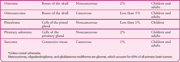
The most common type of primary cancerous brain tumor is a glioma, which has several subtypes. Gliomas account for 65% of all primary brain tumors. However, most cancerous brain tumors are secondary—metastases from cancer that started in another part of the body.
Metastases may grow in a single part of the brain or in several different parts. Many types of cancer—including breast cancer, lung cancer, cancers of the digestive tract, malignant melanoma, leukemia, and lymphoma—can spread to the brain. Lymphomas of the brain are common among people who have AIDS and, for unknown reasons, are becoming more common among people who have a normal immune system.
Brain tumors can cause problems in the following ways:
By directly invading and destroying brain tissue
By directly putting pressure on nearby tissue
By increasing pressure within the skull (intracranial pressure) because the tumor takes up space
By causing fluids to accumulate in the brain
By blocking normal circulation of cerebrospinal fluid through the spaces within the brain
By causing bleeding
Symptoms
Symptoms occur whether a brain tumor is non-cancerous or cancerous. Noncancerous tumors grow slowly and may become quite large before causing symptoms.
A brain tumor can cause many different symptoms, and symptoms may occur suddenly or develop gradually. Which symptoms develop first and how they develop depend on the tumor’s size, growth rate, and location. In some parts of the brain, even a small tumor can have devastating effects. In other parts of the brain, tumors can grow relatively large before any symptoms appear. As the tumor grows, it pushes and stretches but usually does not destroy nerve tissue, which can compensate for these changes very well. Thus, symptoms may not develop at first.
Many symptoms result from increased pressure within the skull. The most common is a headache (see page 648), which is often the first symptom. However, most headaches are not caused by brain tumors. A headache due to a brain tumor usually recurs more and more often as time passes. It eventually becomes constant, without relief. It is often worse when people lie down. The headache may awaken people from sleep. A gradually growing tumor causes a headache that typically is worse when people first awaken. If headaches with these characteristics start in people who have not had headaches before, a brain tumor may be the cause.
Often, increased pressure within the skull also affects mental function and mood. The personality may change. For example, people may become withdrawn, moody, and, often, inefficient at work. They may feel drowsy, confused, and unable to think. Such symptoms are often more apparent to family members and co-workers than to the affected person. Depression and anxiety, especially if either develops suddenly, may be an early symptom of a brain tumor. People may behave bizarrely. They may become uninhibited or behave in ways they never have before. In older people, certain brain tumors cause symptoms that may be mistaken for those of dementia (see page 689).
Common Symptoms of Some Brain Tumors
ASTROCYTOMAS AND OLIGODENDROGLIOMAS
Some astrocytomas and oligodendrogliomas grow slowly and may initially cause only seizures. Others (anaplastic astrocytomas and anaplastic oligodendrogliomas) grow fast and are cancerous. They can cause various symptoms of brain dysfunction. Glioblastoma multiforme, a type of astrocytoma, grows so fast that it increases pressure in the brain, causing headaches and slowed thinking. If the pressure becomes high enough, drowsiness, then coma, may result.
Symptoms vary depending on the tumor’s location.
Frontal lobes (located behind the forehead): Tumors located here can cause weakness and personality changes. If they develop in the dominant frontal lobe (the left lobe in most people and the right lobe in some left-handers), they can cause speech disturbances.
Parietal lobes (located behind the frontal lobes): Tumors located here can cause loss of or changes in sensation. Sometimes vision is partially lost in both eyes so that neither eye can see the side opposite the tumor.
Temporal lobes (located above the ears): Tumors located here can cause seizures and, if they develop on the dominant side, the inability to understand and use language.
Occipital lobes (toward the back of the head): Tumors located here can cause partial loss of vision in both eyes.
In or near the cerebellum (above the back of the neck): Tumors located here, especially medulloblastomas in children, can cause alterations in eye movements, incoordination, unsteadiness in walking, and sometimes hearing loss and vertigo. They can block the drainage of cerebrospinal fluid, causing fluid to accumulate in the spaces within the brain (ventricles). As a result, the ventricles enlarge (a condition called hydrocephalus), and pressure within the skull increases. Symptoms include headaches, nausea, vomiting, difficulty turning the eyes upward, and lethargy. In infants, the head enlarges. Greatly increased pressure can cause herniation of the brain, which can result in coma and death.
MENINGIOMAS
Meningiomas are usually noncancerous but may recur after they are removed. They occur more often in women and usually appear in people aged 40 to 60, but they can begin growing during childhood or later life. Symptoms depend on where the tumor develops. They may include weakness or numbness, seizures, an impaired sense of smell, changes in vision, and impaired mental function. In older people, a meningioma may cause dementia.
PINEAL TUMORS
Pineal tumors usually develop during childhood and often cause early puberty. They can block the drainage of cerebrospinal fluid around the brain, leading to hy-drocephalus. The most common type of pineal tumor is a germ cell tumor. Symptoms include the inability to look up and drooping eyelids.
PITUITARY GLAND TUMORS
The pituitary gland, located at the base of the skull, controls much of the body’s endocrine system. Tumors of the pituitary gland (pituitary adenomas) are usually non-cancerous. They may secrete abnormally large amounts of pituitary hormones or block production of hormones. When large amounts of hormones are secreted, effects vary depending on which hormone is involved.
For growth hormone, extreme height (gigantism) or disproportionate enlargement of the head, face, hands, feet, and chest (acromegaly)
For corticotropin, Cushing’s syndrome
For thyroid-stimulating hormone, hyperthyroidism
For prolactin, the cessation of menstrual periods (amenorrhea) in women, production of breast milk in women who are not breastfeeding (galactorrhea), and, in men, loss of libido, erectile dysfunction, and enlargement of the breasts (gynecomastia)
Pituitary gland tumors can block hormone production by destroying the tissues in the pituitary gland that secrete hormones, eventually resulting in insufficient levels of these hormones in the body. Headaches commonly occur. If the tumor enlarges, peripheral vision in both eyes is lost.
Other common symptoms of a brain tumor include vertigo, loss of balance, and incoordination. Later, as the pressure within the skull increases, nausea, vomiting, lethargy, increased drowsiness, intermittent fever, and even coma may occur. Vision may blur suddenly when people change positions. Some brain tumors, usually primary tumors, cause seizures.
Depending on which area of the brain is affected (see page 677), a tumor can do any of the following:
Cause an arm, a leg, or one side of the body to become weak or paralyzed
Impair the ability to feel heat, cold, pressure, a light touch, or sharp objects
Make people unable to express or understand language.
Increase or decrease the pulse and breathing rates if the tumor compresses the brain stem
Reduce alertness
Impair the ability to hear, smell, or see (causing such symptoms as double vision and loss of vision)
For example, a pituitary tumor may press on the nearby optic nerves (2nd cranial nerve), which are involved in vision, and thus impair peripheral vision. Any of these symptoms suggests a serious disorder and requires immediate medical attention.
If a tumor blocks the flow of cerebrospinal fluid through the spaces within the brain (ventricles), fluid may accumulate in the ventricles, causing them to enlarge (a condition called hydrocephalus). As a result, pressure within the skull increases. In addition to other symptoms of increased pressure, hydrocephalus makes turning the eyes upward difficult. In infants and very young children, the head enlarges.
If the pressure within the skull is greatly increased, the brain may be pushed downward because the skull cannot expand. Herniation of the brain (see art on page 735) may result. There are two main types:
Transtentorial herniation: The upper part of the brain (cerebrum) is forced through the narrow opening (the tentorial notch) in the relatively rigid tissue that separates the cerebrum from the lower parts of the brain (cerebellum and brain stem). In people with this type of herniation, consciousness is reduced. The side of the body opposite the tumor may be paralyzed.
Tonsillar herniation: A tumor that originates in the lower part of the brain pushes the lowest part of the cerebellum (cerebellar tonsils) through the opening at the base of the skull (foramen magnum). As a result, the brain stem, which controls breathing, heart rate, and blood pressure, is compressed and malfunctions. If not diagnosed and treated immediately, a tonsillar herniation rapidly results in coma and death.
People with metastases to the brain may also have symptoms related to the original cancer. For example, if the cancer originated in the lungs, people may cough up bloody mucus. With metastases, weight loss is common.
Symptoms worsen over time unless the tumor is treated. With treatment, particularly for benign tumors, some people completely recover. For others, life span is shortened, sometimes greatly. The outcome depends on the type and location of the tumor.
Diagnosis
Doctors consider the possibility of a brain tumor in people who have had a seizure for the first time or who have the characteristic symptoms. Although doctors can often detect brain dysfunction during a physical examination, other procedures are needed to diagnose a brain tumor.
Standard x-rays of the skull can detect tumors that erode bone (such as a meningioma or pituitary adenoma). However, magnetic resonance imaging (MRI) and computed tomography (CT) are more useful because they can detect all types of brain tumors. They can also show the tumor’s size and exact position in great detail. When a brain tumor is detected, more diagnostic procedures are done to determine the particular kind.
Sometimes a spinal tap (lumbar puncture—(see art on page 635) is done to obtain cerebrospinal fluid for examination under a microscope. This procedure is done when doctors suspect that the tumor has invaded the layers of tissues that cover the brain (meninges), often compressing the cranial nerves, blocking the absorption of cerebrospinal fluid, or both. The procedure may also help when the diagnosis or the type of tumor is unclear. Cerebrospinal fluid may contain cancer cells. However, a spinal tap cannot be done in people who have a large tumor that is increasing pressure within the skull. In these people, removing cerebrospinal fluid during a spinal tap may cause the tumor to move, resulting in herniation of the brain.
A biopsy of the tumor (removal of a sample of the tumor for examination under a microscope) is usually needed to identify the type of tumor, including whether it is cancerous. A biopsy may be done during surgery in which all or part of the tumor is removed. If a tumor is difficult to reach, a biopsy may be done using three-dimensional needle placement (stereotactic biopsy) with CT, which enables doctors to precisely locate the tumor.
Treatment
Treatment of a brain tumor depends on its location and type. When possible, the tumor is removed surgically in a procedure called craniotomy (which involves opening the skull). Some brain tumors can be removed with little or no damage to the brain. However, many grow in an area that makes removal by traditional surgery difficult or impossible without destroying essential structures.
Traditional surgery sometimes causes brain damage that can lead to symptoms such as partial paralysis, changes in sensation, weakness, and impaired mental function. Nevertheless, removing a tumor—whether cancerous or noncancerous—is essential if its growth threatens important brain structures. Even when a cure is impossible, surgery may be useful to reduce the tumor’s size, relieve symptoms, and help doctors determine whether other treatments, such as radiation therapy or chemotherapy, are warranted.
Noncancerous Tumors: Surgical removal is often safe and cures the person. However, very small tumors and tumors in older people may be left in place as long as they are not causing symptoms. Sometimes radiation therapy is given after surgery to destroy any remaining tumor cells. Radiosugery focuses the radiation and is effective in treating noncancerous tumors such as meningiomas and acoustic neuromas. Therefore, radiosurgery often is used instead of traditional surgery for these tumors.
Cancerous Brain Tumors: Usually, a combination of surgery, radiation therapy, and chemotherapy is used. As much of the tumor as can be removed safely is removed, and then radiation therapy is begun. Radiation therapy is given over a course of several weeks. Radiosurgery is used when traditional surgery cannot be, especially for the treatment of metastases.
For very aggressive tumors, chemotherapy is given with radiation therapy. Radiation therapy plus chemotherapy rarely cures but may shrink a tumor enough to keep it under control for many months or even years.
After radiation therapy, ongoing chemotherapy is used to treat some types of cancerous brain tumors. Chemotherapy appears to be particularly effective in treating anaplastic oligodendrogliomas.
Increased Pressure Within the Skull: This extremely serious condition requires immediate medical attention. Drugs such as mannitol and corticosteroids are usually given by injection to reduce the pressure and prevent herniation. They reduce swelling around the tumor. Within days or sometimes hours, corticosteroids can often restore functions lost because of the tumor and relieve headache, even if the tumor is large.
If the tumor is blocking the flow of cerebrospinal fluid through the spaces within the brain, a device may be used to drain the cerebrospinal fluid and thus reduce the risk of herniation. The device consists of a small tube (catheter) connected to a gauge that measures the pressure within the skull. The tube is inserted through a tiny opening drilled in the skull. A local anesthetic (usually plus a sedative) or a general anesthetic may be used. The tube is removed or converted to a permanent drain (shunt) after a few days. During this time, doctors surgically remove all or part of the tumor or use radiosurgery or radiation therapy to reduce the size of the tumor and thus relieve the blockage.
Metastases: Treatment depends largely on where the cancer originated. Radiation therapy directed at the metastases in the brain is often used. Surgical removal may benefit people who have only a single metastasis. Sometimes radiosurgery is used.
Some experimental treatments, such as implantation of pellets containing chemotherapy drugs or of radioactive pellets in the tumor, are being tried.
End-of-Life Issues: People with cancerous brain tumors have a limited life expectancy and are likely to become unable to make decisions about medical care. Consequently, establishing advance directives is advisable (see page 69). Advance directives can help a doctor determine what kind of care people want if they become unable to make decisions about medical care.
Many cancer centers, especially those with hospice facilities, provide counseling and home health services.
Spinal Cord Tumors
A spinal cord tumor is a noncancerous (benign) or cancerous (malignant) growth in or around the spinal cord.
People may have weak muscles, lose sensation in particular areas of the body, or become unable to control bowel and bladder function.
Magnetic resonance imaging can usually detect spinal cord tumors.
Treatment may involve surgical removal, radiation therapy, or both.
Spinal cord tumors are much less common than brain tumors. Spinal cord tumors may be primary or secondary.
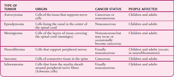
Primary spinal cord tumors may be cancerous or noncancerous. They may originate in the cells within or next to the spinal cord. Only about 10% of primary spinal cord tumors originate in the cells within the spinal cord. These tumors can extend within the cord and cause a fluid-filled cavity (syrinx) to form.
The other 90% of primary spinal cord tumors originate in cells next to the spinal cord, such as those of the spinal nerve roots—the parts of spinal nerves that emerge from the spinal cord (see art on page 626). Meningiomas and neurofibromas, which originate in cells next to the cord, are the most common primary spinal tumors. They are noncancerous.
Understanding Tumor Treatment
Craniotomy: After part of the scalp is shaved, an incision is made through the skin. A high-speed drill and a special saw are used to remove a small piece of bone above the tumor. The tumor is located and removed using one of the following:
A scalpel may be used to cut out the tumor.
A laser may be used to vaporize the tumor.
A device that emits ultrasound waves may be used to break the tumor apart, so that the pieces can be suctioned out (aspirated).
Lasers and ultrasound devices are used to remove tumors that would be difficult to cut out. Usually, the bone is then replaced, and the incision stitched closed.
Stereotactic techniques: These techniques are ways to localize tumors very precisely. Computers are used to produce a three-dimensional image. The three-dimensional image can be obtained by attaching a light-weight metal imaging frame with a series of rods to the person’s skull. A local anesthetic is given, and the pins are attached to the skull, piercing the skin. The rods appear as dots on a CT scan, providing reference points, which help locate the tumor. Other devices, such as a viewing wand or compass system, do not involve attaching a frame and may be used instead. Stereotactic techniques can be used to biopsy or remove tumors or to insert implants containing a chemotherapy drug or radioactive pellets.
Radiosurgery: Radiosurgery is not really surgery because no incision is required. Focused radiation is used to destroy a tumor. Several machines, including a gamma knife and a linear accelerator, can produce this type of radiation.
When a gamma knife is used, an imaging frame is attached to the person’s skull. The person lies on a sliding bed, and a large helmet with holes in it is placed over the frame. The head of the bed is then slid into a globe that contains radioactive cobalt. Radiation passes through the holes in the helmet and is aimed precisely at the tumor.
A linear accelerator circles the head of the person, who lies on a sliding bed. The linear accelerator rotates around the person and aims radiation precisely at the tumor from different angles.
Implants: After a tumor is removed and before the skull and incision are closed, wafers soaked with a chemotherapy drug may be placed in the space where the tumor was. As the wafers gradually dissolve, they release the drug to destroy any remaining cancer cells.
A thin tube (catheter) may be inserted through an incision and used to place radioactive implants directly into the tumor. The implants may be removed after a few days or months or may be left in place. Unlike people who have externally applied radiation therapy, people who have radioactive implants are radioactive for a time (that is, they give off radiation). They thus need to take precautions as advised by their doctor. After this procedure, surgery may be necessary to remove dead cancer cells.
Shunts: If a tumor causes pressure within the skull to increase, a shunt may be surgically placed. A shunt is a thin piece of tubing that is inserted into one of the spaces of the brain (ventricles) or sometimes into the space around the spine that contains cerebrospinal fluid (subarachnoid space). The other end of the tubing is threaded under the skin from the head usually to the abdominal cavity. Excess cerebrospinal fluid is drained from the brain into the abdominal cavity, where it is absorbed. The shunt contains a one-way valve that opens when there is too much fluid in the brain. Shunts may be temporary (until the tumor is removed) or permanent.
Secondary spinal cord tumors, which are more common, are metastases of cancer originating in another part of the body and thus are always cancerous. Metastases most commonly spread to the vertebrae from cancers that originate in the lungs, breasts, prostate gland, kidneys, or thyroid gland. Metastases compress the spinal cord or nerve roots from the outside. Lymphomas may also spread to the spine and compress the spinal cord.
Symptoms
Symptoms are caused by pressure on the spinal cord and nerve roots. Pressure on the spinal cord may cause the following:
Back pain that progressively worsens, is unrelated to activity, and is worse when people lie down
Decreased sensation, progressive weakness, or paralysis in areas controlled by the parts of the spinal cord below the part that is compressed
Erectile dysfunction
Loss of bladder and bowel control
Pressure on the spinal cord may also block the blood supply to the cord, resulting in death of tissue, fluid accumulation, and swelling. Fluid accumulation may block more of the blood supply, leading to a vicious circle of damage. Symptoms due to pressure on the spinal cord can worsen quickly.
Pressure on spinal nerve roots can cause pain, numbness, tingling, weakness in areas supplied by the compressed nerve root. Pain may radiate along the nerve whose root is compressed. If compression continues, the affected muscles may waste away. Walking may become difficult.
TUMORS THAT ORIGINATE IN OR NEAR THE SPINAL CORD
Diagnosis
Compression of the spinal cord by a tumor must be diagnosed and treated immediately to prevent permanent damage.
Doctors consider the possibility of a spinal cord tumor in people who have certain cancers in other parts of the body, who develop pain in a specific area of the spine, and who have certain patterns of weakness or tingling. Because the spinal cord is organized in a specific way, doctors can locate the tumor by determining which parts of the body are not functioning normally (see art on page 794).
Doctors must rule out other disorders that can affect the function of the spinal cord, such as sore back muscles, bone bruises, an inadequate blood supply to the spinal cord, fractured vertebrae, compression by a collection of pus (abscess), a blood clot, or a herniated disk.
Several procedures can help doctors diagnose a spinal cord tumor. Magnetic resonance imaging (MRI) is considered the best procedure for examining all the structures of the spinal cord and spine. When MRI is unavailable, myelography with computed tomography (CT) may be done instead. X-rays of the spine can show only changes in the bones, and many tumors do not affect the bone when they are in an early stage.
A biopsy is usually needed to diagnose the precise type of tumor, especially primary spinal cord tumors. However, a biopsy is not needed for spinal cord tumors that result from metastases if cancer has been diagnosed elsewhere in the body. Often, a biopsy requires surgery, but sometimes it can be done using a needle with CT or MRI to guide doctors as they place the needle in the tumor.
Treatment
If symptoms suggest that the tumor is compressing the spinal cord, corticosteroids (such as dexamethasone) are immediately given in high doses to reduce the swelling. Such tumors are treated as soon as possible, often surgically.
Many tumors of the spinal cord and spine can be removed surgically. If tumors cannot be removed, radiation therapy is used, sometimes after surgery to relieve the pressure on the spinal cord is done.
Recovery generally depends on how quickly treatment begins and how much damage was done. Removal of meningiomas, neurofibromas, and some other primary spinal cord tumors may be curative.
Radiation Damage
Radiation therapy is one component in the treatment of tumors of the nervous system. It is directed at the general area (such as the whole head) when people have several tumors or a tumor that does not have distinct borders. When the tumor has distinct borders, therapy can be directed specifically at the tumor.
Radiation from these treatments sometimes damages the nervous system, despite the best efforts to prevent damage (see page 1989).
Whether damage occurs and how severe it is depend on several factors:
How much radiation is given over the entire course of treatment (total cumulative dose)
How much radiation is given in each dose
How long the treatments are given
How much of the body is exposed to radiation
How susceptible the person is
Symptoms of radiation damage can develop in the first few days (acute) or months of treatment (early-delayed) or several months or years after treatment (late-delayed). Symptoms can remain the same or worsen and can be temporary or permanent.
Acute encephalopathy can result from radiation to the brain. Fluid accumulates within the cells of the brain, causing the entire brain to swell (called cerebral edema). Symptoms include headaches, nausea, vomiting, drowsiness, and confusion. Acute encephalopathy usually begins shortly after the first or second dose of radiation is given, but sometimes it begins 2 to 4 months after radiation therapy is completed. Usually, symptoms diminish during the radiation treatments, and corticosteroids such as dexamethasone may help prevent or reduce cerebral edema.
Early-delayed radiation damage causes symptoms similar to those of acute encephalopathy. Symptoms usually diminish on their own over several days to weeks, sometimes more rapidly if corticosteroids are used.
If radiation is directed at the spine in the neck or upper back, early-delayed radiation myelopathy may develop. This disorder sometimes causes a sensation similar to an electric shock. The sensation begins in the neck or back, usually when the neck is bent forward, and shoots down to the legs. This disorder usually resolves without treatment.
 Did You Know…
Radiation therapy, used to treat brain and spinal cord tumors, can damage the brain and spinal cord.
Late-delayed radiation damage causes symptoms many months or years after radiation therapy. Many children and adults who receive whole-head brain radiation therapy develop late-delayed toxicity if they survive long enough. The most common cause in children is radiation therapy to prevent leukemia or to treat a type of brain tumor called medulloblastoma (see table on page 741). Symptoms include progressively worsening dementia, memory loss, difficulty thinking, mistaken perceptions, personality changes, and, in adults, unsteadiness in walking.
After radiation therapy for tumors near the spine, late-delayed myelopathy may develop. This disorder causes weakness, loss of sensation, and sometimes the Brown-Séquard syndrome. In this syndrome, one side of the spinal cord is damaged, resulting in weakness on one side of the body and loss of pain and temperature sensation on the other side. On the weak side of the body, people may be unable to sense where their hands and feet are without looking at them (position sense). Late-delayed radiation myelopathy usually does not subside and often results in paralysis.
Nerves near the site of the radiation therapy may also be damaged. For example, radiation to a breast or lung may damage nerves in the arms, and radiation to the groin may damage nerves in the legs. Weakness or loss of sensation may result.