CHAPTER 125
Cranial Nerve Disorders
Twelve pairs of nerves—the cranial nerves—lead directly from the brain to various parts of the head, neck, and trunk. Some of the cranial nerves are involved in the special senses (such as seeing, hearing, and taste), and others control muscles in the face or regulate glands. The nerves are named and numbered (according to their location, from the front of the brain to the back).
A cranial nerve disorder may affect the connections between cranial nerve centers within the brain. An example is internuclear ophthalmoplegia. Or, a disorder may affect only one cranial nerve. Examples are trigeminal neuralgia, Bell’s palsy, hemifacial spasm, and glossopharyngeal neuralgia.
 Did You Know…
Some cranial nerve disorders cause problems with eye movement.
Others cause brief, intermittent attacks of excruciating facial pain.
Symptoms depend on which nerves are damaged. For example, damage often occurs to nerves that control eye movement. If both eyes have trouble moving in the same direction, people may not be able to look in that direction. If only one eye can look in a certain direction, people may have double vision (two images seen side by side) when they look in that direction.
When doctors suspect a cranial nerve disorder, they test the function of a cranial nerve by asking the person to do simple tasks, such as to follow a moving target with the eyes.
Internuclear Ophthalmoplegia
Internuclear ophthalmoplegia is impairment of horizontal eye movements caused by damage to certain connections between nerve centers in the brain stem.
In internuclear ophthalmoplegia, the nerve fibers that coordinate both eyes in horizontal movements—looking from side to side—are damaged. These fibers connect collections of nerve cells (centers or nuclei) that the 3rd cranial nerve (oculomotor nerve) and the 6th cranial nerve (abducens nerve) originate from. In older people, the disorder usually results from a stroke, and only one eye is affected. In younger people, it usually results from multiple sclerosis, and both eyes are often affected. Less common causes include Lyme disease, tumors, and toxicity due to a drug (such as tricyclic antidepressants).
Horizontal eye movements are impaired, but vertical ones are not. The affected eye cannot turn inward, but it can turn outward. When a person looks to the side opposite the affected eye, the following happens:
 The affected eye, which should turn inward, cannot move past the midline. That is, the affected eye looks straight ahead.
As the other eye turns outward, it often makes involuntary, repetitive fluttering movements called nystagmus. That is, the eye rapidly moves in one direction, then slowly drifts in the other direction.
People with internuclear ophthalmoplegia may have double vision.
One-and-a-half syndrome results when the disorder that causes internuclear ophthalmoplegia also damages the center that coordinates and controls horizontal eye movements (horizontal gaze center). When the person tries to look the either side, the affected eye remains motionless in the middle. The other eye can turn outward but not inward. As in internuclear ophthalmoplegia, vertical eye movements are not affected.
In internuclear ophthalmoplegia and one-and-a-half syndrome, the eyes can turn inward when the person looks inward (as when focusing on a nearby object) even though the eyes cannot turn inward when the person looks to the side.
For internuclear ophthalmoplegia or one-and-a-half syndrome, treatment and outlook (whether the disorder abates or eventually resolves) depends on the disorder that caused it.
Conjugate Gaze Palsies
In conjugate gaze palsies, the two eyes cannot move in one direction (side to side, up, or down) at the same time.
Conjugate gaze palsies affect horizontal gaze (looking to the side) most often. Upward gaze is affected less often, and downward gaze is affected even less often. People may notice that they cannot look in certain directions.
There are no specific treatments.
Viewing the Cranial Nerves
Twelve pairs of cranial nerves emerge from the underside of the brain, pass through openings in the skull, and lead to parts of the head, neck, and trunk.

Horizontal Gaze Palsy: The most common cause is damage to the brain stem, often by a stroke. Often, the palsy is complete and severe. That is, the eyes cannot move to the side at all. Palsies can also be caused by damage to the front part of the cerebrum, usually by a stroke. The resulting palsy may not be as severe as that caused by damage to the brain stem, and symptoms often lessen with time.
Vertical Gaze Palsy: Vertical gaze decreases gradually with aging, but vertical gaze palsy is more severe than age-related changes. Usually, upward gaze is affected. The most common cause is damage to the top part of the brain stem (midbrain), usually by a stroke or tumor.
The pupils are usually large (dilated). In response to light, they may constrict more slowly and less completely than normal. When people attempt to look up, the eyes rapid move in one direction, then slowly drift in the other direction. These involuntary, fluttering eye movements are called nystagmus.
If downward gaze but not upward gaze is impaired, the cause is usually progressive supranuclear palsy (see page 778).
Palsies of Cranial Nerves That Control Eye Movement
These disorders involve paralysis of cranial nerves that control eye movement (the 3rd, 4th, or6th nerves), impairing the ability to move the eyes. How eye movement is affected depends on which nerve is affected.
The eye is moved by three pairs of muscles, controlled by the 3rd, 4th, and 6th cranial nerves. These muscles move the eye up and down, right and left, and diagonally. People may have double vision when they look in certain directions.
THIRD CRANIAL NERVE (OCULOMOTOR NERVE) PALSY
A palsy of the 3rd cranial nerve can be caused by brain disorders—such as a head injury, a bulge (aneurysm) in an artery supplying the brain, a hemorrhage, or a tumor—or by diabetes.
Symptoms
The affected eye turns outward when the unaffected eye looks straight ahead, causing double vision. The affected eye can move only to the middle when looking inward and cannot move up and down. Because the 3rd cranial nerve also raises the eyelids and controls the pupils, the eyelid droops, and the pupil may be widened (dilated). It may not narrow (constrict) in response to light.
The disorder causing the palsy may worsen, resulting in a serious, life-threatening condition. For example, a severe headache may occur suddenly, or a person may become increasingly drowsy or less responsive. In such cases, the cause may be a ruptured aneurysm, which then bleeds. Dilation of and lack of response to light (fixation) by both pupils indicates deep coma and possibly brain death (see box on page 708).
Diagnosis and Treatment
The diagnosis is based on results of a neurologic examination and computed tomography (CT) or magnetic resonance imaging (MRI). If the pupil is affected or if symptoms suggest a serious underlying disorder, CT is done immediately. If a ruptured aneurysm is suspected and CT does not detect blood, a spinal tap (lumbar puncture—see art on page 635), magnetic resonance angiography, CT angiography, or cerebral angiography is done.
Treatment depends on the cause. Emergency treatment is required if a life-threatening disorder is the cause.
FOURTH CRANIAL NERVE (TROCHLEAR NERVE) PALSY
Often, the cause cannot be identified. The most common identified cause is a head injury, often due to a motorcycle accident. Occasionally, diabetes causes this palsy. Rarely, the cause is a tumor, an aneurysm, or multiple sclerosis.
One or both eyes may be affected. The affected eye cannot turn inward and down. As a result, people see double images, one above and slightly to the side of the other. Thus, going down stairs, which requires looking inward and down, is difficult. However, tilting the head to the side opposite the affected muscle can compensate and eliminate the double images. This position can eliminate the double images because people use eye muscles that are unaffected by the palsy to focus both eyes on an object.
Did You Know…
Palsy of the 4th cranial nerve causes double vision, but tilting the head to one side eliminates it.
Usually, the diagnosis is suspected if a person has characteristic abnormal eye movements. CT or MRI may be done.
The disorder causing the palsy, if identified, is treated. Eye exercises may help. Sometimes surgery is necessary to eliminate double vision.
SIXTH CRANIAL NERVE (ABDUCENS NERVE) PALSY
Many disorders can cause this palsy:
Head injuries
Tumors
Multiple sclerosis
Aneurysms
Brain infections, such as meningitis, a brain abscess or a parasitic infection
Complications of an ear or eye infection
Blockage of an artery supplying the nerve, as can result from diabetes, a stroke, a transient ischemic attack, or vasculitis
Wernicke’s encephalopathy (commonly due to chronic alcoholism)
Benign intracranial hypertension (pseudotumor cerebri—see page 655)
Respiratory infections (in children)
Some of these disorders put pressure on the nerve by causing nearby swelling or by increasing pressure within the skull. Others interfere with blood flow to the nerve.
If this palsy occurs alone (without other cranial nerve palsies), its cause is often never identified.
Symptoms
The affected eye cannot turn fully outward and may turn inward when people look straight ahead. Double vision occurs when people look toward the side of the affected eye. Other symptoms depend on the cause. They include severe headache, accumulation of fluid (edema) in the conjunctiva, numbness in the face and mouth, loss of vision, and inability to move the eye in other directions.
Diagnosis and Treatment
Usually, doctors can easily identify a 6th cranial nerve palsy, but the cause is less obvious. An ophthalmoscope is used to look into the eye and check for evidence of tumors, increased pressure, and abnormalities in blood vessels. CT or, preferably, MRI is done to exclude tumors and other abnormalities. If the results are unclear, a spinal tap (lumbar puncture) is done to determine whether pressure within the skull is increased and whether a tumor or swelling due to an infection is compressing the nerve. If symptoms suggest vasculitis, blood is withdrawn to check for signs of inflammation, such as certain abnormal antibodies (antinuclear antibodies and rheumatoid factor) in the blood and an abnormal erythrocyte sedimentation rate (ESR—how quickly red blood cells settle to the bottom of a test tube containing blood). After all tests are done, the cause may remain unknown.
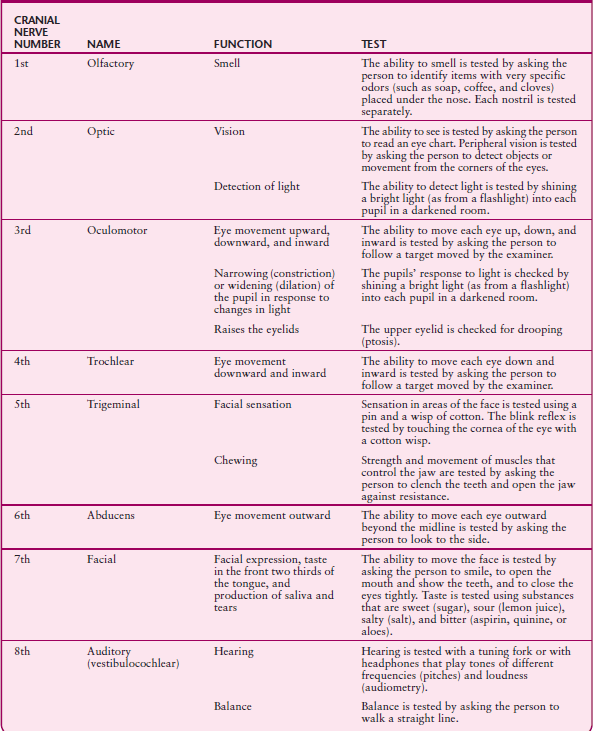
TESTING CRANIAL NERVES

Treatment depends on the cause. When the cause is treated, the palsy usually resolves. Palsies with no identifiable cause usually resolve without treatment within 2 months, as do those due to a blocked blood vessel.
Trigeminal Neuralgia
Trigeminal neuralgia (tic douloureux) is severe facial pain due to malfunction of the 5 th cranial nerve (trigeminal nerve). This nerve carries sensory information from the face to the brain and controls the muscles involved in chewing.
The cause is usually unknown but sometimes is an abnormally positioned artery that compresses the trigeminal nerve.
People have repeated short, lightning-like bursts of excruciating stabbing pain in the lower part of the face.
Doctors base the diagnosis on the characteristic pain.
Certain anticonvulsants or antidepressants, baclofen, or a local anesthetic may relieve the pain, but surgery is sometimes needed.
Trigeminal neuralgia usually occurs in middle-aged and older people, although it can affect adults of all ages. It is more common among women.
In most cases, the cause is unknown. A common known cause is an abnormally positioned artery that compresses the trigeminal nerve near where it exits the brain. Occasionally in younger people, trigeminal neuralgia results from nerve damage due to multiple sclerosis. Rarely, trigeminal neuralgia results from damage due to herpes zoster (a viral infection) or compression by a tumor.
Symptoms
The pain can occur spontaneously but is often triggered by touching a particular spot (called a trigger point) on the face, lips, or tongue or by an action such as brushing the teeth or chewing. Repeated short, lightning-like bursts of excruciating stabbing pain can be felt in any part of the lower portion of the face but are most often felt in the cheek next to the nose or in the jaw.
Usually, only one side of the face is affected. The pain usually lasts seconds but may last up to 2 minutes. Recurring as often as 100 times a day, the pain can be incapacitating. Because the pain is intense, people tend to wince, and thus the disorder is sometimes called a tic. The disorder commonly resolves on its own, but bouts of the disorder often recur after a long pain-free interval.
Diagnosis
Although no specific test exists for identifying trigeminal neuralgia, its characteristic pain usually makes it easy for doctors to diagnose. However, doctors must distinguish trigeminal neuralgia from other possible causes of facial pain, such as disorders of the jaw, teeth, or sinuses and trigeminal neuropathy (which is often due to compression of the trigeminal nerve caused by a tumor, stroke, an aneurysm, or multiple sclerosis). Trigeminal neuropathy can be distinguished because it causes loss of sensation and often weakness in parts of the face and trigeminal neuralgia does not.
Treatment
Because the bouts of pain are brief and recurrent, typical analgesics are not usually helpful, but other drugs, especially certain anticonvulsants (which stabilize nerve membranes), may help. The anticonvulsant carbamazepine is usually tried first. Gabapentin or phenytoin, also anticonvulsants, may be prescribed if carbamazepine is ineffective or has intolerable side effects. Baclofen (a drug used to reduce muscle spasms) or a tricyclic antidepressant (such as amitriptyline—see table on page 868) may be used instead. A local anesthetic may be injected into or around the nerve (a nerve block) to provide temporary pain relief.
If the pain continues to be severe, surgery may be done. If the cause is an abnormally positioned artery, a surgeon separates the artery from the nerve and places a small sponge between them. This procedure (called vascular decompression) usually relieves the pain for many years. If the cause is a tumor, the tumor can be surgically removed.
If people have pain unrelieved by drugs and surgery seems too risky, a test can be done to determine whether other procedures would help. For the test, alcohol is injected into the nerve to temporarily block its function. If alcohol relieves the pain, disrupting the nerve may relieve the pain, sometimes permanently. The nerve may be cut surgically or with a radiofrequency probe (using heat) or gamma knife (using radiation), or it may be destroyed by injecting a drug such as glycerol into it. However, these treatments are used as a last resort. They often provide only temporary relief—for months to a few years—and afterward, discomfort in the face returns and is even more severe.
Bell’s Palsy
Bell’s palsy is sudden weakness or paralysis of muscles on one side of the face due to malfunction of the 7th cranial
nerve (facial nerve). This nerve moves the facial muscles, stimulates the salivary and tear glands, and enables the front part of the tongue to detect tastes.
The disorder may result from a herpes simplex viral infection.
People may feel pain behind the ear, then one side of the face may become weak or completely paralyzed.
Doctors usually base the diagnosis on symptoms.
Corticosteroids may be used to reduce swelling of the nerve.
With or without treatment, most people recover completely within several months.
Bell’s palsy affects about 23 of 100,000 people at some time. Bell’s palsy may result from herpes simplex virus type 1, which causes herpes mouth infections. But the cause is sometimes unknown. Lyme disease can cause facial nerve paralysis, which is similar to Bell’s palsy. In blacks, sarcoidosis (see page 511) is a common cause of facial nerve paralysis.
Symptoms
Pain behind the ear may be the first symptom. It may develop several hours or even a day or two before the facial muscles weaken. Facial weakness occurs suddenly. The effect ranges from mild weakness to complete paralysis. By 48 hours, the weakness is as severe as it will be. Only one side of the face is affected. It becomes flat and expressionless. However, people often feel as though the face is twisted because the muscles on the unaffected side tend to pull the face to that side every time they make a facial expression. Wrinkling the forehead, blinking, and grimacing may be difficult or impossible. For most people, the face feels numb or heavy, even though sensation remains normal.
Closing the eye on the affected side may be difficult. People may be unable to close the eye completely, and they blink less frequently. The eye also tends to turn upward when it is closed.
Did You Know…
Bell’s palsy is often caused by the same virus that causes herpes mouth infections.
Lyme disease can cause facial nerve paralysis, similar to Bell’s palsy.
Bell’s palsy may interfere with the production of saliva and tears. People may have dry eyes and mouth, or they may drool. Because fewer tears are produced and the eye blinks less often (blinking helps moisten the eye’s surface), the eye becomes dry, resulting in pain and eye damage. Eye damage is usually minor but can be serious if the eye is not moistened and protected another way. People may be unable to taste with the front part of the tongue on the affected side. The ear on the affected side may perceive sounds as abnormally loud (a condition called hyperacusis) because the muscle that stretches the eardrum is paralyzed. This muscle is located in the middle ear.
Occasionally, as the facial nerve heals, it forms abnormal connections, resulting in unexpected movements of some facial muscles or in watering of the eyes (“crocodile tears”) during salivation. Because the facial muscles are not used for a long time, permanent tightening of the muscles (contractures) occasionally occurs.
Diagnosis
There is no specific test for Bell’s palsy, but it can usually be diagnosed based on symptoms. Bell’s palsy (and other types of facial nerve paralysis) can be distinguished from a stroke because a stroke usually causes weakness only in the lower part of the face rather than in the entire face. People who have had a stroke can close the eyes tightly and wrinkle the brow. Also, a stroke typically causes weakness of an arm and a leg.
Doctors can distinguish Bell’s palsy from other, rare disorders that cause facial nerve paralysis (such as tumors, infections, and skull fractures) because the other disorders cause different symptoms and symptoms usually develop slowly. Usually, doctors can exclude these disorders on the basis of the person’s history and results of x-rays, magnetic resonance imaging (MRI), or computed tomography (CT). A blood test may be done to check for Lyme disease, and a blood test and a chest x-ray may be done to check for sarcoidosis.
Treatment and Prognosis
An antiviral drug that is effective against herpes simplex virus (such as acyclovir, famciclovir, or valacyclovir) is usually given by mouth even when the cause is unknown. These drugs prevent the virus, if present, from replicating. If symptoms have been present less than 48 hours, a corticosteroid, such as prednisone, is given by mouth to reduce swelling of the nerve. Taking a corticosteroid may slightly speed and improve the recovery of movement.
If the eye cannot close completely, it must be protected from dryness to reduce the risk of eye damage. Eye drops consisting of artificial tears or a salt (saline) solution are applied to the eye until it can close completely. People may need to wear an eye patch some of the time, particularly during sleep. Rarely, in severe cases, the upper and lower eyelids are sewn together.
When facial paralysis is partial, most people recover completely within several months whether they are treated or not. When the paralysis is total, the outcome varies. Tests (nerve conduction studies and electromyography—see page 636) can be done to help predict the likelihood of recovery. Many people do not recover completely. The facial muscles may remain weak, causing the face to droop.
Hemifacial Spasm
Hemifacial spasm is painless involuntary twitching of one side of the face due to malfunction of the 7th cranial nerve (facial nerve). This nerve moves the facial muscles, stimulates the salivary and tear glands, and enables the front part of the tongue to detect tastes.
Hemifacial spasm affects men and women but is more common among middle-aged and older women.
The spasms may be caused by an abnormally positioned artery or loop of an artery that compresses the 7th cranial nerve where it exits the brain stem.
Muscles on one side of the face twitch involuntarily, usually beginning with the eyelid, then spreading to the cheek and mouth. Twitching may be intermittent at first but may become almost continuous. The disorder is essentially painless but can be embarrassing.
The diagnosis is made when doctors see the spasms. Magnetic resonance imaging (MRI) should be done to check for a tumor, other structural abnormalities, and evidence of multiple sclerosis. Usually, MRI can detect the abnormal loop of artery pressing against the nerve.
Botulinum toxin is the drug of choice. It is injected into the affected muscles. The same drugs used to treat trigeminal neuralgia—carbamazepine, gabapentin, phenytoin, baclofen, and tricyclic antidepressants (see table on page 868)—may help. If drug treatment is unsuccessful, surgery may be done to separate the abnormal artery from the nerve by placing a small sponge between them (see art on page 842).
Glossopharyngeal Neuralgia
Glossopharyngeal neuralgia consists of recurring attacks of severe pain in the back of the throat, the area near the tonsils, the back of the tongue, and part of the ear. The pain is due to malfunction of the 9th cranial nerve (glossopharyngeal nerve), which moves the muscles of the throat and carries information from the throat, tonsils, and tongue to the brain.
The cause is usually unknown but sometimes is an abnormally positioned artery that compresses the glossopharyngeal nerve.
Taking the Pressure Off a Nerve
When pain results from an abnormally positioned artery pressing on a cranial nerve, the pain can be relieved by a surgical procedure called vascular decompression. This procedure may be done to treat trigeminal neuralgia, hemifacial spasms, or glossopharyngeal neuralgia.
If the trigeminal nerve is compressed, an area on the back of the head is shaved, and an incision is made. The surgeon cuts a small hole in the skull and lifts the edge of the brain to expose the nerve. Then the surgeon separates the artery from the nerve and places a small sponge between them. A general anesthetic is required, but the risk of side effects from the procedure is small. Side effects include facial numbness, facial weakness, double vision, infection, bleeding, alterations in hearing and balance, and paralysis. Usually, this procedure relieves the pain, but in about 15% of people, pain recurs.

People have brief attacks of excruciating pain, affecting one side of the tongue or throat and sometimes an ear.
Doctors diagnose the disorder if a local anesthetic applied to the back of the throat eliminates the pain.
Certain anticonvulsants or antidepressants, baclofen, or a local anesthetic may relieve the pain, but surgery is sometimes needed.
Glossopharyngeal neuralgia, a rare disorder, usually begins after age 40 and occurs more often in men. Often, its cause is unknown. But sometimes glossopharyngeal neuralgia results from an abnormally positioned artery that compresses the glossopharyngeal nerve near where it exits the brain stem. Rarely, the cause is a tumor in the brain or neck.
Symptoms
Attacks are brief and occur intermittently, but they cause excruciating pain. Attacks may be triggered by a particular action, such as chewing, swallowing, talking, coughing, or sneezing. The pain usually begins at the back of the tongue or back of the throat. Sometimes pain spreads to the ear. The pain may last several seconds to a few minutes and usually affects only one side of the throat and tongue. In 1 to 2% of people, the heartbeat is affected. It slows so much that it stops temporarily, causing fainting.
Diagnosis and Treatment
Glossopharyngeal neuralgia is distinguished from trigeminal neuralgia (which causes similar pain) based on the pain’s location or results of a specific test. For the test, a doctor touches the back of the throat with a cotton-tipped applicator. If pain results, the doctor applies a local anesthetic to the back of the throat. If the anesthetic eliminates the pain, the glossopharyngeal neuralgia is diagnosed. Magnetic resonance imaging (MRI) is done to check for tumors.
The same drugs used to treat trigeminal neuralgia—carbamazepine, gabapentin, phenytoin, baclofen, and tricyclic antidepressants (see table on page 868)—may help. If these drugs are ineffective, applying a local anesthetic (such as cocaine) to the back of the throat may provide temporary relief. However, for permanent relief, surgery may be needed. The glossopharyngeal nerve is separated from the artery that is compressing it by placing a small sponge between them.
Hypoglossal Nerve Disorders
Disorders of the 12th cranial nerve (hypoglossal nerve) cause weakness or wasting (atrophy) of the tongue on the affected side. This nerve moves the tongue.
Causes include a tumor or bone abnormality at the base of the skull, a stroke, infection of the brain stem, or an injury to the neck, such as that due to surgical removal of a blockage from an artery in the neck (endarterectomy). Amyotrophic lateral sclerosis (Lou Gehrig’s disease) can also damage the hypoglossal nerve.
The tongue becomes weak on the affected side and eventually wastes away (atrophies). As a result, people have difficulty speaking, chewing, and swallowing. Damage due to amyotrophic lateral sclerosis causes tiny, subtle twitching movements (fasciculations) on the surface of the tongue.
Magnetic resonance imaging (MRI) is usually done to look for a tumor or evidence of a stroke. A spinal tap (lumbar puncture) may be necessary if cancer or infection is possible. Treatment depends on the cause.