
It is very difficult to begin a book on the brain without sounding like a cliché. The brain is certainly the most complex known entity in the universe. It is more complex than all of quantum physics (which was created by someone’s brain), all of the laws of the universe, and any other phenomenon that you can think of. This first chapter will look at some fundamental concepts that need to be addressed before discussing the brain in more detail. It will start with very basic information so that you can develop an overall understanding of how the brain functions.
There are two major divisions of the nervous system: the central nervous system and the peripheral nervous system. The central nervous system (CNS) consists of the brain and the spinal cord. The peripheral nervous system (PNS) consists of nerves outside of the brain and the spinal cord. The peripheral nervous system is traditionally divided further into two divisions: the somatic nervous system (generally considered to be under voluntary control, such as the skeletal muscles) and the autonomic nervous system (generally considered not to be under voluntary control, such as digestion). The autonomic nervous system is further divided into the sympathetic nervous system (which functions to speed up your body’s organs) and the parasympathetic nervous system (which functions to slow down your body’s organs). This book will concentrate on the brain and its interaction with other nervous systems and the body.
The enteric nervous system is a third division of the autonomic nervous system not often mentioned in many texts on the brain. The enteric nervous system is a network of nerves that innervate the viscera, organs in the body cavities, especially in the abdominal cavity (e.g., the gastrointestinal tract, pancreas, gall bladder, etc.).
The average human brain weighs about three pounds and is the consistency of Jell-O (the pickled brains you see in jars are actually hardened). However, there is quite a bit of variation in brain size just like there is variation in body size. A person with a bigger brain is not necessarily smarter than one with a smaller brain, all other things being equal. For instance, Albert Einstein’s brain, which he donated to science after his death, is reported to have weighed only 1,230 grams or about 2.71 pounds, which is slightly smaller than average.

A large number of brain cells are lost through attrition, programmed cell death, and other methods. However, claims that 5,000–10,000 or more brain cells are lost daily are unfounded. No one really knows how many brain cells there are and certainly no one knows how many get “lost.”
Many texts report that the human brain contains about 100 billion nerve cells (neurons) and trillions of support cells (e.g., glial cells). However, more recent estimates have suggested that this figure is somewhat overstated. Neurons are nerve cells that are specific to the CNS and are connected in a number of intricate pathways and networks. The actual number of these connections may exceed 100 trillion! It is the connections between the neurons (the nerve cells in the brain) that allow neurons to communicate with each other, and this activity is responsible for all of your actions.
For most of the voluntary actions that people make (and a good number of involuntary ones), these initial behaviors begin in the brain where they are formulated. The message is then sent down the spinal cord into the peripheral nervous system allowing one to take action. Your central nervous system operates as a type of body control center and complex communication system that is composed of a sophisticated network operating both chemically and electrically. Your brain also responds to information that is transmitted from your sense organs through your spinal cord and relayed to your brain.
Incoming information is transmitted via afferent (incoming) nerve cells in sense organs to afferent neurons on the underside of your spinal cord (the ventral, or belly, side). This information is sent through the spinal cord to your brain. Your brain then interprets this information and the appropriate action is decided on. This response is sent via outgoing (efferent) nerve cells or neurons back down your spinal cord to your muscles (or whatever part of the body that is appropriate) via the dorsal (back) side of your spinal cord.
So for instance, if you are touching a soft fur, the information about the feel of the fur is sent from your skin to your spinal cord (via afferents) to your brain. Suppose you decide that it is pleasing and that you want to stroke it further (this decision takes place in your brain). That information is sent from your brain (via efferent nerve cells) to your spinal cord and then to the muscles in your arm and hand that allow you to stroke it. Your nervous system integrates, detects, and processes countless bits of information at any given moment.
There are situations when the brain is not involved in movement. Certain reflexes like the patellar reflex, when the doctor strikes your knee with a rubber mallet and your knee extends, do not involve your brain. These occur via a loop from the receptors in your body to your spinal cord and back again. However, for the vast majority of actions, the brain is in control.
The main architect of everything that happens in your brain is a very special nerve cell called the neuron. Neurons come in many different shapes and sizes and there will be more concerning them in subsequent chapters of this book. The first order of business is to take a look at a typical neuron, discuss its parts, and how it basically works. Figure 1-1 is a depiction of a typical neuron.
Neurons consist of several parts: At the top part of the neuron in Figure 1-1 there are several structures known as dendrites. Dendrites receive chemical messages from other neurons. Moving down the neuron is the soma or cell body. Here all the functions needed to maintain the health and integrity of the neuron occur, such as metabolic functions and so forth. Moving further down the neuron leads to the axon, which is the signaling part of the neuron. Most axons are covered with a fatty sheath known as the myelin sheath; however, the entire axon is not covered with myelin and there are small areas where the axon is uncovered. The myelin sheaths resemble elongated pillows running down the length of the axon (these spaces in between the myelinated areas are termed the nodes of Ranvier). At the end of the axon there is a bulb where the axon terminates (the terminal bulb) and a space called the synapse that separates the axon of one neuron (the sending part) from the dendrites of another neuron (the receiving part). The neuron depicted in Figure 1-1 is a prototype; there are several different types of neurons. In Appendix B you will find a link that allows you to view actual neurons.
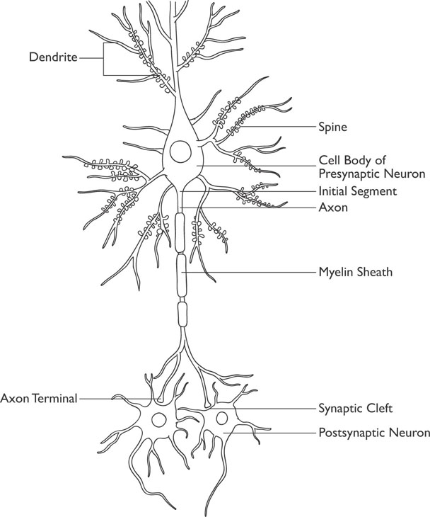
Figure 1-1: A Typical Neuron
Excitatory neurons stimulate neuronal firing, whereas inhibitory neurons reduce the rate of neuronal firing. Motor neurons are involved in motor functioning, whereas sensory neurons are involved in detecting and interpreting sensory stimulation. An interneuron connects other neurons together and is neither sensory nor motor in its functioning.
The process of signaling between neurons is quite complicated and will be simplified for this discussion. Basically what happens is that stimulation from sensory systems or from your thoughts results in a neuron being “activated.” Typically this consists of chemical substances known as neurotransmitters attaching themselves to the dendrites of a neuron. If a sufficient amount of neurotransmitters attach themselves to the neuron, this will result in the activation of an electric charge (an actual signal) known as an action potential being sent down the axon of the neuron.
The process of the action potential in a neuron depends on its capacity to react to a stimulus with an electrical discharge. This process is quite complicated but it involves changes in the electrical charges of the ions within the neuron compared to the electrical charges of the ions outside of the neuron’s cell wall. When the neuron is activated, the negative-based inner charge of the neuron becomes more positive due to an exchange of positively charged ions through the cell membrane via special gates. This results in a loss of negatively charged ions, which move out of the cell (when the neuron is not stimulated, this process is stabilized to maintain a relatively consistent balance). This change in the charge of the neuron results in the action potential, or electrical charge, being generated and traveling down the axon of the neuron.
The myelin sheath on the axon acts as a sort of insulator to facilitate the transmission of the electric charge. The electric charge literally jumps from space to space (the unmyelinated nodes of Ranvier) as it moves across the axon. When the electric charge reaches the end of the axon (the presynaptic terminals), it results in the release of neurotransmitters into the synaptic space. These neurotransmitters will attach themselves to the dendrites of adjoining neurons and the process will continue. Once the neuron has released its load of neurotransmitters, it is in a refractory period for a short time until the cell normalizes.
Most neurons communicate in this manner, but there are a very small number of neurons that communicate with one another via electrical charges. These neurons are called gap junctions.

When a neuron connects with another neuron, it is said to synapse at that site. Most of the neurons in the brain communicate chemically with each other, as this allows for a wide variation in the types of messages that can be sent and a greater potential to mediate the signal firing and signal strength than if neurons communicated via an electrical impulse.
Every neuron is only activated by a specific neurotransmitter or a specific set of neurotransmitters. Thus, the signaling between neurons is accomplished via a chemical process (neurotransmitter), whereas the messaging within the neuron is accomplished via an electric charge (action potential). These chemical communications between neurons and the communications they deliver result in your thoughts, feelings, and actions.
Neurotransmitters are chemical substances that the brain uses for communication between neurons and are created within neurons. There are hundreds of chemical substances that qualify as neurotransmitters. These substances are not used exclusively in the brain, although when they are utilized in the brain, they are called neurotransmitters. Outside of the brain when the same substances travel through the bloodstream, they are referred to as hormones. For example, the neurotransmitter serotonin, known to be important in mood, is also important in the process of digestion and is found in the gut. Most neurotransmitters have multiple functions. The neurotransmitter dopamine has important functions regarding your mood, movements, and memory.
The movement of the action potential down the axon of a neuron is one-directional. An action potential travels down the neuron toward the synapse—never the other way around. Once an action potential traverses the nodes of Ranvier, the upstream portion of the cell enters a refractory period and cannot generate a new action potential for a short period of time.
Neurons fire on an “all or none” basis; that is, there is no such thing as a neuron firing halfway (there is no such thing as half of an action potential). Neurotransmission is regulated by how often, how fast, or by the pattern of neuronal firing. Some neurotransmitters excite the system (cause a more rapid firing of neurons); some inhibit the system (cause neurons to fire at a slower pace); and others modulate how neurons fire. It is through these patterns of firing that the different signals are sent back and forth in the brain and to the body. The following table lists several of the more common neurotransmitters. You can also find a link to a site in Appendix B that can provide you with more information about neurotransmitters.
 SEVERAL WELL-KNOWN NEUROTRANSMITTERS
| Neurotransmitter | Basic Function |
| Dopamine | Responsible for arousal levels, mood; important in motivation; involved in voluntary movements |
| Serotonin | Effects on mood and anxiety, appetite, sleep, memory and learning, temperature regulation, and other functions |
| Acetylcholine | Controls activity in brain areas associated with attention, learning and memory, movement, and other functions |
| Epinephrine (Adrenaline) | Effects on attentiveness and mental focus; outside the CNS, it is involved in the “fight-or-flight response” |
| Glutamate | The major excitatory neurotransmitter in the brain |
| Enkephalins and Endorphins | Modulate pain, reduce stress, and promote a sensation of calmness; related to opiate drugs like heroin, they also decrease physical functions such as respiration |
| GABA (Gamma-aminobutyric acid) | The major inhibitory neurotransmitter in the CNS, it helps neurons recover after transmission, reduces anxiety, and reduces stress |
Neurons are not the only cells in the brain and spinal cord. There are other cells such as glial cells, which perform a number of functions in the brain. At one time it was believed that glial cells were just support cells; however, it is now known that glial cells perform other functions. These functions include surrounding and holding neurons in place, supplying nutrients and oxygen to neurons, insulating one neuron from another neuron, forming myelin, and destroying and removing dead neurons. Three types of CNS glial cells are astrocytes, oligodendrocytes, and microglia. While there are billions of neurons in the brain, there are one to five trillion glial cells in the brain, or nearly ten to fifty times more glial cells in the brain than neurons!
Astrocytes perform numerous functions. They support the endothelial cells (a thin layer of cells lining the interior of blood vessels) that form the blood-brain barrier, provide nutrients, and help repair the brain and spinal cord following trauma. The major function of the oligodendrocytes is to provide support for axons and to produce the myelin sheath that insulates them. Microglia are the immune defenses in the CNS constantly hunting for damaged neurons and infectious agents.
An extremely important part of the CNS is the spinal cord. Without the spinal cord your brain would be totally useless. It is important to remember that the brain cannot transmit information or receive any information without the spinal cord. The human spinal cord is about 18 inches in length (about 44 to 45 cm, but it is typically slightly longer in men than in women). The spinal cord is housed by the spinal vertebrae that offer some protection from injury. Neurons project from the spinal cord to other areas of the body, and nerves from the body project to the spinal cord.
Cranial nerves emerge strictly from the brain, as opposed to spinal nerves that project from segments of the spinal cord. In humans, there are twelve pairs of cranial nerves that are involved in different functions. Ten of the pairs of cranial nerves project from the brain stem, whereas two pairs project from the cerebrum.
When the spinal cord is transected (cut) or damaged, the brain cannot communicate with regions of the body below the site of the cut. There are three major functions of the spinal cord:
Acting as an agent for transmitting motor information, which travels from the brain, down the spinal cord, and to the body
Acting as an agent for the relay of sensory information, which travels from the body to the brain
Acting as a center for coordinating some types of reflexes
Structural and functional brain imaging has revolutionized the fields of neuroscience and medicine. The American neurosurgeon Walter Dandy first introduced ventriculography and later developed pneumoencephalography, both imaging methods in the early 1900s that often required the pumping of air into the brain; however, both procedures carried significant risks and could be quite painful. The technique of cerebral angiography was introduced in the late 1920s by neurologist Egas Moniz. This is a technique that takes pictures of the veins and arteries in the brain. This technique became refined and is still an important tool that is used today in neurosurgery.
Brain imaging can be noninvasive, such that no penetration of tissue occurs (CT or MRI), or invasive, where there are such tissue penetrations (PET, fMRI). The brain is never penetrated. Invasive images sound threatening, but there typically is no more than minimal discomfort involved as only an IV is used.
Further advancements, such as computerized tomography (CT), magnetic resonance imaging (MRI), and positron emission tomography (PET), have led to researchers and physicians being able to visualize the brain and other areas of the body for diagnostic, treatment, and research purposes. The advancements in neuroimaging have led to many remarkable discoveries. CT scans literally take x-rays of brain tissue, whereas MRI scans use a completely different technique that results in a much more detailed image of the brain tissue. These two techniques are known as structural imaging techniques in that they take a static picture of the brain and can be used to determine changes in brain structure. PET scans are a method whereby researchers can view the metabolic changes in the brain, thus PET scans and other scans like them such as functional MRI (fMRI) are known as functional imaging techniques. During a PET scan, a harmless radioactive isotope is injected into the bloodstream and tracked to allow researchers to study brain metabolism.
Electroencephalography (EEG) is the recording of electrical activity of the brain by placing electrodes along one’s scalp. Physicians who specialize in performing and interpreting neuroimaging techniques are called neuroradiologists.
There are a number of other advanced neuroimaging techniques and new developments in the field of imaging the brain being made all the time. There are several links provided in Appendix B to allow you to see how these techniques appear.