In the following two chapters, we will see that organisms exchange information that does not depend on sensory organs. Cells and organisms communicate through molecules and signals generated by the variations in the field. It is likely that these signals play a primary role in governing the physiology of the organisms. As in the inorganic world, cellular signals allow things to communicate with each other; they control the metabolism and rapid transmissions (in real time) between cells of any part of the organism. From top to bottom, cells are in constant radio contact with each other, and everything is always informed about everything.
We will see how the information networks involve all bodies, cellular and noncellular, and how the relationships between cells and organisms are the same as those between individuals and groups. We will see how the emotional component is a part of the biological universe and how it can influence the extrasensorial communications between plant and animal organisms. Let us start from the very beginning.
In the Beginning, There Was the Onion
In the Soviet Union in 1922, the Russian biologist Alexander Gavrilovic Gurwitsch, director of the Institute of Histology at Moscow University, was carrying out research on cell division; he was convinced that the multiplication of cells could also be stimulated by natural oscillation. To verify the hypothesis he used a simple plant substrate: onion bulbs. Later he also used onion roots in a very long series of experiments showing how the cells transmit signals, which had never been attempted before. Gurwitsch continued until reproducibility was obtained. Then he announced that plant organisms emit mitogenetic radiations capable of increasing the speed of mitosis (cell division).1 In other words, they enable the cell to multiply faster.
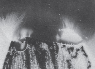
Here is his experiment: he placed two young onion bulbs with roots attached very close to but not touching one another, perpendicularly (fig. 9.1). The horizontal one was placed only 5 mm away from the vertical one. He placed the horizontal root in a glass cylinder covered with a metallic sheath with the ends left open. The vertical root was screened in exactly the same way, except for a small window corresponding to the area made by proliferating cells, called the meristem. The window provided a target for the other root, which was placed so that its base pointed to the meristem. After a few hours, the onions were examined under the microscope. Gurwitsch noticed that in the target section of the vertical root with the cells were more numerous compared with the shielded cells. The incidence of mitosis was at least 25 percent higher.
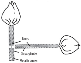
Figure 9.1. Gurwitsch’s onion bulbs. The root of the onion in the horizontal position spontaneously irradiates the vertical one.
What does this mean? The tip of the root emitted something that stimulated the cellular growth of the other root, but only in the uncovered area. Gurwitsch found that this experiment could be reproduced if a quartz filter was placed between the two roots, but not if gelatin or glass was placed between them. From this he concluded that the waves radiating from the bulbs were in the ultraviolet band (UV). Since UV rays are intensively absorbed by the tissues, he thought that the mitogenetic radiation would not act directly, but by inducing the cells to a second radiation, which would be the true cause of the increased growth. Apparently it was able to transmit from one cell to another in the same organism.
The first releases published by Gurwitsch in 1923 caused such a stir that the experiments were repeated in France, Germany, England, Italy, and the Soviet Union, and (despite numerous instances of failure) many researchers confirmed the results. It was concluded that cells are able to stimulate the growth of other cells by sending very weak frequencies.
According to Gurwitsch, all organisms have sources of self-radiation, which stimulate the homeostatic mechanism that regulates the growth rates of the cells. This prevents the cells from multiplying beyond the number established by natural laws and ensures they maintain their natural intended form. But this brings us back to the lingering questions about what shapes matter and what is meant by “natural laws”? Once again, we can think of a basic code, the guardian of the boundaries of form.
Dennis Gabor (who was awarded the Nobel Prize in Physics forty years after his discovery of holography) successfully recreated the Gurwitsch phenomenon, together with T. Reiter, and they published their results in 1928.2 They confirmed that embryonic tissues and malignant tumors have a high degree of irradiation, more intense in younger tissues and quickly growing. Reiter and Gabor—who were then scientists at Siemens and Halske Electric Company in Berlin—managed to modify the development of microorganisms by subjecting them to a source of ultraviolet radiation selected through a system of prisms and lenses. By exposing eggs from sea urchins to UV radiations, abnormal larvae were obtained. This also happened when they were subjected to frequencies emitted by certain organisms (for example the Bacterium tumefaciens).3 The German biophysicist Rajewsky also confirmed the existence of mitogenetic radiations.4 The Italian professor G. Cremonese in 1929 managed to imprint (after a selection for filtration) photographic plates with frequencies emitted by living organisms.5 The variety of results suggested that these cellular emissions could belong to a range of very different frequencies.
Thus, in the twenties, something important was happening that caught the attention of the scientific community. A new frontier was outlined with the discovery that cells not only build molecules, but they also generate impulses like tiny radio stations. With the discoveries of mitogenetic waves and those able to imprint photographic plates, there was good hope of establishing some new approaches to cellular physiology. Scientists realized that to gain a correct understanding of the cell, it was necessary to explore the physics of frequencies in areas that normally had been explained only by molecular chemistry. However, it was still just the beginning of a great adventure.
In the thirties, Italian doctor Giocondo Protti, inspired by Gurwitsch’s work, discovered that there is a radiating power in human blood similar to that observed in roots and plant bulbs.6 In fact, the energy coming from a drop of blood is so intense that it can stimulate proliferation (reproduction) in yeast cultures; it can also transfer this energy to another drop of blood lacking these properties. Just as with the mitogenetic waves, the energy from blood can even go through quartz; it is therefore in the ultraviolet range (fig. 9.2).
Protti noticed that hemoradiation (that is, the emission frequencies of blood) tends to increase during pregnancy and in situations where functions are at a maximum, while it decreases during fasting and in cancer cases.7 The Gurwitsch School observed that cancer sufferers have lost much of the radiant properties within their blood, while the tumor itself is very rich in mass, which could be the reason for the stimulation of its cell multiplication. According to Gurwitsch, cancer occurs when hematic radiation is concentrated at a single point in the organism, which accelerates the proliferation of the cells. Protti discovered that human serum has an oncologic power, that is, it can destroy tumor cells. He also found that by increasing the radiating power of the blood, temporal cells could be killed by means of light radiation (cytophotometric).8 Treated blood can in turn radiate and make non-radiating blood begin to radiate again. Injections of blood with a high radiating power increase the vital tone of animals whose blood has been deprived of radiating power by prolonged fasting: they brought about a reduction in tissue acidity, with obvious improvements in the course of many chronic pathologies, even degenerative ones.9 What wonderful things came out during the years of research on the “light of the blood,” as Protti defined it. And to think, all this from an onion!
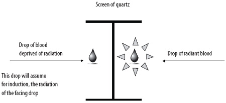
Figure 9.2. Transfer of ultraviolet hemoradiation through quartz to a drop of blood lacking that radiation (from G. Protti)
And Then There Was Yeast
The Gurwitsch School also discovered that small quantities of fresh yeast added to the blood of a cancer sufferer could make it radiate once again, even after the removal of the yeast. Protti carried out many experiments with a strain of Saccharomyces in which the high power of radiation was able to destroy growing tumor cells. The experiment was reproduced with both glass and quartz diaphragms separating the yeast from the tumor tissue, which once again proved that these are ultraviolet waves, since the radiating power of fresh yeast is only transmitted if the diaphragm is made of quartz and not of glass (fig. 9.3).
The cancer cells are not destroyed if the yeast is dry.10 Protti writes that the yeast, like the tumor, is also “anxious to live”: it multiplies rapidly, emits intense radiations, and has a high glycolytic power and other characteristics similar to the cancer cells. The ancient homeopathic principle of simila simillibus curentur seems to be respected: the tumor can be cured by something similar to it. In fact, particular quartz needles filled with yeast and injected into tumor masses proved to be able to kill the malignant cells by “colliquative necrosis,” compared with empty quartz needles.11 In other words, the radiating power of the blood and certain fresh yeasts, in certain conditions, could destroy tumors. Protti measured the radiation emitted by these yeasts, with waves measuring between 1,900 and 2,400 Å; once again, we are in the ultraviolet area.
All these researchers, from Gurwitsch to Gabor and Protti, first communicated their results to the International Congress of Electro-Radio-Biology held in Venice in 1934 under the high presidency of Guglielmo Marconi. That congress was an extraordinary and unrepeatable event! Pier Luigi Ighina was also there; he was already conducting experiments on the ability of information to modify matter. The days in Venice were a great event and were a clear indication that they were close to concluding something very important. But then war broke out, and the research was completely forgotten about.
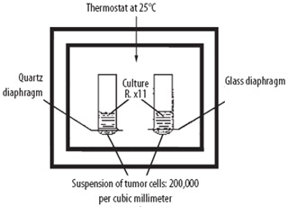
Figure 9.3. Using a setup similar to that shown here, in which quartz and glass diaphragms were both used to separate yeast cultures from suspensions of tumor cells, Protti found that the destruction due to the radiating power of fresh yeast is only transmitted if the diaphragm is made of quartz and not of glass. (From G. Protti)
Everything was resumed in the 1950s, when scientists started to talk about waves emitted by cells. A group of Italian physicists guided by Ugo Facchini from Milan resumed the experiments of Gurwitsch, who at the time was dying in Moscow, aged 80. The physicists wanted to verify the hypothesis that living matter emitted radiation, using photomultipliers to analyze the seeds of cereals and pulses. Photomultipliers are able to capture and amplify very weak fluxes of light, making them capable of measuring the weakest luminous radiations from living organisms that have been exposed to photonic stimulation. They were able to affirm that radiation from the analyzed seeds presented a band of around 500 nanometers in the optical range, with characteristic bioluminescence of a very weak intensity, millions of time weaker than that of a glowworm.12 In other words, cells emit light.
In the 1960s, some tissues and other cellular cultivations were used to irradiate different biological systems, and it was confirmed that blood, yeasts, bacteria, the eggs of sea urchins, and other organisms emit and receive mitogenetic waves, amplifiable by being passed through nutritive solutions with bacteria. In the same period, the Soviets resumed their research into ultrafine luminescence. Alexandra Gurwitsch, replacing her father at the Academy of Medical Science in Moscow, together with A. S. Agaverdiyev and others, was certain that the Gurwitsch radiation exists in all forms of plant or animal life, from the simplest to the more complex, and that the spectrum and the intensity of these waves varies according to species. Particularly, they thought the emissions could increase noticeably when a biological system is at a dying point.13
Also in those years, in Los Angeles, the engineer George Lawrence wanted to attempt Gurwitsch experiments; for that purpose he invented a piece of high-impedance equipment with which he studied different stimulations of cells in slices of onion one-half centimeter thick and connected to an electrometer. He discovered that they reacted not just to certain irritating agents (such as smoke) but also to the mental image of their destruction. Yes—every time the scientist manifested an intention to destroy them, the cells reacted in different ways according to the psychic qualities of the person having the thought.14 Just the scientists’ imagination of dramatic events for the cell seemed sufficient to induce variations in cellular response. Intention is a very powerful form of energy, as we will also see with plants. Lawrence’s experiment seems to be the first in scientific literature to present the idea that imagination, guided by a real intention, can influence cell metabolism. We will come back to this point.
Only in the 1970s did scientists finally start to recognize that such radiations are useful to the cells for communication.15 Alexander Dubrov was the first one to note what was then called mirror image. The researcher studied the behavior of two cells in glass tubes placed next to each other; he assessed the electrical potentials and biochemical parameters, hoping to get a glimpse of new impulses in their cellular growth, but he did not discover anything new. He injected one with a poisonous substance, with the intention of observing whether the distress was transmitted to the nontreated cell, but there was still nothing. However, when he substituted quartz tubes (which allow the passage of ultraviolet waves) for the glass ones, the symptoms of distress manifested themselves in the nontreated cell. This was the first step toward a great discovery that carries the signatures of a group of Siberian researchers.
In 1972, at the Institute of Clinical and Experimental Medicine in Novosibirsk in Siberia, some researchers, inspired by the observations of Dubrov, started a long series of experiments on cell cultures. Two glass balls containing cells of the same human tissue were joined, separated only by a glass diaphragm. In the first culture, a poison was introduced to kill cells. The second, not being infected, remained intact. By repeating the experiment using a diaphragm of quartz instead of glass, the cells of the second culture suffered and died even if not infected (fig. 9.4). This happened with different types of viruses and poisons inoculated only on the first culture and without the passage of molecules from one to another. The death of the cells of the second culture seemed to have been induced by electromagnetic signals very close to the ultraviolet band.16
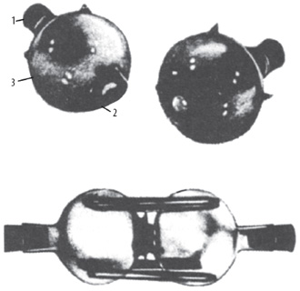
Figure 9.4. The Novosibirsk experiment: two glass balls separated by a quartz diaphragm, containing cellular cultivations. The poisoning of the cells of the first ball induces death also in the second (from F. A. Popp)
The Novosibirsk experiment was repeated (with 80 percent reproducibility) thousands of times in the 1970s. After five thousand experiments, in 1981 the prestigious Soviet science magazine Nauka published the work authored by V. P. Kaznachev and L. P. Mikhailova titled “Ultraweak Radiation in Intercellular Interactions.” The response to this event was so big that some risked using the term cellular consciousness to signify a form of control mediated by electromagnetic waves emitted by cells. In other words, this work indicated that forms of communication outside of biochemical paradigms could be possible, suggesting structures and mechanisms of action different from those previously known. The Novosibirsk experiment opened the doors to a new world of transmissions via invisible waves, all too often interpreted in a more esoteric sense than in a scientific one, with serious damage to research. Other discoveries like that of electricity, magnetism, and radioactivity experienced a similar journey.
With Novosibirsk we entered a new era. We acquired the knowledge that if we expose healthy cells to signals emitted by dying cells it can be as dangerous as exposing them directly to a virus or a chemical poison. This is the power of information in the organic world, information that was being transmitted as invisible waves. We came to understand that cells constantly emit photons and that their emissions can increase rapidly in situations of suffering. The experiments of Novosibirsk were repeated in the 1980s in many countries, in Australia,17 Brazil,18 China, Japan, Austria,19 Poland, and the Soviet Union,20 in the United States,21 and in Germany.22
Some researchers23 speak of “waves of degradation” emitted by dying cells, which appear to be caused because of the loss of their internal balance. These could stimulate other cells to enter into mitosis so that the number of cells in the organism tends to remain constant. Even if barely reproducible, these experiments are interesting because, if it is true that cells can emit contrasting signals (apoptotic and mitogenetic waves), it means that there is a homeostatic control even in small cellular groups, which presupposes that the system of intrinsic regulation could be the field connected to the cells themselves.
The Light of Things
Dying cells emit a lot more photons, a fact photographed in the Kirlian pictures of dying organisms. Surely you have seen images of leaves, fingertips, or other objects surrounded by luminous halos, like those of the saints. This is the Kirlian effect: a picture obtained by putting the object against a photographic plate that receives a high-frequency and high-voltage current.
Discovered in the nineteenth century by the Czech professor Barthélemy Navratil, electro-photography was perfected by the Polish doctor Jodko Narkiewicz, who was the first to photograph what the local press called “electric radiation of the human body.”24 In the twenties Mr. and Mrs. Kirlian from the Soviet Union set up a device that is used today all over the world. If the intensity of the charge is sufficient, the image of the object is surrounded by a luminescence that in noncellular bodies and things is uniform and constant, while in organisms it varies in intensity and quality (fig. 9.5).
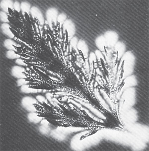
Figure 9.5. Kirlian photograph of a leaf. The leaf form is clearly visible in the luminescent halo. (From L. Galateri)
Unfortunately, the nonscientific usage of Kirlian photography by prana therapists, parapsychologists, and healers in order to document a mysterious halo, the so-called aura, and to quantify a person’s therapeutic power, discredited Kirlian photography in the eyes of the scientific community, and the effect ending up being considered esoteric and sometimes even fraudulent.
In 1970, the Russian physicist Viktor Adamenko compared the Kirlian effect to the phenomenon of St. Elmo’s fires: flames and blue flares (described also by Shakespeare in The Tempest) that can accompany thunderstorms when the high tension causes the air to ionize. Some people think that the Kirlian halo is the bioplasma of the photographed object; other people say it is electron beams. Adamenko discovered that in the human body, the Kirlian light is concentrated in the Chinese acupuncture points. This suggested to the German Peter Mandel the possibility of a diagnostic system based on studying the acupuncture points of the hands and feet photographed with the Kirlian method.25
Presman argues that the luminescence expresses a form of communication with the environment,26 a hypothesis that is not far from ours. The studies carried out by Adamenko in the Soviet Union and by others27—particularly Stanley Krippner at the Maimonides Medical Center in New York28—have registered the following observations:
- The so-called lie detector exploits the power of the emotions to vary the conductivity of the skin: telling a lie will result in minimal variations of the measured values. The same happens with the Kirlian effect; it can vary in intensity, shape, and color when there is emotional stress. For example, the images of the fingertips of relaxed subjects are different from those of anxious ones.
- Electro-photography is so sensitive that simply touching the photographed subject is sufficient to alter the chromatic values being recorded.
- Being in a state of relaxation induced by hypnosis, meditation, or the intake of cannabis seems to increase the dimensions and luminosity of the halo, which is in turn reduced when the subject is tense.29 The images of the fingertips may also vary due to illness, and this can be used for diagnosis.30
- The Kirlian effect seems to express a field of energy that surrounds and permeates the object and is sensitive to emotional moods. Could this vibrating light be information exchanges with the environment?
- Certain luminous halos certainly imply a field informed by the basic code of a thing that preserves identity and shape; for example, in the Kirlian photograph of a stemless leaf, the luminescence also includes the area of the missing stem (fig. 9.6). This is called the ghost effect: the halo also surrounds what is “not there” or, better, “what cannot be seen, that was expressed as the stem.” Although not perceptible by the senses, the stem is still present because the unity of the shape includes it.
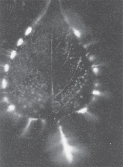
Figure 9.6. “Ghost effect”: in this Kirlian photograph of a stemless leaf, the luminescence also includes the area of the missing stem. (From L. Galateri)
Here we have an instance of shape without substance. It is plausible that the luminous image of the stem indicates that the field of the leaf—which contains the information of the entire design and image of the leaf—remains intact despite the amputation, exactly because it has to maintain the unity of the shape. Remember that, according to Harold Saxton Burr, the electric field of a bud already has the shape of the adult plant. Perhaps we can think of it as a hologram over time.
In other Kirlian photographs of plants, such as photographs of leaves with some missing parts, the luminous halo tends always to recreate the original structure (fig. 9.7).
Figure 9.7. Despite the apex of this leaf being torn, the luminescence tends to preserve the shape of the whole leaf, as guided by a field that maintains the shape. (From L. Galateri)
Another observation is that the halo of a leaf cut twenty-four hours before is much less intense than that of one that has just been cut. However, if two such leaves are photographed together, the field of the more vital one includes the weaker one until they become one unit, as if they put their energy together (fig. 9.8). All this data sustains the hypothesis of a field informed by its code.
Another experiment that has been done is to place a leaf in a glass for examination, then to remove it. If a photo is taken of the empty glass after a few hours, a luminous halo will still be visible, just as if the leaf were still there.31 This is a similar phenomenon to that observed by electro-acupuncture doctors with phials of tested homeopathic remedies: even after the content evaporated, the phial could still be used for testing. The information of the substance was preserved in the glass as a sort of an imprint. Is this a residue of waves or sounds that still vibrate in the glass’s ultrastructure? About the imprint left by things, Ighina writes that he has noticed that objects leave prints of themselves; organisms leave a memory of themselves on things or in their field.
Figure 9.8. Interaction between the fields of a leaf of privet just cut (on the right) and a dying one (on the left). The tendency is to form a unique field. (From L. Galateri)
Another Soviet scientist, Lepeshkin, poisoned yeast with sublimate ether inside a quartz tube. When the cells died, the photons they emitted in the moment of death were sufficient to blacken a photographic emulsion solution. Lepeshkin measured the wavelength of this radiation, and it was about 2,000 Angstrom, and the caloric emission of the dying yeast was about two calories per gram.32 A leaf taken from a tree, when examined with the Kirlian method, also showed an increase in the intensity of photonic emission shortly before dying, visually confirming what the American researcher Cleve Backster (whose research we will explore in the next chapter) identified in the twenties as the “cry of pain” of plants and other organisms at the point of death.
The Light of DNA
The work of Gurwitsch and others on the photonic signal emissions from cells was resumed in the 1970s by Fritz Albert Popp, then professor at the University of Kaiserslautern and later director at the Institute of Biophysics of the same city. In the years between 1971 and 1973, Popp carried out research on the genesis of cancer by studying a model of the isomers of benzopyrene and aromatic hydrocarbons taken from tar (isomers are two or more compounds with the same formula but with different properties due to different arrangements of the atoms in the molecule). The 3,4-benzopyrene causes cancer, the 1,2-benzopyrene does not. Unlike the 3,4-benzopyrene, the 1,2-benzopyrene does not absorb light in between the visible and the ultraviolet bands. Popp concluded that the carcinogenic effect of the 3,4-benzopyrene could be linked to its capacity of absorbing light.33
Popp also did research into biophotons, and in 1975 he proved their existence. They are tiny amounts of light that all living beings are able to store and radiate. When a photon (which is a quanta of light) imprints an atom, it arouses it, causing an electron to jump to the external orbit; when the electron returns to its previous level the photon is re-emitted by radiation (visible or not). By using sophisticated photomultipliers (fig. 9.9), Popp discovered that all cells emit this type of ultrafine electromagnetic radiation in the range between ultraviolet and long radio waves. It is manifested in the visible as a very weak luminescence, whose intensity is 1018 times weaker than daylight, comparable—if you want—to a lighted candle seen from twelve miles away. This is not to be confused with the bioluminescence emitted by fireflies or certain species of fish or bacteria.34
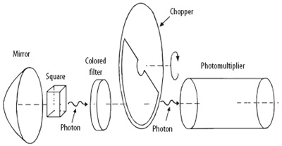
Figure 9.9. Diagram of a photomultiplier. In the darkroom, the little curve of the quartz containing the cells placed in front of a mirror is focused by a photomultiplier. The colored filter allows the selection of different colors of light. The chopper is a dark-light rotating disk that makes it possible to select the photonic signals actually emitted by the cells, distinguishing them from the background noise. (From F. A. Popp)
It is worth remembering that the luminescent properties of certain substances that emit electromagnetic waves (infrared, ultraviolet, X rays, or gamma waves) come from energy absorbed as electromagnetic radiation. If the phenomenon lasts less than eight to ten seconds it is described as fluorescence; if it lasts longer then it is phosphorescence.
Popp observed that stress, disease, and suffering are accompanied with increased biophotons. Initially he interpreted the bioluminescence as deriving from cell metabolism, something like the sparkles that come out of the electric wire of a tram. Later one of his students called Rattemeyer thought that the biophotons came from the cells’ DNA, the controller of all of the cells’ vital information. He was proved right. In 1981 Popp and Rattemeyer published their conclusion that biophotons are emitted (though not exclusively) from cellular DNA and that, during mitosis, when the chromatin (a combination of nucleic acids, mostly DNA, and protein found in the cellular nucleus) is dissolved, that is, when it passes from phase G0 to phase G1, cells release massive photon emissions. At that point the bioluminescence was not anomalous, but formed signals of great significance to the cells.35
Then it was understood that biophotons have a guiding role in the regulation of all cellular physiological functions; by exciting the electron layers of the molecules, they are responsible for the initiation of biochemical reactions. In other words, the orders are given by the DNA, but it is the biophotons that carry the messages and ignite the metabolic fuse. Once again, it is possible that molecular events are the consequence of physical reactions that are based on the energy quanta. Without the ongoing work of the biophotons, cellular metabolism would end up in chaos and would take life with it. Popp’s studies suggest that the intrinsic mechanism of existence must be sought in physical and chemical models that describe effects, not causes. Popp was convinced that biophotons were emitted by the heterochromatin, part of the DNA (about 98 percent of the molecule) that is not expressed in genes and seems meaningless. It could be, instead, the most important part of the DNA, storing light to regulate the cellular biochemical reactions.36
Popp noticed that this is possible only if the biophotons constitute an electromagnetic field of high coherence. In fact, the more numerous the frequencies are, the greater the degree of information and the lower the possibility that other photons, even intense ones, can interfere with its processes. A weak radiation but with a high informative order will never be disturbed by a stronger radiation that is lacking order. The cell always tries to defend itself from chaotic external radiations that could confuse its communications. Nature makes cells resistant to great electromagnetic impulses, but they are susceptible to others, even very weak ones, if they are coherent, like those of homeopathic oscillations or other techniques such as electro-acupuncture and the acupuncture itself.
According to Popp, DNA behaves as a photoaccumulator: it is activated by absorbing light. It then immediately emits it in the form of coherent photons that act like laser rays. As mentioned earlier, in a laser all the waves oscillate in phase, making its light monochromatic and extremely pure. Unlike the light of a bulb that radiates in all directions (chaotic), a laser light becomes a unique unidirectional beam (coherent). Once again consistency is the key word for understanding how very weak intensity is sufficient to govern complex cellular functions. The energy absorbed by the DNA is compensated for by ultrafine cellular radiation. The DNA thus works like a radio station that regulates not only the cellular biochemical processes but also the communications between cells.
Let us try to gather these results together: according to Popp DNA acts like a radio station; Preparata and Del Giudice see molecules as antennas that emit and receive signals; and the apparatus of the TFF works like a radio station where medicines emit signals. Together they indicate that the human body and the world of things are governed by radio transmissions, which build a well-known network of communications. Prigogine wrote that it is communication that elevates life above chaos, enabling it to evolve. Cells are open systems, dissipative structures that communicate by means of countless light beams that originate from each nucleus. Even more than the membranes, it is photonic resonance that unites the cells. This has consequences for the phenomenon of recognition, immune response, and susceptibility to tumor degeneration. It is obvious that biophotons are responsible for the communication between the cells; the chemical transmitters are only the instruments.
In 1982 at the Max Planck Institute of Astronomy in Heidelberg, Popp and Beetz made the biophotons visible using a residual light amplifier with a fluorescent screen.37 By first using cress and then another type of cell more sensitive to photons (the unicellular green algae, Acetabularia acetabulum), Popp managed to demonstrate that the photonic emissions periodically fluctuate according to cellular biorhythms; they are intense when the cells are developing more rapidly, and they disappear at death. In accordance with Mr. and Mrs. Kirlian and the researchers from Novosibirsk, Popp noticed an increase of luminescence in the case of strong cellular suffering: poisoning of a cell increased its photonic production prior to death, which was reproduced on the computer screen as luminous explosions and as spikes on the graph showing emissions (fig. 9.10).
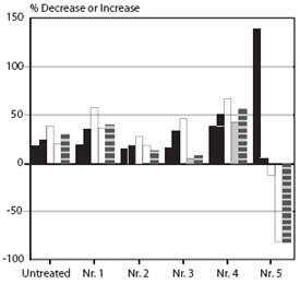
Figure 9.10. Reduced photonic emission of Acetabularia poisoned with atrazine (Nr. 5) compared with the nontreated control, a clear expression of cellular suffering preceded by a peak of strong photonic emission (“the cry of pain”).
Popp was the first to demonstrate that two cells communicate with each other at short distances through coherent light emitted by DNA, which also controls the functions of cells. In fact, he showed that if an Acetabularia in a bowl of quartz in the darkroom of a photomultiplier is stimulated with white or colored light for a few seconds, its luminous emissions will appear on the screen in the shape of pulsating explosions. After a little time the intensity of light weakens, but if we put seaweed emitting similar luminous signals near it for a second, the first will be reactivated with new emissions of light; the cells seem to dialogue with each other. The time used by the cell to release the absorbed light from its accumulator (the DNA) is important. The more rapidly it is emitted the less good is the accumulator.38
DNA represents the priming and control system for cellular biochemical functions, it plays a role in maintaining homeostasis (the ability of a system to self-regulate itself to maintain its balance), and it is responsible for communications, even at great distances between cells. Through the light protected in its deep spaces, the DNA transmits information at a very high speed throughout a whole organism. This would explain the reason why the majority of the biochemical reactions in a cell take place at least a billion times quicker compared with the same systems in vitro. The biophotons are far more numerous in living beings than in in vitro systems, confirming the presence of informative networks operating in vivo; the network’s effectiveness increases with the complexity of the organism.39 This explains why in the more organized systems the response to stimulations, as for example to the TFF, is more rapid and effective, according to the Kaiserslautern postulate (that the more complex the system, the more complete the decoding of the signals).
It has been observed that there is a balance between photons and growth: when cells develop, many photons will curb proliferation, while growth increases when the photons are reduced. It is possible that in cancers this regulation is disturbed if carcinogens damage the DNA or interfere with the coherence of the emitted light. Popp thinks that cancer can be associated with (and may be caused by) the loss of coherence of the radiation field with uncontrolled cellular proliferation.40 In general, each disease may be linked to perturbations of the cellular fields; perhaps the accumulation of disordered oscillations within the organism produces distorted regulations. The theory of the biophotons shows that weak impulses, well below the threshold of perception, have no value for their energy content but have an important significance for information conveyance. This points to the importance of an electromagnetic type of system of intrinsic regulation in organisms; it may be that every chemical reaction is preceded by something physical.
Water, Cells, and Light
At this point, we have to go back to water and its relationship with the light frequencies emanating from the DNA. Among its many functions, water grants stability to the DNA: if the water percentage around the double helix falls below 30 percent, it results in the denaturation (structural change in macromolecules caused by extreme conditions) of the nucleic acids.41 Biological systems, being open, have a high degree of order because, contrary to the closed systems, they are far from the thermodynamic balance. This allows them to exchange a lot of energy and produce coherent behaviors. Life needs to be as far as possible from the thermodynamic balance.
If we warm liquid from below, its molecules get agitated in a chaotic way, but a certain amount of heat triggers convection currents that become more and more stable until they form so-called Benard cells: ordered microscopic structures like little cells in rows composing a mosaic of a dissipative type. In other words, if we move away from the thermodynamic balance (for example, with a boiling action), beyond the chaos, the molecules restructure in an orderly manner. A system can be ordered only when it intersects with a great flux of energy, which must not stay there for long: an energetic jam would destroy the coherence.
Frölich proposed the model of coherent oscillations.42 Water molecules are powerful magnets (we have already seen that they have high dipole values), and they tend to couple their oscillations under the influence of an electromagnetic field: they start to dance all together, like in a ballet. In this way, energy is transmitted to the entire system, as in the model of dissipative structures. At certain stages of field intensity the molecules start to oscillate in a coherent way, emanating a particular wave that behaves like a laser. Think about it: a laser produced by water! These are long-range interactions, able to control the entire system. Thanks to them, the molecules inside of cells are able to interact instantaneously. And it is also thanks to these laser beams that water can receive information and release it, as in homeopathy, in the TFF, or in other methods of water activation.
Biophotons are cellular messengers, waves with high coherence that rightly fall within the models of Prigogine and Frölich. They are not information, but they are carriers. They are not generated by the cell; they are light captured from the environment, which is then emitted in a coherent way in order to transport information. The biophotons suggest a regulatory control principle in the cell identifiable with the field itself. But let us leave the cells and start to explore the complex organisms and their non-sensorial communications. At this level of organization there is a new element we have to take into account: the emotions.
 COMMUNICATION BETWEEN CELLS