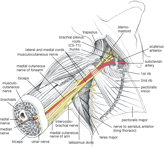
Plate 1 Axilla, to expose the neurovascular bundle; the axillary vein and most of pectoralis major has been removed and pectoralis minor has been divided. To show the origins of the plexus from between the scalene muscles, the clavicle has been divided and sternomastoid refl ected.
Reproduced from Mackinnon, Pamela and Morris, John, Oxford Textbook of Functional Anatomy, vol 1, p112 (Oxford, 2005). With permission of OUP.
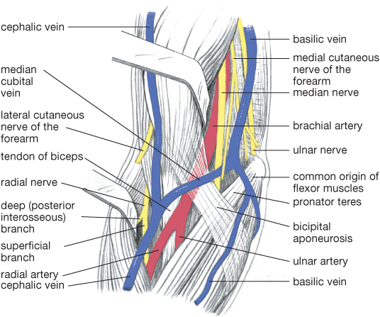
Plate 2 Anterior aspect of elbow; ‘cubital fossa’. Note the retraction of biceps and brachioradialis.
Reproduced from Mackinnon, Pamela and Morris, John, Oxford Textbook of Functional Anatomy, vol 1, p202 (Oxford, 2005). With permission of OUP.

Plate 3 Deep dissection of gluteal region; gluteus maximus and medius largely removed.
Reproduced from Mackinnon, Pamela and Morris, John, Oxford Textbook of Functional Anatomy, vol 1, p142 (Oxford, 2005). With permission of OUP.
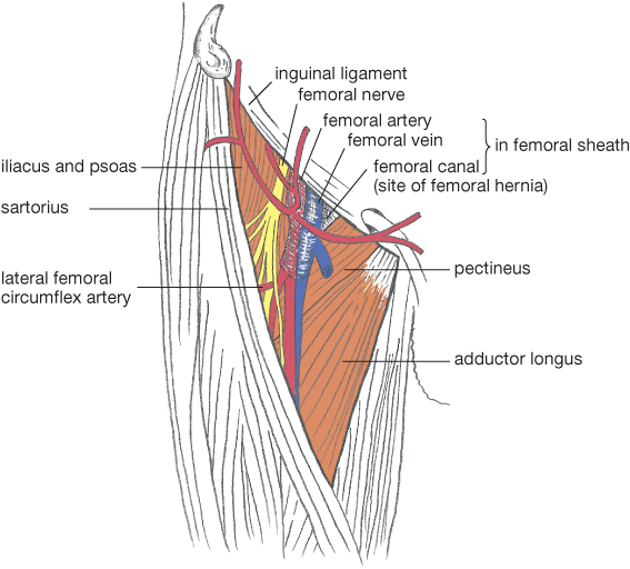
Plate 4
Femoral triangle.
Reproduced from Mackinnon, Pamela and Morris, John, Oxford Textbook of Functional Anatomy, vol 1, p179 (Oxford, 2005). With permission of OUP.
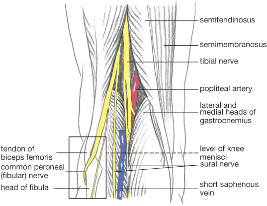
Plate 5 Popliteal fossa; popliteal vein removed above entrance of short saphenous vein. In the boxed area, the muscles have been removed to show the relationship of the common peroneal nerve to the neck of the fi bula.
Reproduced from Mackinnon, Pamela and Morris, John, Oxford Textbook of Functional Anatomy, vol 1, p180 (Oxford, 2005). With permission of OUP.
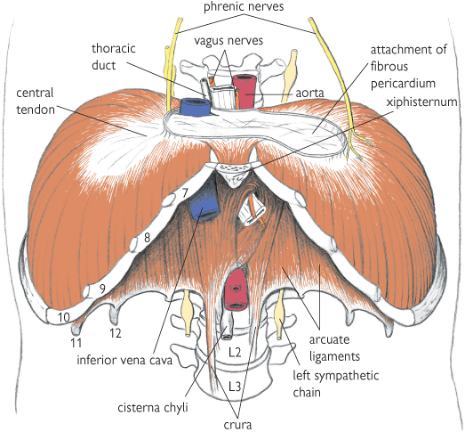
Plate 6 The diaphragm and structures passing through it.
Reproduced from Mackinnon, Pamela and Morris, John, Oxford Textbook of Functional Anatomy, vol 1, p98 (Oxford, 2005). With permission of OUP.
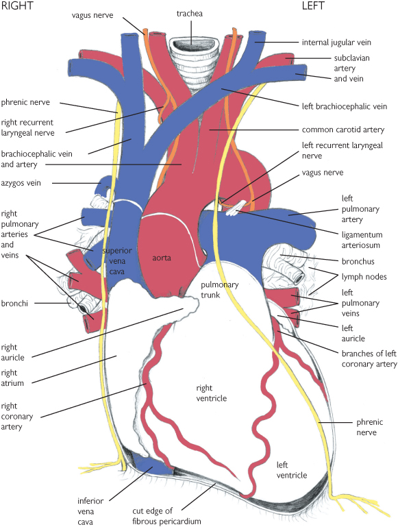
Plate 7 The heart and great vessels viewed from the front.
Reproduced from Mackinnon, Pamela and Morris, John, Oxford Textbook of Functional Anatomy, vol 2, p73 (Oxford, 2005). With permission of OUP.
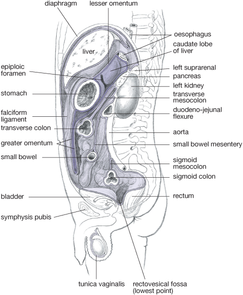
Plate 8 Arrangement of peritoneum of the greater and lesser sacs in a parasagittal section of the abdominal cavity.
Reproduced from Mackinnon, Pamela and Morris, John, Oxford Textbook of Functional Anatomy, vol 2, p134 (Oxford, 2005). With permission of OUP.
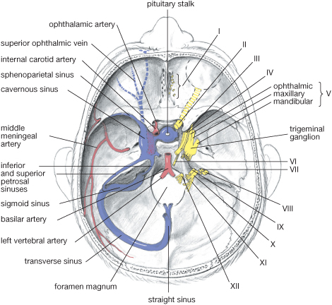
Plate 9 Interior of skull base: vessels and nerves.
Reproduced from Mackinnon, Pamela and Morris, John, Oxford Textbook of Functional Anatomy, vol 3, p48 (Oxford, 2005). With permission of OUP.

Plate 10 Deep dissection of neck, showing prevertebral muscles, brachial plexus, root of neck, and sympathetic chain.
Reproduced from Mackinnon, Pamela and Morris, John, Oxford Textbook of Functional Anatomy, vol 3, p190 (Oxford, 2005). With permission of OUP.
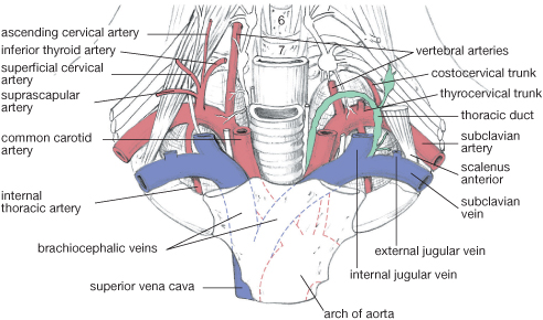
Plate 11 Vessels in the root of neck.
Reproduced from Mackinnon, Pamela and Morris, John, Oxford Textbook of Functional Anatomy, vol 3, p154 (Oxford, 2005). With permission of OUP.
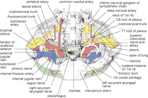
Plate 12 Root of neck.
Reproduced from Mackinnon, Pamela and Morris, John, Oxford Textbook of Functional Anatomy, vol 3, p191 (Oxford, 2005). With permission of OUP.