CHAPTER 8
Understanding Organismal Biology
Lesson 8-1. Support and Movement
Vocabulary
• homeostasis
• exoskeleton
• endoskeleton
• ligaments
• cartilage
• hydrostatic skeleton
• cardiac muscle
• smooth muscle
• skeletal muscle
• tendons
• flexor
• extensor
• actin
• myosin
With the evolution of multicellular organisms, specialization of structure and function became possible. The structure and function of a group of cells, organized into tissues and organs, could be focused on one task. For example, they could be specialized for locomotion or reproduction. As a result, these processes would function more efficiently. However, specialization of structure and function also posed a challenge. Somehow, the workings of all the various tissues and organs had to be coordinated so that all the functions carried out by a multicellular organism proceeded in a controlled and coordinated manner.
Animals have mechanisms to regulate their biological processes within very narrow limits. This regulation results in a stable internal environment. The maintenance of a stable internal environment is known as homeostasis. Homeostasis is essential if an animal is to survive in the face of a constantly changing environment.
An understanding of homeostasis involves an understanding of the various organ systems that make up a multicellular organism. These include systems for support, movement, transport, digestion, protection, defense, reproduction, development, and communication. The coordination of all these systems in animals is the responsibility of still another two systems—the endocrine and nervous systems.
Support
The bodies of some animals are protected by a hard covering. For example, a lobster is completely covered by a shell made of protein and chitin. This external covering is called an exoskeleton. An exoskeleton provides both support and protection for the animal. Muscles attached to the exoskeleton enable the animal to move. Animals that belong to the phylum Arthropoda have exoskeletons. In addition to lobsters, these animals include horseshoe crabs, insects, spiders, and crustaceans.
Although an exoskeleton protects and supports these animals, it does pose one limitation. An animal with an exoskeleton cannot grow very large, especially if it flies. Large animals then must have some other means of support. This support is provided by an endoskeleton, which is found inside an organism’s body. All vertebrates (fish, amphibians, reptiles, birds, and mammals) have an endoskeleton. An endoskeleton provides not only support but also protection. Bones that make up the endoskeleton protect delicate internal organs such as the heart and lungs.
The Human Skeletal System
More than 200 bones make up the human skeleton. Some bones are fused together, such as those that make up an adult’s skull. Most of the bones, however, are connected at joints. Bones are connected to one another by ligaments. Ligaments allow these bones to move freely. As they move, cartilage prevents bones from rubbing against one another. Cartilage is a tissue that is firm but much softer than bone.
Cartilage is firm enough to form the endoskeleton of some animals, such as sharks and rays. In fact, the long bones of the human skeleton, such as those found in the arms and legs, are first made of cartilage. This cartilage is slowly replaced by bone as the organism develops. However, this process does not occur in the center of each long bone. Instead, this area becomes a hollow cavity filled with bone marrow, where new blood cells are formed.
The cartilage in some structures is never replaced by bone, no matter how old a person gets. These structures include the tip of the nose and the external ear. Cartilage makes these structures flexible and less likely to break when struck.
In addition to support, protection, and production of blood cells, the skeletal system has another function. It is involved in homeostasis by helping to regulate the mineral level in the blood. Consider what happens when the calcium level in the blood begins to drop. Calcium that is stored in bones is released into the blood. The calcium level in the blood must be maintained so that the muscles can function.
Movement
Animals use their muscles to move their bodies and also move substances within their bodies. Even animals that lack a skeleton can move with just the help of their muscles. These animals include flatworms and annelids. The contraction of their muscles puts pressure on the fluids inside their bodies. The fluids cannot be compressed. As a result, the fluids flow along the length of the body and cause it to lengthen. When the muscles relax, the fluids retreat and the animal’s body shortens. This type of motion is brought about by muscles and fluids which form a hydrostatic skeleton.
The Human Muscular System
The muscular system works with the skeletal system to support, protect, and move the body. There are three types of muscular tissue in the human body. One type is called cardiac muscle. This type of muscle is found only in the heart. Cardiac muscle is responsible for heart contractions that pump blood to all parts of the body. Cardiac muscle is involuntary. This means that a person does not have conscious control over the muscle’s contraction.
The second type of muscular tissue in humans is called smooth muscle. This type of muscle lines the walls of internal organs and structures, such as the intestines, stomach, blood vessels, bladder, and uterus. Like cardiac muscle, smooth muscle is involuntary.
The third type of muscle is called skeletal muscle. As its name suggest, this type of muscle makes up the muscles that are attached to bones. Skeletal muscle is voluntary, meaning that a person can consciously control its contractions. Skeletal muscles are attached to bones by tendons.
Two opposing skeletal muscles work together to move a bone. One is known as a flexor. The contraction of a flexor brings two bones together. For example, contraction of the biceps muscles brings the forearm and upper arm together. The opposing muscle is called an extensor. The contraction of an extensor straightens the two bones. In this case, contraction of the triceps muscle straightens the arm. When an extensor contracts, a flexor relaxes, and vice versa.
When viewed under a microscope, skeletal muscle is seen to have alternating dark and light bands. These bands form a pattern or striations. As a result, skeletal muscle is also called striated muscle. These bands are formed by two muscle proteins—actin and myosin. Muscle contraction involves the sliding of these two proteins past one another. To get an idea of how this works, take the fingers on one hand and slide them between the fingers on your other hand. This action simulates what happens when a muscle contracts. Now move your fingers apart. This action simulates what happens when a muscle relaxes.
Lesson Summary
• Homeostasis is the maintenance of a stable internal environment despite changes in the external environment.
• An external skeleton is known as an exoskeleton; an internal skeleton is called an endoskeleton.
• Both an exoskeleton and an endoskeleton provide protection, support, and help in movement.
• Bones of the human endoskeleton also produce blood and regulate the mineral level in the blood.
• A hydrostatic skeleton enables some animals to move through the interaction of muscles and fluids.
• Three types of muscles are found in humans: cardiac, smooth, and skeletal.
• Cardiac and smooth muscle are involuntary, while skeletal muscle is voluntary.
• Skeletal muscle contracts and relaxes when actin and myosin protein molecules slide past one another.
REVIEW QUESTIONS
1. Muscles are attached to bones by
(A) ligaments
(B) tendons
(C) actin
(D) myosin
(E) flexors and extensors
2. Which of the following organisms has an exoskeleton?
(A) shark
(B) frog
(C) snake
(D) crayfish
(E) protist
3. The human skeletal system
(A) protects internal organs
(B) is involved in body movement
(C) produces blood cells
(D) regulates the blood calcium level
(E) all of the above
4. Which human muscle types are classified as involuntary?
I. skeletal
II. smooth
III. cardiac
(A) I and III only
(B) I and II only
(C) II and III only
(D) II only
(E) I, II, and III
5. Examine the following illustration of human muscle tissue.
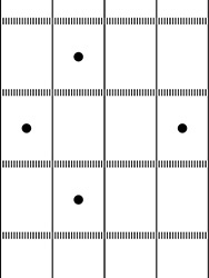
Figure 8-1
Where in the body would you expect to find this type of muscle tissue?
(A) face
(B) stomach
(C) intestines
(D) heart
(E) bladder
6. Sensorimotor coordination for balance and complex muscle movements is associated with the
(A) cerebellum
(B) cochlea
(C) nervous system
(D) cardiac muscles
(E) cerebrum
ANSWERS AND EXPLANATIONS
1. (B) Tendons attach a muscle to a bone, while ligaments attach one bone to another bone.
2. (D) Like all arthropods, a crayfish has an exoskeleton.
3. (E) The human skeletal system provides support and protection, helps move the body, produces blood cells, and regulates blood calcium levels.
4. (C) Smooth and cardiac muscles cannot be consciously controlled.
5. (A) The illustration shows skeletal (striated) muscles which are found in the muscles of the head where they control facial expressions, chewing, and eye movements.
6. (A) The cerebellum in the brain coordinates voluntary movements such as those that maintain balance and complex muscle movements.
Lesson 8-2. Transport
Vocabulary
• open circulatory system
• closed circulatory system
• atrium
• ventricle
• septum
• vein
• artery
• pulmonary circulation
• systemic circulation
• capillaries
• plasma
• erythrocytes
• leukocytes
• platelets
• pharynx
• larynx
• trachea
• bronchus
• alveolus
• diaphragm
Transport of materials in some organisms simply involves the processes of diffusion and osmosis. For example, unicellular organisms such as protozoa depend on diffusion of nutrients from their environment and the diffusion of waste products out of their cells. Water moves in and out of these organisms by osmosis. Even some multicellular organisms depend only on diffusion and osmosis for the transport of substances. Hydra, for example, consist of two cell layers. Both layers are in direct contact with the environment, allowing for nutrients and wastes to enter and leave the cells by diffusion and osmosis. In contrast to protozoa and hydra, most animals need a specialized transport system. Only then can cells not in direct contact with their environment get the nutrients they need and eliminate the wastes they produce.
Open and Closed Systems
Transport of nutrients and wastes is mainly the responsibility of the circulatory system. Some organisms have an open circulatory system. In such a system, blood flows within vessels only some of the time. At other times, blood seeps through open spaces called sinuses. Both mollusks and arthropods have an open circulatory system.
Other multicellular organisms have a closed circulatory system. In such a system, blood continuously flows within vessels. For example, annelids have a closed circulatory system where blood flows toward the head in a dorsal vessel. The blood then flows to five vessels called aortic arches, which pump the blood to a ventral vessel that transports it toward the posterior. By pumping the blood, the aortic arches act like hearts that are found in vertebrates.
Fish, for example, have a two chambered heart, consisting of one atrium and one ventricle. The atrium is the chamber that collects the blood returning from various parts of the body. The atrium pumps the blood to the ventricle, which is the chamber that then pumps the blood to all parts of the body. Amphibians have a three-chambered heart. Birds and mammals have a four-chambered heart, consisting of two atria and two ventricles.
The Human Circulatory System
Figure 8-2 illustrates how blood flows through the human heart.

Figure 8-2
Be sure to refer to the above illustration when reading about the human circulatory system. Notice that the illustration shows how the heart appears in a person looking at you. Locate the septum, which is a muscular wall that separates the left side of the heart from the right side. Now locate the structures labeled anterior (superior) vena cava and posterior (inferior) vena cava. These are the blood vessels that return blood from all parts of the body except the lungs. A blood vessel that returns blood from a part of the body to the heart is called a vein. Notice that a pulmonary vein returns blood from the lungs to the left atrium.
Blood is then pumped from the atria into the ventricles. Valves in the heart prevent the blood from flowing backward. Locate the right ventricle. Notice that it pumps blood to the lungs through the pulmonary artery. A blood vessel that transports blood from the heart to all parts of the body is called an artery. Arteries are thicker and more muscular than veins. What is the name of the artery that transports blood from the right ventricle to the lungs? From the left ventricle to the body?
Blood flow between the heart and lungs is known as pulmonary circulation. Blood flow between the heart and the rest of the body is called the systemic circulation. Use the illustration shown above to trace with your finger how these two systems are related. Do this by tracing how blood flows from the leg through the posterior vena cava and eventually flows back to the leg.
Blood returning from the body to the right atrium is low in oxygen and high in carbon dioxide. This blood has a purplish color is known as deoxygenated blood. Deoxygenated blood is pumped to the right ventricle and then out the pulmonary artery. The pulmonary artery splits, sending a branch to each lung. At the lung, the blood gives up its carbon dioxide and picks up oxygen. This exchange takes place in blood vessels called capillaries. A capillary is the blood vessel where materials are exchanged between the blood and cells. Substances, such as oxygen and carbon dioxide, are exchanged by diffusion. The wall of a capillary is only one-cell thick so that these processes can occur easily.
Blood that leaves the lungs is called oxygenated blood, which has a red color. Oxygenated blood flows from each lung through a pulmonary vein. These two veins join to form the large pulmonary vein that enters the left atrium. The septum prevents oxygenated blood from mixing with deoxygenated blood in the heart.
Blood, whether oxygenated or deoxygenated, consists of a liquid called plasma. Plasma is mostly water containing dissolved substances such as nutrients, hormones, and gases. Plasma makes up about 50% of the blood volume. The rest is made up of solids that are suspended in the plasma. These solids include erythrocytes, or red blood cells. These cells make up about 45% of the blood volume. Red blood cells contain a protein called hemoglobin that transports oxygen. In contrast, carbon dioxide is transported in the plasma. The remaining 5% of the blood volume is made up of leukocytes and platelets. Leukocytes are white blood cells, which play a major role in protecting the body against disease. Platelets are needed for blood clotting.
The Human Respiratory System
The various systems in the human body work closely together. This is especially true of the circulatory and respiratory systems. You read that the pulmonary circulation is responsible for supplying oxygen to the blood and removing carbon dioxide. The respiratory system must get oxygen to the lungs and take away the carbon dioxide that is brought there by the circulatory system.
Air containing oxygen enters the nose and then passes to the pharynx, which is an area located at the back of the throat. Next, air travels to the larynx, or voice box. From here, air passes into the trachea or windpipe. Then air flows to a bronchus, which divides into two bronchi with one going into each lung.
The air passage in each lung continues to branch and branch. Eventually, each tiny branch ends in an alveolus, or tiny air sac. The lungs contain millions of alveoli. These alveoli are the sites where gases are exchanged between the circulatory system and the respiratory system. The following illustration shows how these gases are exchanged.
Like a capillary, an alveolus is surrounded by a wall that is only one cell thick. Oxygen, which is in higher concentration in the alveolus, can easily diffuse into the blood in the capillary. Carbon dioxide can easily diffuse in the opposite direction. Notice in Figure 8-3 that water vapor also diffuses from the capillary into the alveolus. This is why you breathe out water vapor that can condense on a mirror when you exhale.
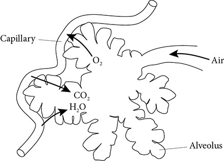
Figure 8-3
Breathing is controlled by a muscle called the diaphragm. The diaphragm forms the bottom wall of the chest cavity. When you inhale, the diaphragm contracts and moves downward. This increases the volume of the chest cavity and reduces the air pressure inside. As a result, air rushes into the nose and begins its journey to the lungs. When you exhale, the diaphragm relaxes and moves upward. This decreases the volume of the chest cavity and increases the air pressure inside. As a result, air is forced out the lungs and begins it journey to the nose.
Lesson Summary
• Most multicellular organisms require a transport system to deliver nutrients and remove wastes from cells not in direct contact with the environment.
• Transport systems include the circulatory and respiratory systems.
• Some organisms possess an open circulatory system where blood at times seeps through open spaces.
• Most organisms possess a closed circulatory system where blood is always confined to a vessel or heart.
• The human circulatory system consists of a four-chambered heart, arteries, veins, and capillaries that transport blood throughout the body.
• The human circulatory system consists of pulmonary and systemic circulations.
• Blood consists of plasma, erythrocytes, leukocytes, and platelets.
• The human respiratory system consists of the pharynx, larynx, trachea, bronchus, and alveoli.
• Oxygen and carbon dioxide gases are exchanged between capillaries of the circulatory system and alveoli of the respiratory system.
REVIEW QUESTIONS
Each set of lettered choices below refers to the numbered statement immediately following it. Select the lettered choice that best answers each statement.
(A) left ventricle
(B) right atrium
(C) pulmonary vein
(D) pulmonary artery
(E) aorta
1. Receives blood returning from the arm
2. Blood vessel that contains oxygenated blood
3. Blood vessel that contains deoxygenated blood
(A) diaphragm
(B) alveolus
(C) bronchus
(D) pharynx
(E) larynx
4. The voice box
5. Controls inhalation and exhalation
6. Site of diffusion of gases
ANSWERS AND EXPLANATIONS
1. (B) The right atrium receives blood from the arm through the anterior vena cava.
2. (C) The pulmonary vein carries oxygenated blood from the lungs to the left atrium.
3. (D) The pulmonary artery carries deoxygenated blood that is on its way to the lungs.
4. (E) The larynx is the voice box.
5. (A) The diaphragm contracts during inhalation and relaxes during exhalation.
6. (B) Gases are exchanged between the alveolus and a capillary.
Lesson 8-3. Nutrition
Vocabulary
• intracellular digestion
• extracellular digestion
• alimentary canal
• mechanism digestion
• salivary glands
• amylases
• chemical digestion
• epiglottis
• esophagus
• peristalsis
• proteases
• bile
• lipase
• villi
• nephron
• glomerulus
• Bowman’s capsule
• proximal convoluted tubule
• loop of Henle
• distal convoluted tubule
• collecting tube
• ureter
• bladder
• urethra
Every cell of an organism needs nutrients. Organisms whose cells are in direct contact with the environment obtain nutrients by diffusion. Some unicellular organisms, such as paramecium, ingest tiny food particles. A food vacuole forms around these particles. Enzymes are secreted into the vacuole to break down the food. Digestion that takes place within a cell is known as intracellular digestion.
Some organisms, such as hydra, use their tentacles to sweep food into their body’s cavity. Enzymes are released into the cavity where they break down the food. Digestion that takes place outside a cell is called extracellular digestion. As a result of extracellular digestion, the food particles are small enough to diffuse into the cells. The food particles are then further digested through intracellular digestion. Undigested materials are then expelled through the same opening through which the food particles entered.
More complex multicellular animals have more specialized mechanisms for nutrition. For example, annelids, such as the earthworm, have what is sometimes called a “tube-within-a-tube” digestive system. This consists of a digestive tract (one tube) that extends the length of its body (second tube). The digestive tract is also known as an alimentary canal. Food enters through a mouth, and wastes pass out through an anus. Specialized structures aid in the digestive process as the food passes through the alimentary canal.
The Human Digestive System
Figure 8-4 illustrates the various parts that make up the human digestive system.

Figure 8-4
As you read about the human digestive system, follow the process in the illustration. Food enters through the mouth. Here the food is chewed. Teeth break down the food into smaller pieces in a process known as mechanical digestion. Mechanical digestion is the process by which nutrients are physically changed into smaller pieces. However, these pieces have not been changed chemically.
The human digestive system also consists of accessory organs that contribute to the digestive process taking place in the alimentary canal. The salivary glands in the mouth are examples of the accessory organs. The salivary glands secrete enzymes known as amylases that travel through ducts to enter the mouth. Amylases begin the process of chemical digestion. Chemical digestion is the process by which nutrients are chemically changed into new substances. For example, if you chewed a piece of bread long enough, amylases in your mouth would break down the polysaccharides into simple sugars. The digested bread would taste sweet.
The partially digested food then passes to the pharynx. Swallowing causes a tiny flap called the epiglottis to close off the trachea. As a result, the food passes into the esophagus and not the trachea where it would cause choking. The esophagus is a narrow tube that connects the pharynx to the stomach. Smooth muscles in the esophagus move the food along through waves of contractions called peristalsis. No digestion occurs in the esophagus.
Food next enters the stomach. Smooth muscles churn the stomach walls, continuing the process of mechanical digestion. Specialized cells in the stomach release digestive enzymes called proteases. With the help of hydrochloric acid, proteases begin the chemical digestion of proteins.
Food next enters the small intestine. Bile produced by the liver and stored in the gall bladder enters the small intestine through a duct. Bile is not an enzyme. Rather, the bile helps to break down the lipids so that they mix with water. Chemical digestion can only proceed in the presence of water. Lipids do not mix well with water. Bile sees to it that they do.
The pancreas secretes digestive enzymes that also enter the small intestine. Specialized cells in the small intestine also secrete digestive enzymes. These enzymes include amylases, proteases, and lipases. A lipase is an enzyme that speeds the chemical digestion of lipids. The digestive process is completed in the small intestine. Carbohydrates have been digested to simple sugars, proteins into amino acids, and lipids into fatty acids and glycerol. These products of digestion are now small enough to be absorbed by the small intestine. Tiny structures called villi greatly increase the surface area so that as many nutrients as possible are absorbed.
Undigested materials pass from the small intestine to the large intestine. If you examine the illustration of the human digestive system closely, you will see a tiny structure that is attached to the alimentary canal where the small and large intestine meet. This is the appendix. The large intestine stores the undigested wastes until they are eliminated through the rectum and anus. In addition to serving as a storage area, the large intestine also removes any excess water from the solid wastes before they are eliminated.
Excretion
Elimination is the term used for the removal of undigested materials by the digestive system. Excretion is the term used for the removal of metabolic wastes produced by an organism. You read that the respiratory system excretes carbon dioxide, a waste product of aerobic respiration. In addition to carbon dioxide, organisms also produce nitrogenous wastes from the metabolism of proteins and nucleic acids. Again, organisms, such as protozoa and hydra, can excrete these wastes directly into the environment because their cells are in direct contact with the environment. These organisms excrete nitrogenous wastes in the form of ammonia.
Most organisms, however, need specialized structures and even entire systems to deal with their nitrogenous wastes. Arthropods excrete nitrogenous wastes in the form of solid uric acid crystals through specialized structures called Malphigian tubules. Annelids secrete nitrogenous wastes in the form of urea through nephridia. Urea is also excreted by the human excretory system.
The Human Excretory System
Urea is made by the liver and transported by the blood to the kidneys. The functional unit of the kidney is the nephron. Each kidney contains about one million nephrons. The illustration on the following page shows the structure of a nephron.
Use Figure 8-5 to trace how urine is formed. Blood carrying wastes is delivered to the afferent arteriole, which is a small artery. This arteriole forms a tuft of capillaries known as a glomerulus. Surrounding the glomerulus is Bowman’s capsule, which is part of the nephron. Notice how Bowman’s capsule leads to the proximal convoluted tubule, then to the loop of Henle, next to the distal convoluted tubule, and finally to the collecting duct. Both urea and nutrients pass by diffusion from the glomerulus into Bowman’s capsule. This process is known as filtration.
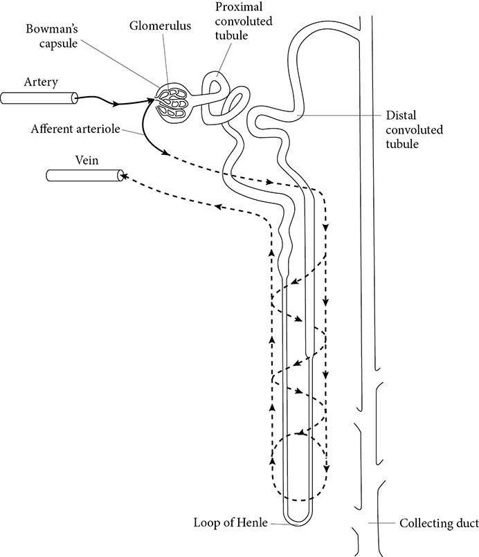
Figure 8-5
As the nutrients and wastes pass through the nephron, the nutrients are removed and returned by active transport to a second capillary network that surrounds the nephron. This process is known as reabsorption. If needed by the body, some water is also removed by osmosis before the fluid passes to the collecting tubule. Additional water can also be removed as the fluid passes through the collecting tube.
The collecting tubes from all the nephrons in a kidney merge and lead to a ureter. Each ureter leads to the bladder where the urine is stored until it is excreted through the urethra.
Lesson Summary
• Digestion within a cell is called intracellular digestion, while digestion occurring outside a cell is known as extracellular digestion.
• Mechanical digestion breaks down food physically, while chemical digestion changes it into new substances.
• Carbohydrate digestion begins in the mouth and is completed in the small intestine. Amylases change carbohydrates into simple sugars. The pancreas contributes by secreting amylases into the small intestine.
• Protein digestion begins in the stomach and is completed in the small intestine. Proteases change proteins into amino acids. The pancreas contributes by secreting proteases into the small intestine.
• Lipid digestion begins and is completed in the small intestine. Lipases change lipids into fatty acids and glycerol. The liver contributes by producing bile that mechanically breaks down lipids. The pancreas contributes by secreting lipases into the small intestine.
• Nutrients are absorbed by villi lining the small intestine.
• Undigested wastes are stored in the large intestine.
• Nitrogenous wastes are excreted by the kidneys as urea.
• The functional unit of a kidney is a nephron.
REVIEW QUESTIONS
1. Which structure plays a role in mechanical digestion?
(A) pancreas
(B) large intestine
(C) salivary gland
(D) stomach
(E) appendix
2. Which structure contributes three types of enzymes that aid in the digestion of carbohydrates, lipids, and proteins?
(A) pancreas
(B) esophagus
(C) salivary gland
(D) stomach
(E) liver
3. Bile
(A) breaks down lipids into fatty acids and glycerol
(B) contributes to digestion by allowing lipids and water to mix
(C) is produced in the gall bladder
(D) is an enzyme involved in lipid digestion
(E) passes from the gall bladder into the large intestine
Base your answers to questions 4 and 5 on Figure 8-6.
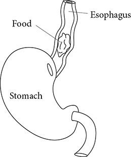
Figure 8-6
4. Notice where the food is found. What nutrient can be found in the food at this point?
(A) amino acids
(B) fatty acids
(C) glycerol
(D) partially digested proteins
(E) partially digested carbohydrates
5. What process is being shown in this illustration?
(A) absorption
(B) diffusion
(C) peristalsis
(D) elimination
(E) ingestion
6. Which structure is part of a nephron?
(A) bladder
(B) villi
(C) ureter
(D) urethra
(E) loop of Henle
ANSWERS AND EXPLANATIONS
1. (D) As the stomach muscles contract and relax, they aid in mechanical digestion.
2. (A) The pancreas secretes amylases, proteases, and lipases.
3. (B) Bile “emulsifies” lipids by breaking them down mechanically so that they can mix with water.
4. (E) Amylases secreted by the salivary glands would have partially digested the carbohydrates in the mouth.
5. (C) Food passes through the esophagus by peristalsis.
6. (E) The loop of Henle is part of the nephron where nutrients are reabsorbed.
Lesson 8-4. Protection
Vocabulary
• ectothermic
• homeothermic
• thermoregulation
• epidermis
• dermis
• hypodermis
• phagocyte
• phagocytosis
• lymphocytes
• B-cell
• pathogens
• T-cell
• lymph nodes
• lymphatic system
Some organisms are unable to regulate their body temperature. Instead, their body temperature changes as the external temperature changes. These animals are ectothermic, or cold-blooded animals. Among the vertebrates, these animals include fishes, amphibians, and reptiles. In contrast, homeothermic, or warm-blooded, animals can maintain a fairly stable internal temperature despite fluctuations in their external environment. Among vertebrates, these animals include birds and mammals. In humans, one organ that plays a role in thermoregulation is the skin.
The Skin
The skin consists of three layers. The outermost layer is the epidermis. The epidermis helps protect the body from disease by preventing microbes from entering the body. The epidermis also contains specialized cells called melanocytes. These cells produce a pigment called melanin that protects the body from the damage caused by ultraviolet light from the sun.
Beneath the epidermis is the dermis. This layer of the skin contains sweat glands and oil glands. Sweat glands secrete a mixture of water, salts, and a small amount of urea. Tiny ducts carry these materials to the epidermis where they are excreted. The evaporation of water from the epidermis also helps in thermoregulation. In colder weather, capillaries in the dermis constrict. As a result, blood flow to the surface is restricted. This restriction retains heat and helps in thermoregulation. The opposite happens in warm weather when the capillaries dilate to allow more heat to escape from the body.
The deepest layer of the skin is called the hypodermis. Here fat cells help insulate the body against the cold. The thickness of the hypodermis layer can vary greatly between individuals.
The Immune System
You learned that white blood cells play a role in protecting the body against disease. White blood cells are part of the immune system, a major player in keeping the body healthy.
There is only one type of red blood cell, but there are many types of white blood cells. One type of white blood cell is called a phagocyte. As its name suggests, this type of white blood cell carries out phagocytosis. This is a process by which a cell engulfs a tiny object, such as a bacterium, takes it inside the cell, and then digests or destroys it. There are several types of phagocytes, each specialized for a different role in protection. Some of these phagocytes present their digested components to other white blood cells in the immune system. These cells, in turn, carry out certain protective functions. These cells include lymphocytes.
Two main types of lymphocytes are involved. One is known as a B-cell. B-cells produce antibodies that destroy invading microbes, including viruses, bacteria, and parasites. Organisms that can cause disease are known as pathogens. Each pathogen brings about the response of a particular B-cell. Because there are millions of different pathogens, there are millions of different B-cells circulating in the blood that protect the body.
Another type of lymphocyte is called a T-cell. There are two different types of T-cells. One type of T-cell helps B-cells and other T-cells multiply and coordinate their actions. These are known as helper T-cells. Another type of T-cell is called a killer T-cell. These killer T-cells directly destroy a pathogen.
Lymphocytes travel in the blood stream. They also make up structures called lymph nodes. Lymph nodes are part of the lymphatic system, which is closely associated with the circulatory system. Unlike the circulatory system, the lymphatic system transports materials in only one direction—from body tissues back to the blood through lymphatic vessels. Lymph nodes are scattered along the way where lymphocytes can destroy pathogens that pass by. When confronted by a large number of pathogens, the lymphocytes will multiply. As a result, the lymph node gets larger. Swollen lymph nodes are a sign of infection.
Lesson Summary
• The skin acts as a barrier to protect the body against pathogens and helps in homeostasis through thermoregulation.
• The immune system includes several types of white blood cells that protect the body against pathogens.
• Phagocytes engulf and destroy pathogens.
• B-cell lymphocytes produce antibodies to destroy pathogens.
• Helper T-cells coordinate the immune response, while killer T-cells directly destroy pathogens.
• Lymph nodes contain lymphocytes that destroy pathogens.
REVIEW QUESTIONS
1. The skin helps to maintain body temperature
(A) by synthesizing melanin
(B) by acting as a barrier against pathogens
(C) through the action of its sweat glands
(D) when capillaries in the dermis constrict in warm weather
(E) when oil glands release their secretion onto the surface
2. Which type of white blood cell protects the body by making antibodies?
(A) B-cells
(B) helper T-cells
(C) killer T-cells
(D) phagocytes
(E) all type of lymphocytes
3. The HIV virus that causes AIDS destroys helper T-cells. As a result, the body
(A) is more prone to infections
(B) has a compromised immune system
(C) has a weakened defense system against pathogens
(D) cannot produce enough B-cells to mount a proper immune response
(E) all of the above
4. Millions of different kinds of B-cells are found in the blood because they
(A) are needed for phagocytosis of pathogens
(B) transport oxygen in addition to fighting disease
(C) each form in response to a particular pathogen
(D) are needed to attack the bodies various types of tissues
(E) will no longer form after a certain age
5. Skin protects the body from harmful ultraviolet radiation by producing a dark-colored pigment called melanin. In which layer of skin is melanin produced?
(A) dead epidermis
(B) living epidermis
(C) dermis
(D) subcutaneous layer
(E) epithelial
6. Which of the following are involved in producing antibodies?
(A) regulatory (suppressor) T cells
(B) B cells
(C) phagocytes
(D) killer T cells
(E) histamines
ANSWERS AND EXPLANATIONS
1. (C) Sweat glands in the dermis release water to the surface that evaporates in warm weather to keep the body cool.
2. (A) Antibodies are made by B-cells.
3. (E) Without helper T-cells, the body’s immune system is compromised. All types of white blood cells are affected.
4. (C) Each pathogen triggers a particular type of B-cell. With millions of different pathogens, millions of different B-cells exist.
5. (B) Melanin production takes place in the living epidermis. The melanin protects the underlying tissues from damage by UV radiation.
6. (B) B cells produce antibodies.
Lesson 8-5. Coordination
Vocabulary
• neuron
• dendrite
• cell body
• axon
• resting potential
• action potential
• myelin sheath
• nodes of Ranvier
• synapse
• neurotransmitters
• central nervous system
• peripheral nervous system
• cerebrum
• cerebellum
• medulla oblongata
• hypothalamus
• somatic nervous system
• autonomic nervous system
• sympathetic division
• parasympathetic division
• reflex action
• endocrine gland
• hormone
• peptide hormones
• steroid hormones
• pituitary
• pineal
• thyroid
• parathyroid
• insulin
• glucagon
• adrenal gland
• testis
• ovary
For all the various systems to work well and maintain homeostasis, they must function in a coordinated manner. Coordination of the various systems is controlled by two specialized systems. These are the nervous system and the endocrine system.
The nervous system allows an organism to receive, process, and respond to stimuli. These stimuli arise in both the external and internal environments. For example, consider what happens when a hydra is touched on its body. The entire body contracts to protect the organism from danger. Nerve cells are scattered throughout the hydra’s body to form what is called a nerve net. Obviously, humans have a much more complex nervous system.
The Human Nervous System
The basic unit of the nervous system is a neuron, or nerve cell. Billions of neurons make up the human nervous system. Their structure depends on their function. However, all neurons have three basic parts. One part is a dendrite. A dendrite is an extension of the cell that receives signals either from the environment or other neurons. The signal next travels to the cell body, where the nucleus and organelles are located. Then the signal continues its journey on to an axon, which is an extension that transmits the signal to a muscle, gland, or other neurons.
A neuron that is not conducting a signal exhibits a resting potential. This is a small electrical potential that is established by an unequal concentration of ions on either side of the cell membrane. At rest, more positive ions than negative ions are located on the outside of the cell membrane of a neuron. These include sodium ions. In contrast, more negative ions than positive ions are located inside the cell. Potassium ions are included among those positive ions inside a neuron.
When a neuron is stimulated, a signal, or impulse, travels along its length, moving in only one direction: dendrite → cell body → axon. The conduction of an impulse is known as an action potential. At the point of an action potential, the permeability of the membrane changes so that the outside of the cell has a net negative charge, while the inside has a net positive charge. The action potential travels across the neuron. After the action potential has passed a particular point, the ion concentrations are returned to the resting potential state. The neuron is then ready to conduct another impulse.
Some neurons are specialized to conduct impulses much faster than others. The axons of these neurons are wrapped with special cells that form a myelin sheath. The myelin sheath does not cover the entire axon. Rather, gaps exist along the axon. These gaps are called nodes of Ranvier. As the action potential moves along these neurons, the action potential jumps across the nodes of Ranvier. As a result, the impulse travels at a faster rate across the axon. Figure 8-7 shows how a neuron transmits an impulse.
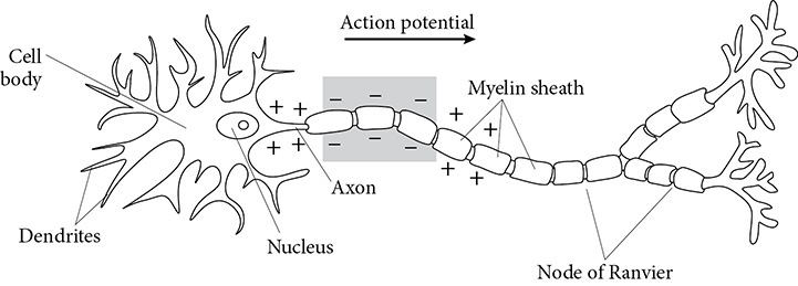
Figure 8-7
Neurons do not come in direct contact with one another. Rather, a small gap, known as a synapse, is present between the axon of one neuron and the dendrite of another neuron or a muscle or gland cell. Chemical messengers, called neurotransmitters, carry the impulse across the synapse. Once the neurotransmitters have crossed the synapse, they are destroyed. If they were not destroyed, the neuron would continue to “fire” or transmit impulses. Not all neurotransmitters stimulate a neuron. In fact, some neurotransmitters inhibit a neuron by raising the action potential that is necessary for the neuron to transmit an impulse. A dendrite, muscle, cell, or gland cell actually can receive several different neurotransmitters. Some stimulate, while others inhibit. Whatever is present in a greater concentration determines what happens.
Neurons are found everywhere in the body. They make up the two basic divisions of the nervous system. One is called the central nervous system. This division consists of the brain and spinal cord. The other division is called the peripheral nervous system.
The brain can be divided into four major parts. The cerebrum is the largest portion of the brain. The cerebrum is the part of the brain that consciously directs the body’s responses to stimuli, such as answering the door when someone knocks or putting on a coat when the weather is cold. The cerebrum also is the center of learning, memory, and all higher cognitive skills associated with human behavior.
The cerebellum coordinates balance and muscle movements. The medulla oblongata controls vital functions like breathing and heart rate. The hypothalamus controls functions such as response to hunger, thirst, pain, and temperature changes.
The peripheral nervous system is further subdivided into the somatic nervous system and the autonomic nervous system. The somatic nervous system is under voluntary control and directs the actions of skeletal muscles. The autonomic nervous system involves involuntary actions. The autonomic system is divided into the sympathetic division that directs so-called “fight-or-flight” responses. For example, the sympathetic division acts when a person is suddenly startled, such as when confronted by a mean-looking dog. The sympathetic activation brings about such changes in your body as an increase in heart rate, respiration and blood pressure. Another division of the autonomic nervous system is the parasympathetic division. This division directs the so-called “rest-and-digest” responses by slowing down the activities of muscles and glands. For example, the parasympathetic system division acts after a person has eaten by dilating the blood vessels in the villi of the small intestine that transport nutrients.
Another function carried out by the nervous system is a reflex action. An example of a reflex action would be the response a person has after touching a hot stove. The person responds without thinking by quickly removing his or her hand from the stove. In this case, an impulse travels from neurons in the finger that response to heat, to the spinal cord, and then back out to muscles in the finger and hand. At the same time, the impulse travels up the spinal cord to the brain where the cerebrum interprets the signals as pain. Traveling to the brain takes a little longer. This is why the person feels the pain after the hand has been withdrawn from the hot stove.
The Human Endocrine System
You learned that the autonomic nervous system controls involuntary actions. Among these actions are the functions of the endocrine glands. An endocrine gland is a structure that produces and secretes a hormone directly into the bloodstream. A hormone is a chemical substance that is produced by a gland that travels in the blood to work on another structure in the body. The structure affected by a hormone is called the target organ.
Hormones are classified into two groups. One group includes the peptide hormones. Peptide hormones are large protein compounds that are too large to diffuse across a cell membrane. As a result, peptide hormones attach to receptors on the cell membrane where they initiate their action. A second group of hormones includes the steroid hormones. These hormones are lipid molecules. Lipids are also present in the cell membrane. As a result, steroid hormones can diffuse across the cell membrane. Steroid hormones enter the nucleus where they bind to regions in the genes to control transcription. Compared to peptide hormones, steroid hormones may take longer to produce a response. However, their effects may last longer.
You read that the hypothalamus is a part of the brain. The hypothalamus is also an endocrine gland. It is not unusual for an organ to be involved in more than one system. The hypothalamus secretes hormones known as releasing factors. These releasing factors stimulate another endocrine gland called the pituitary. The pituitary was once known as the “master gland” because it controls the functions of many of the other endocrine glands. However, this term is rarely used for the pituitary after scientists discovered that it is under the control of the hypothalamus.
The pituitary produces many different hormones. One of its hormones is prolactin, which stimulates milk production and secretion in a female. In fact, several of the pituitary hormones are involved in reproduction and development, which is discussed in the next lesson.
Located at the base of the brain is an endocrine gland called the pineal. The pineal gland produces a hormone called melatonin, which regulates our day–night cycles.
Another endocrine gland is the thyroid. The thyroid secretes a hormone called thyroxin, which stimulates metabolism. Attached to the thyroid is another endocrine gland called the parathyroid. The parathyroid secretes a hormone that regulates the calcium and phosphate levels in the bone and blood.
The pancreas is not only involved in digestion but also in hormone production. The pancreas secretes two hormones that regulate the sugar level in the blood. One hormone is insulin, which lowers blood sugar by stimulating glucose uptake by cells. The other hormone is glucagon, which raises blood sugar by changing glycogen into glucose.
Located on each kidney is an adrenal gland. The most familiar hormone that this endocrine gland produces is epinephrine, more commonly known as adrenalin. Adrenalin is the hormone that is most closely associated with the “fight-or-flight” responses that are controlled by the sympathetic division.
The testis of a male is another endocrine gland. The testes produce steroid hormones known as androgens. Testosterone is the most familiar androgen. The ovary of a female is still another endocrine gland. The ovary produces steroid hormones called estrogens. Figure 8-8 shows the location of the major endocrine glands in the body. Looking just at this illustration, can you identify what each endocrine gland does?

Figure 8-8
Lesson Summary
• The functional unit of the nervous system is the neuron, which consists of an axon, cell body, and dendrite.
• A nerve impulse is an action potential that proceeds in only one direction along a neuron.
• Neurotransmitters are responsible for impulses crossing synapses.
• The nervous system is divided into the central nervous systems, which consists of the brain and spinal cord, and the peripheral nervous system.
• Major portions of the brain include the cerebrum, cerebellum, medulla oblongata, and hypothalamus.
• The peripheral nervous system is divided into the somatic nervous system and the autonomic nervous system. In turn, the autonomic system is divided into the sympathetic and parasympathetic divisions.
• The major glands of the endocrine system include the hypothalamus, pituitary, pineal, thyroid, parathyroid, adrenal, pancreas, testis, and ovary.
• The nervous system and endocrine system play a major role in homeostasis by coordinating the actions of the other body systems.
REVIEW QUESTIONS
Each set of lettered choices below refers to the numbered statement immediately following it. Select the lettered choice that best answers each statement.
(A) central nervous system
(B) somatic nervous system
(C) autonomic nervous system
(D) sympathetic division
(E) parasympathetic division
1. controls “fight-or-flight” responses
2. controls skeletal muscles
3. stores information
4. spinal cord
(A) parathyroid
(B) pineal
(C) pancreas
(D) ovary
(E) adrenal
5. involved in “flight-or-fight” responses
6. controls daily rhythm cycles
7. regulates blood calcium level
8. located on the kidney
ANSWERS AND EXPLANATIONS
1. (D) The sympathetic division of the autonomic nervous system helps prepare the body for stressful situations.
2. (B) Nerves of the somatic nervous system stimulate skeletal muscle to contract.
3. (A) The brain, which is part of the central nervous system, stores information.
4. (A) The spinal cord is also part of the central nervous system.
5. (E) The adrenal gland secretes epinephrine, a hormone involved in the “fight-or-flight” reactions.
6. (B) Night–day rhythms are controlled by the pineal gland.
7. (A) A hormone produced by the parathyroid regulates the calcium level in the blood stream.
8. (E) The adrenal gland is located on the kidney. The word renal refers to kidney.
Lesson 8-6. Reproduction and Development
Vocabulary
• asexual reproduction
• sexual reproduction
• seminiferous tubules
• vas deferens
• Fallopian tube
• uterus
• cervix
• vagina
• menstrual cycle
• fertilization
• menstruation
• follicular phase
• ovulation
• corpus luteum
• luteal phase
• negative feedback
• zygote
• inner cell mass
• blastocyst
• embryo
• placenta
• gastrula
• ectoderm
• mesoderm
• endoderm
Reproduction is the key to survival of a species. There are two types of reproduction. Asexual reproduction involves the division of either a single cell or an entire organism to form a new individual. Asexual reproduction involves only one parent. For example, bacteria and protozoa undergo binary fission in which the single cell divides to form two new cells. Starfish can also undergo asexual reproduction. New starfish can regenerate from one of their arms cut off along with a small portion of their central body.
In contrast sexual reproduction involves two parents, one male and the other female. Because two parents are involved, sexual reproduction results in greater genetic diversity among the offspring. Keep in mind that some organisms can carry out both asexual and sexual reproduction, depending on conditions. For example, two bacteria can exchange genetic material in a process called conjugation. However, most organisms carry out only sexual reproduction. Such is the case with humans.
The Human Reproductive System
The hormone testosterone is responsible for the maturation of sperm cells. Mature sperm are produced within tiny coiled tubes within the testes. These tubes are called seminiferous tubules. The sperm then exit the testes through the vas deferens, which leads to the urethra. In males, both sperm and urine exit through the urethra and out the penis.
In females, hormones mature an egg cell in the ovary. After maturation, the egg cell is swept by cilia into the Fallopian tube. The Fallopian tube is also called an oviduct. The Fallopian tubes leading from each ovary meet at the uterus. The neck of the uterus is called the cervix, which leads to the vagina.
Hormones control the events that take place in the female reproductive cycle. These events form the menstrual cycle. The menstrual cycle is the period of time, usually lasting about 28 days, in which the female reproductive tract is prepared should fertilization take place. Fertilization is the fusion of an egg with a sperm, a process that usually occurs in the upper portion of the Fallopian tube.
The menstrual cycle involves four stages. Although these four stages form a cycle, it is convenient to begin at some point in the cycle. The first stage is known as menstruation, which starts at day 1. During menstruation, the uterine lining is shed. The lining includes blood vessels. As a result, menstruation results in bleeding as these blood vessels are broken down and discarded along with the uterine lining. Menstruation lasts for about 5 days.
The second stage, called the follicular phase, begins about day 6. The follicular stage is also known as the proliferative phase. During this stage, hormones from the pituitary gland stimulate the maturation of an egg in the ovary. In response to these hormones, the ovary in turn produces a hormone called estrogen. Estrogen from the ovary stimulates the division of cells in the uterus. As a result, the uterine lining thickens. Estrogen also promotes the formation of new blood vessels in the uterus. The follicular stage lasts until about day 13 of the menstrual cycle.
The third stage, ovulation, usually occurs about day 14. Ovulation involves the release of a mature egg from the ovary. This egg cell is swept into the Fallopian tube where it can be fertilized should sperm be present. Ovulation is stimulated by another hormone produced by the pituitary gland. Following ovulation, the structure that housed the egg while it was being matured in the ovary becomes a corpus luteum. The corpus luteum secretes progesterone, a hormone that brings on the fourth and final stage of the menstrual cycle.
The fourth stage, known as the luteal phase, begins on about day 15. The luteal phase is also known as the secretory phase. During this stage, the uterus continues to enlarge and become enriched with more blood vessels. These changes are attributable to progesterone. The uterus is now prepared to receive a fertilized egg descending from the Fallopian tube. However, if no fertilized egg becomes implanted in the uterus, the uterine lining is shed and the menstrual cycle starts again.
Estrogen and progesterone are just two of the hormones involved in the menstrual cycle. The interaction between all the hormones is quite complex. The level of each hormone must be controlled. These levels must also be adjusted, depending on whether or not a fertilized egg implants itself in the uterus. These hormones are controlled by a mechanism known as negative feedback.
Consider how the estrogen level is controlled by negative feedback. Recall that the pituitary gland secretes a hormone that stimulates the ovary to produce estrogen. As a result, the estrogen level continues to rise during the follicular phase.
Also recall that the pituitary is under the control of the hypothalamus. When the estrogen level rises to a certain level, estrogen in the bloodstream inhibits the hypothalamus from stimulating the pituitary. As a result, the pituitary stops secreting the hormone that stimulates the ovary to make estrogen. Therefore, the estrogen level starts to drop. In effect, there is feedback from the ovary to the hypothalamus. Because the estrogen level drops as a result of this feedback, the result is a negative impact on this hormone’s level in the bloodstream.
Development
If fertilization has taken place, then a zygote, or fertilized egg, will implant itself in the uterine lining. The zygote will start to undergo mitosis, usually within 24–36 hours of fertilization. The zygote continues to divide to form a small, solid ball of cells. Mitotic divisions continue. More cells are produced. In addition, a cavity forms inside the ball. The result is an outer ring of cells. Attached to this outer ring on one side is the inner cell mass. Together, the outer ring of cells and the inner cell mass form a structure called a blastocyst.
The inner cell mass will continue to develop and form the embryo. The embryo is the term given to the early stages of the development of an individual. The outer ring of cells will form the embryo’s contribution to the placenta. Uterine tissue will also contribute to the placenta. The placenta is the structure that serves as the site of exchange of materials between the two. For example, nutrients will pass from mother to embryo across the placenta. The nutrients will then pass into the embryo via an umbilical cord that attaches the growing embryo to the placenta.
Mitotic divisions continue in the blastocyst, which now begins to get larger. The next stage of development is called a gastrula. For the first time, groups of cells become distinct and form three different layers. One layer, called the ectoderm, will develop into external structures such as the skin and also form the nervous system. A second layer, called the mesoderm, will form structures such as bones, muscles, blood vessels, and kidneys. The third layer, the endoderm, will develop into the inner linings of the digestive and respiratory systems. Keep in mind that almost all structures develop from more than one of these layers. For example, the epidermis of the skin comes from ectoderm, while the blood vessels in the dermis derive from mesoderm.
Lesson Summary
• Asexual reproduction involves only one parent, whereas sexual reproduction involves two parents of opposite sex.
• In a human male, sperm are matured in the seminiferous tubules in the testes where they next pass into the vas deferens and exit the body through the urethra.
• In a human female, an egg is matured in the ovary and then released into a Fallopian tube. The egg then moves down to the uterus, and if it is not fertilized, will pass out the vagina.
• The menstrual cycle last about 28 days, during which hormones prepare the uterus should an egg be fertilized and implanted.
• The menstrual cycle consists of menstruation, the follicular phase, ovulation, and the luteal phase.
• A fertilized egg is known as a zygote, which develops into a blastocyst that consists of an outer wall of cells and an inner cell mass.
• At the gastrula stage, cells specialize to form ectoderm, mesoderm, and endoderm.
REVIEW QUESTIONS
Each set of lettered choices in Figures 8-9 and 8-10 refers to the numbered statement immediately following it. Select the lettered choice that best relates to each statement.

Figure 8-9
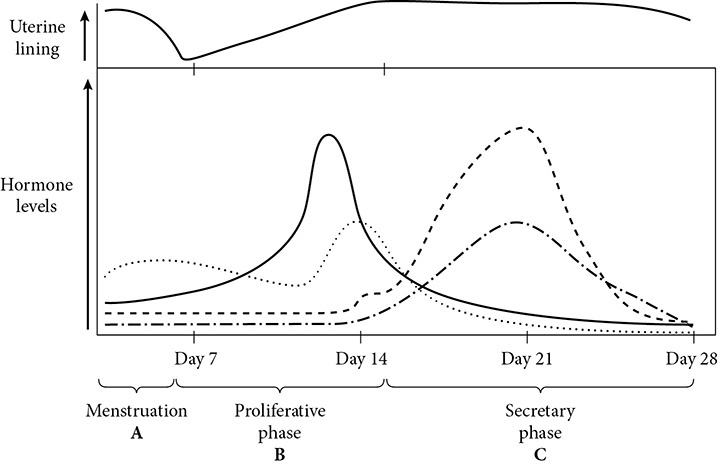
Figure 8-10
1. the usual site of fertilization
2. site of placenta formation
3. site that produces the hormone estrogen
4. part of cycle where uterine lining builds up
5. part of cycle where estrogen level peaks
6. hormone secreted by the corpus luteum after ovulation has occurred
ANSWERS AND EXPLANATIONS
1. (B) A sperm travels up the oviduct to fertilize an egg cell.
2. (D) The placenta forms in the uterus.
3. (C) Estrogen is produced by the ovary.
4. (B) The uterine lining becomes thicker during the proliferative phase as a result of estrogen secreted by the ovary.
5. (B) Estrogen level peaks toward the end of the proliferative phase.
6. (E) Progesterone is released by the corpus luteum that forms in the ovary after the mature egg has been released in ovulation.
Lesson 8-7. Organismal Behavior
Vocabulary
• ethology
• innate
• learned
• reflex
• fixed action pattern
• habituation
• sensitization
• associative learning
• operant conditioning
• concept
• imprinting
• foraging
Behavior is an action performed by an organism that alters the relationship between the organism and its environment. Ethology is the study of behavior. Behaviors can be innate (instinctual) or learned, or sometimes a combination of the two. The action may be performed because of an external stimulus, such as the rising of the sun, or because of an internal stimulus, such as thirst. Many actions, such as mating behavior, are performed because of a combination of external and internal factors.
Innate Behavior
Innate behavior is instinctual. That means that the animal can perform a certain behavior the first time the animal encounters the proper stimulus. Innate behaviors are the result of the survival mechanism in the brain being developed by natural selection over many generations.
One type of innate behavior is reflexive behavior. A reflex is an automatic response to a stimulus by a part of the animal’s body. For example, if you touch something very hot, your withdrawal reflex will cause you to pull your hand away immediately without any conscious thought on your part. Reflexes protect animals from harm by allowing them to respond quickly to a stimulus.
Another type of innate behavior is called a fixed action pattern. This consists of a relatively complex sequence of instinctive behaviors that rarely varies within a species. Once the sequence is begun, it is nearly always completed. For example, a nesting Greylag goose will push a displaced egg back into the nest with its beak. The goose will do the same beak-nudging behavior if a rock takes the place of the egg or even if the egg is taken away. The sight of a missing egg in the nest is the stimulus that provokes the behavior. These behaviors are also part of the natural selection process, because they help the animal adapt to its environment.
Learned Behavior
Learned behavior is just what it sounds like: behavior that an animal develops over time that helps it adapt to its environment. These behaviors include habituation, sensitization, associative learning, operant conditioning, and concepts.
Habituation and sensitization are related simple learning processes. Habituation is the process through which an animal ceases to respond to a stimulus. For example, an animal may grow “used to” a strong odor and stop responding to it. Sensitization is the process through which an animal responds more strongly or more often to a stimulus. For example, if an animal reacts strongly to a loud sound, it may become so averse to the loud sound that it will respond the same way to a sound at a much lower volume.
Associative learning, or conditioned response, is also a simple form of learned behavior. It is the process by which an animal responds to a stimulus and then learns to respond the same way to a different stimulus. Russian physiologist Ivan Pavlov is famous for his experiments in associative learning. Pavlov observed that dogs salivate when food is presented to them. The food, an unconditioned stimulus, triggers a reflex in the salivary glands, an unconditioned response. Pavlov found that if he rang a bell every time he fed the dogs, the dogs eventually salivated any time they heard the bell, even if no food was presented. The bell is a conditioned stimulus that produces the conditioned response of salivating. The dogs learned to associate the bell with eating and responded to the bell in the same way they did to the food itself.
Operant conditioning, or instrumental conditioning, is also called trial-and-error learning because the animal may try various responses before finding the one that is rewarded. Maze problems are a form of operant conditioning because the animal tries many pathways before finding the one that leads to the reward.
Concepts are much more complex learned behaviors in which an animal makes an abstract generalization about a specific problem. For example, a monkey can learn to pick the object in a group that is different from the other objects if choosing the odd object is rewarded each time. The monkey has formed the concept that the odd object in a group should be the one chosen in order to receive food, even if the way in which the object differs from the group is changed.
Blending Innate And Learned Behaviors
Some behaviors seem to be a blend of innate and learned behaviors. The process of imprinting, for example, involves both types of behavior. Imprinting is the process of bonding that takes place at an important period in early infancy. The drive to imprint is innate, so an infant can imprint on an animal that is not its parent, or even on an object. Imprinting is also a learned behavior: infants learn certain behaviors through following the behavior patterns of those they imprint upon. For example, baby geese learn to walk by following their parents around, but if a baby goose imprints on a human, the baby goose will follow the human around instead.
Foraging for food is a crucial survival behavior for animals that involves a blend of the innate drive to eat with the learned behaviors that tend to lead the animal to successful acquisition of food.
Mating behavior is also a blend of innate and learned behaviors. The innate behavior of mating combines with courtship and mating rituals that are often complex, and the animals that learn to perform those behaviors the best are more likely to attract a mate and carry on their genetic line.
Social Behavior
Animal social behavior refers to the interactions between two or more animals. These animals are usually of the same species and cooperate in foraging, parenting, mating, and communication. Social behaviors such as division of labor, cooperation, and altruism are crucial in complex societies. However, social animals are often fiercely competitive and aggressive. They engage in territoriality and dominance rituals. Charles Darwin introduced the idea that it is the best competitors within a species, the fittest individuals, that survive and reproduce. Animal societies are often a complex balance of cooperation and competition.
Lesson Summary
• Behaviors can be innate (instinctual) or learned, or sometimes a combination of the two.
• Innate behavior is instinctual and includes reflexive behavior and fixed action patterns.
• Learned behaviors include habituation, sensitization, associative learning, operant conditioning, and concepts.
• Many behaviors, such as imprinting, foraging, and mating behavior, are a blend of innate instincts and learned behaviors.
• Social behaviors within groups of organisms balance cooperative behaviors such as division of labor and altruism with competitive behaviors such as dominance rituals and territoriality.
REVIEW QUESTIONS
Questions 1–4
Select which one of the following choices best matches the statements below. Some choices may be used once, more than once, or not at all.
(A) reflexive behavior
(B) fixed action pattern
(C) habituation
(D) associative learning
(E) social behavior
1. Whenever it thunders, the dog barks. When the dog barks, the cat runs under the bed. Now the cat runs under the bed any time it thunders, even if the dog is not in the house.
2. A man living near railroad tracks no longer notices the loud sound of the trains.
3. When exposed to cold, a human begins shivering.
4. A dog circles around before lying down, no matter what surface he is on.
5. Which of the following are benefits to living in social groups?
I. ability to avoid infectious disease
II. ability to defend better territory
III. ability to capture larger prey
(A) I only
(B) II only
(C) III only
(D) II and III only
(E) I, II, and III
6. Monarch butterflies in Eastern North America migrate to the Sierra Madre Mountains of Mexico each fall. What is the most likely reason for this behavior?
(A) They are looking for other butterflies to mate with.
(B) Butterflies have a reflexive response to aggressive fall wind patterns.
(C) They are evading predators such as birds.
(D) There is not enough food available in North America to support the monarch population.
(E) Most cannot survive the winter temperatures and must go somewhere warmer.
ANSWERS AND EXPLANATIONS
1. (D) The cat has learned to associate thunder with the dog barking.
2. (C) The man has become habituated to the sound of the trains.
3. (A) Shivering is an uncontrolled innate behavior.
4. (B) The dog is exhibiting a fixed action pattern by performing a ritual that does not change and is always done to completion.
5. (D) Group living provides more individuals who can cooperate in hunting and repelling invaders, but it is a drawback for avoiding infectious disease, which spreads more easily within a group.
6. (E) Their migratory behavior from a cold climate to a warm climate indicates that many or all of the group would not survive the winter temperatures in Eastern North America.