THE ANKLES & FEET
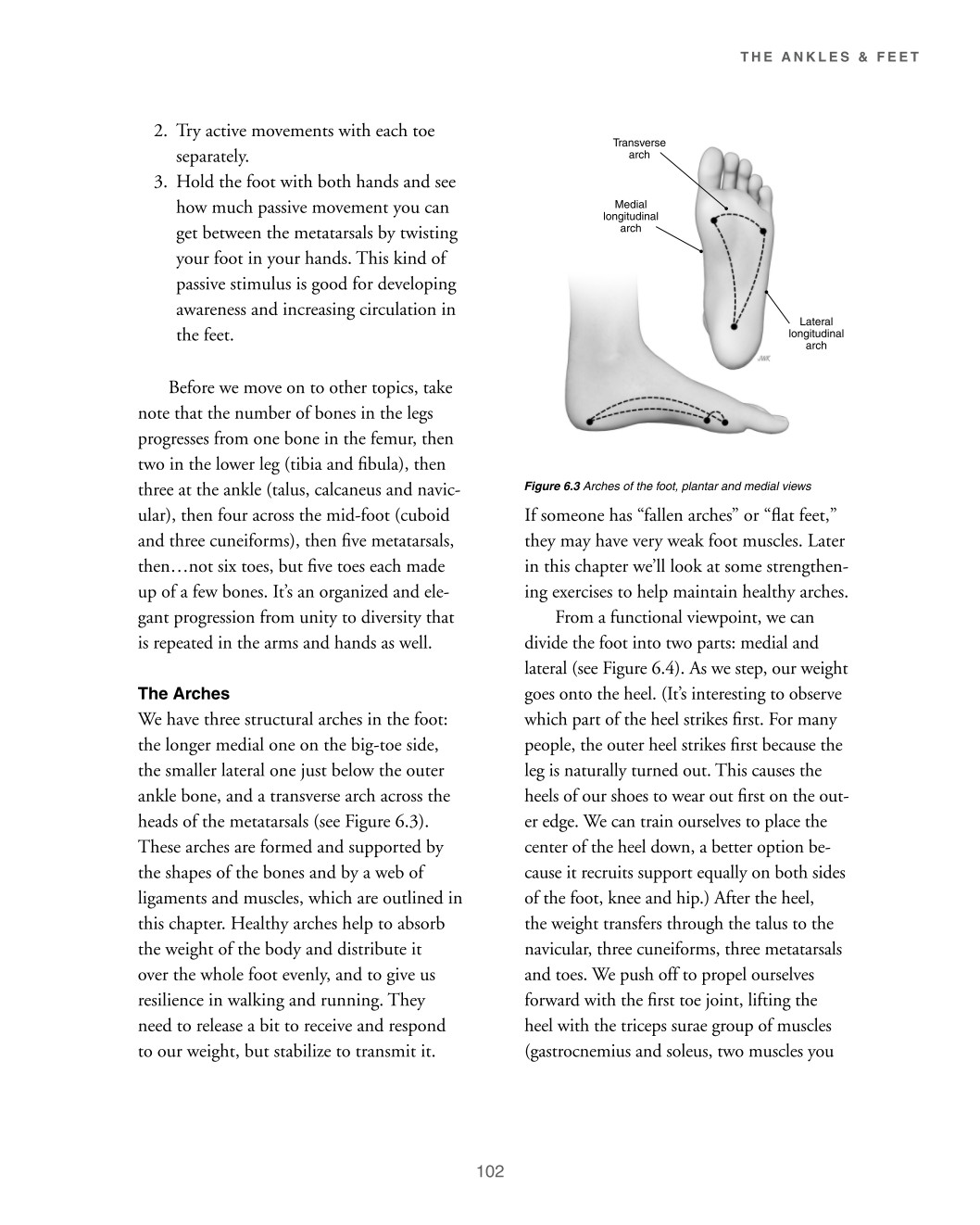
2. Try active movements with each toe separately. Transverse arch 3. Hold the foot with both hands and see how much passive movement you can get between the metatarsals by twisting your foot in your hands. This kind of passive stimulus is good for developing awareness and increasing circulation in the feet. Before we move on to other topics, take note that the number of bones in the legs progresses from one bone in the femur, then two in the lower leg (tibia and fibula), then three at the ankle (talus, calcaneus and navic- ular), then four across the mid-foot (cuboid and three cuneiforms), then five metatarsals, then…not six toes, but five toes each made up of a few bones. It’s an organized and ele- gant progression from unity to diversity that is repeated in the arms and hands as well. The Arches We have three structural arches in the foot: the longer medial one on the big-toe side, the smaller lateral one just below the outer ankle bone, and a transverse arch across the heads of the metatarsals (see Figure 6.3). These arches are formed and supported by the shapes of the bones and by a web of ligaments and muscles, which are outlined in this chapter. Healthy arches help to absorb the weight of the body and distribute it over the whole foot evenly, and to give us resilience in walking and running. They need to release a bit to receive and respond to our weight, but stabilize to transmit it. Medial longitudinal arch
Lateral longitudinal arch
Figure 6.3 Arches of the foot, plantar and medial views If someone has “fallen arches” or “flat feet,” they may have very weak foot muscles. Later in this chapter we’ll look at some strengthen- ing exercises to help maintain healthy arches. From a functional viewpoint, we can divide the foot into two parts: medial and lateral (see Figure 6.4). As we step, our weight goes onto the heel. (It’s interesting to observe which part of the heel strikes first. For many people, the outer heel strikes first because the leg is naturally turned out. This causes the heels of our shoes to wear out first on the out- er edge. We can train ourselves to place the center of the heel down, a better option be- cause it recruits support equally on both sides of the foot, knee and hip.) After the heel, the weight transfers through the talus to the navicular, three cuneiforms, three metatarsals and toes. We push off to propel ourselves forward with the first toe joint, lifting the heel with the triceps surae group of muscles (gastrocnemius and soleus, two muscles you
102