Foreign Bodies and Bezoars
Foreign Bodies in the Stomach and Intestine
Asim Maqbool, Chris A. Liacouras
Once in the stomach, 95% of all ingested objects pass without difficulty through the remainder of the gastrointestinal tract. Perforation after ingestion of a foreign body is estimated to be <1% of all objects ingested. Perforation tends to occur in areas of physiologic sphincters (pylorus, ileocecal valve), acute angulation (duodenal sweep), congenital gut malformations (webs, diaphragms, diverticula), or areas of previous bowel surgery.
Most patients who ingest foreign bodies are between the ages of 6 mo and 6 yr. Coins are the most commonly ingested foreign body in children, and meat or food impactions are the most common accidental foreign body in adolescents and adults. Patients with nonfood foreign bodies often describe a history of ingestion. Young children might have a witness to ingestion. Immediate concerns are what the foreign body is, the location of the foreign body, the size of the foreign body, and the time that the ingestion occurred. Approximately 90% of foreign bodies are opaque. Radiologic examination is routinely performed to determine the type, number, and location of the suspected objects. Contrast radiographs may be necessary to demonstrate some objects, such as plastic parts or toys.
Conservative management is indicated for most foreign bodies that have passed through the esophagus and entered the stomach. Most objects pass though the intestine in 4-6 days, although some take as long as 3-4 wk. While waiting for the object to pass, parents are instructed to continue a regular diet and to observe the stools for the appearance of the ingested object. Cathartics should be avoided. Exceptionally long or sharp objects are usually monitored radiologically. Parents or patients should be instructed to report abdominal pain, vomiting, persistent fever, and hematemesis or melena immediately to their physicians. Failure of the object to progress within 3-4 wk seldom implies an impeding perforation but may be associated with a congenital malformation or acquired bowel abnormality.
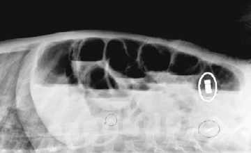
Certain objects pose more risk than others. In cases of sharp foreign bodies, such as straight pins, weekly assessments are required. Surgical removal is necessary if the patient develops symptoms or signs of obstruction or perforation or if the foreign body fails to progress for several weeks. Small magnets used to secure earrings or parts of toys are associated with bowel perforation. Whereas a single magnet in the stomach may not require intervention in an asymptomatic child, a magnet in the esophagus requires immediate removal. When the multiple magnets disperse after ingestion, they may be attracted to each other across bowel walls, leading to pressure necrosis and perforation (Fig. 360.1 ). Inexpensive toy medallions containing lead can lead to lead toxicity. Newer coins can also decompose when subjected to prolonged acid exposure. Unless multiple coins are ingested, the metals released are unlikely to pose a clinical risk.

Ingestion of batteries rarely leads to problems, but symptoms can arise from leakage of alkali or heavy metal (mercury) from battery degradation in the gastrointestinal tract. Batteries can also generate electrical current and thereby cause low-voltage electrical burns to the intestine. If patients experience symptoms such as vomiting or abdominal pain, if a large-diameter battery (>20 mm in diameter) remains in the stomach for longer than 48 hr, or if a lithium battery is ingested, the battery should be removed. Batteries larger than 15 mm that do not pass the pylorus within 48 hr are less likely to pass spontaneously and generally require removal. In children younger than 6 yr of age, batteries larger than 15 mm are not likely to pass spontaneously and should be removed endoscopically. If the patient develops peritoneal signs, surgical removal is required. Batteries beyond the duodenum pass per rectum in 85% within 72 hr. The battery should be identified by size and imprint code or by evaluation of a duplicate measurement of the battery compartment. The National Button Battery Ingestion Hotline (202-625-3333) can be called for help in identification. The Poison Control Center (800-222-1222) can be called as well for ingestion of batteries and caustic materials. Lithium batteries result in more severe injury than a button alkali battery, with damage occurring in minutes. Button batteries in a symptomatic child should be removed, or if there are multiple batteries, they should be removed.
In older children and adults, oval objects larger than 5 cm in diameter or 2 cm in thickness tend to lodge in the stomach and should be endoscopically retrieved. Thin long objects >6 cm in length fail to negotiate the pylorus or duodenal sweep and should also be removed. In infants and toddlers, objects >3 cm in length or >20 mm in diameter do not usually pass through the pylorus and should be removed. An open safety pin presents a major problem and requires urgent endoscopic removal if within reach. Razor blades can be managed with a rigid endoscope by pulling the blade into instrument. The endoscopist can alternatively use a rubber hood on the head of the endoscope to protect the esophagus. Other sharp objects (needles, bones, pins) usually pass the stomach, but complications may be as high as 35%; if possible, they should be removed by endoscope if in the stomach or proximal duodenum. If sharp objects are not able to be removed but no progress is observed in location during 3 days, surgical removal is indicated. Drugs (aggregated iron pills, cocaine) may have to be surgically removed; initial management can include oral polyethylene glycol lavage. Drug body packing (heroin, cocaine) is usually seen on kidneys-ureters-bladder or CT imaging and often passes without incident. Endoscopic procedures may rupture the material, causing severe toxicity. Surgery is indicated if toxicity develops, if the packages fail to progress, or if there are signs of obstruction.
Ingestion of magnets poses a danger to children. The number of magnets is thought to be critical. If a single magnet is ingested, there is the least likelihood of complications. If 2 or more magnets are ingested, the magnetic poles are attracted to each other and create the risk of obstruction, fistula development, and perforation. Endoscopic retrieval is emergent after films are taken when multiple magnets are ingested. Abdominal pain or peritoneal signs require urgent surgical intervention. If all magnets are located in the stomach, immediate endoscopic removal is indicated. If the ingestion occurred greater than 12 hr prior to evaluation, or if the magnets are beyond the stomach and the patient is symptomatic, general surgery should be consulted. If the patient is asymptomatic, endoscopic or colonoscopic removal may be considered, along with a surgical evaluation.
Lead-based foreign bodies can cause symptoms from lead intoxication. Early endoscopic removal is indicated of an object suspected to contain lead. A lead level should be obtained.
Water-absorbing polymer balls (beads) can expand to approximately 400 times their starting size and if ingested may produce intestinal obstruction. Initially of a small diameter, they pass the pylorus only to rapidly enlarge in the small intestine. Surgical removal is indicated.
Children occasionally place objects in their rectum. Small blunt objects usually pass spontaneously, but large or sharp objects typically need to be retrieved. Adequate sedation is essential to relax the anal sphincter before attempting endoscopic or speculum removal. If the object is proximal to the rectum, observation for 12-24 hr usually allows the object to descend into the rectum.
Bibliography
ASGE Standards of Practice Committee. Management of ingested foreign bodies and food impactions. Gastrointest Endosc . 2011;73:1085–1091.
Centers for Disease Control and Prevention (CDC). Injuries from batteries among children aged <13 years—United States, 1995–2010. MMWR Morb Mortal Wkly Rep . 2012;61:661–666.
Cerri RW, Liacouras CA. Evaluation and management of foreign bodies in the upper gastrointestinal tract. Pediatr Case Rev . 2003;3:150–156.
Denney W, Ahmad N, Dillard B, et al. Children will eat the strangest things, a 10-year retrospective analysis of foreign body and caustic ingestions from a single academic center. Pediatr Emerg Care . 2012;28(8):731–734.
Dutta S, Barzin A. Multiple magnet ingestion as a source of severe gastrointestinal complications requiring surgical intervention. Arch Pediatr Adolesc Med . 2008;162:123–125.
Green SS. Ingested and aspirated foreign bodies. Pediatr Rev . 2015;36(10):430–437.
Hussain SZ, Bousvaros A, Gilger M, et al. Management of ingested magnets in children. J Pediatr Gastroenterol Nutr . 2012;55(3):239–242.
Karjoo M, Kader H. A novel technique for closing and removing an open safety pin from the stomach. Gastrointest Endosc . 2003;57:627–629.
Litovitz T, Whitaker N, Marsolek M, et al. Emerging battery-ingestion hazard: clinical implications. Pediatrics . 2010;125:1168.
Mallon PT, White JS, Thompson RL. Systemic absorption of lithium following ingestion of a lithium button battery. Hum Exp Toxicol . 2004;23:193–195.
Naji H, Isacson D, Svensson JF, et al. Bowel injuries caused by ingestion of multiple magnets in children: a growing hazard. Pediatr Surg Int . 2012;28(4):367–374.
Puig S, Scharitzer M, Cengiz K, et al. Effects of gastric acid on euro coins: chemical reaction and radiographic appearance after ingestion by infants and children. Emerg Med J . 2004;21:553–556.
Schalamon J, Haxhija EQ, Ainoedhofer H, et al. The use of a hand-held metal detector for localisation of ingested metallic foreign bodies—a critical investigation. Eur J Pediatr . 2004;163:257–259.
VanArsdale JL, Leiker RD, Kohn M, et al. Lead poisoning from a toy necklace. Pediatrics . 2004;114:1096–1099.
Zamora IJ, Vu LT, Larimer EL, et al. Water-absorbing balls: a “growing” problem. Pediatrics . 2012;130:e1011–e1014.
Bezoars
Asim Maqbool, Chris A. Liacouras
A bezoar is an accumulation of exogenous matter in the stomach or intestine. They are predominantly composed of food or fiber. Most bezoars have been found in females with underlying personality problems or in neurologically impaired persons. Patients who have undergone abdominal surgery are at higher risk for the development of bezoars. The peak age at onset of symptoms is the 2nd decade of life.
Bezoars are classified on the basis of their composition. Trichobezoars are composed of the patient's own hair. It is most frequently a complication of the psychiatric disorder trichotillomania, and the most severe form is known as Rapunzel syndrome (hair bezoar extending beyond the stomach to the small intestine). Phytobezoars are composed of a combination of plant and animal material, and gastric phytobezoars are the most common in patients with poor motility. Lactobezoars were previously found most often in premature infants and can be attributed to the high casein or calcium content of some premature formulas. Swallowed chewing gum can occasionally lead to a bezoar.
Trichobezoars can become large and form casts of the stomach; they can enter into the proximal duodenum. They manifest as symptoms of gastric outlet or partial intestinal obstruction, including vomiting, anorexia, and weight loss. Patients might complain of abdominal pain, distention, and severe halitosis. Physical examination can demonstrate patchy baldness and a firm mass in the left upper quadrant. Patients occasionally have iron-deficiency anemia, hypoproteinemia, or steatorrhea caused by an associated chronic gastritis. Phytobezoars manifest in a similar manner. Detached segments of the bezoar or trichobezoar can migrate to the small intestine as a “satellite masses” and result in small bowel obstruction.
An abdominal plain film can suggest the presence of a bezoar, which can be confirmed on ultrasound or CT examination. On CT a bezoar appears a nonhomogeneous, nonenhancing mass within the lumen of the stomach or intestine. Oral contrast circumscribes the mass.
Bezoars in the stomach can usually be removed endoscopically. If endoscopy is unsuccessful, surgical intervention may be needed. Lactobezoars usually resolve when feedings are withheld for 24-48 hr. Coca-Cola has been used as a dissolution therapy for gastric phytobezoar and has been shown to be effective when used with endoscopy. Trichobezoars almost always require surgical removal.
Sunflower seed bezoars are reported to cause rectal pain and constipation as a result of the seed shells being associated with fecal impaction. Endoscopic removal is indicated, as these bezoars are refractory to enema or lavage management.
Bibliography
Fallon S, Slater B, Lopez M, et al. The surgical management of rapunzel syndrome: a case series and a literature review. J Pediatr Surg . 2013;48(2):830–834.
Kaneko H, Tomomasa T, Kubota Y, et al. Pharmacobezoar complicating treatment with sodium alginate. J Gastroenterol . 2004;39:69–71.
Ladas SD, Kamberoglou D, Karamanolis G, et al. Systemic review: Coca-cola can effectively dissolve gastric phytobezoars as a first-line treatment. Aliment Pharmacol Ther . 2013;37:169–173.
Lynch KA, Feola PG, Guenther E. Gastric trichobezoar: an important cause of abdominal pain presenting to the pediatric emergency department. Pediatr Emerg Care . 2003;19(5):343–347.
Palanivelu C, Rangarajan M, Senthilkumar R, et al. Trichobezoars in the stomach and ileum and their laparoscopy-assisted removal: a bizarre case. Singapore Med J . 2007;48(2):e37–e39.
Purcell L, Gremse DA. Sunflower seed bezoar leading to fecal impaction. South Med J . 1995;88:87–88.