Thyrotoxicosis
Jessica R. Smith, Ari J. Wassner
Although the terms hyperthyroidism and thyrotoxicosis are often interchanged in the literature, they are not synonymous. Hyperthyroidism specifically refers to the synthesis and secretion of excess thyroid hormone from the thyroid gland; in contrast, thyrotoxicosis refers to any state of excess circulating thyroid hormone (and its clinical manifestations) regardless of its source. This distinction is physiologically and clinically relevant because different therapies may be indicated depending on the mechanism of thyroid hormone excess.
Graves disease is the most common cause of hyperthyroidism in children (Table 584.1 ). Graves disease is an autoimmune disorder that results in the production of thyrotropin (TSH) receptor–stimulating antibodies (TRSAbs) that bind and activate the G protein–coupled TSH receptor (TSHR ) to cause increased thyroid hormonogenesis and diffuse glandular growth. Etiologies of nonautoimmune hyperthyroidism include hyperfunctioning thyroid nodules and germline gain-of-function mutations in the TSHR (either autosomal dominant or sporadic). Hyperthyroidism can also occur in patients with McCune-Albright syndrome as a result of an activating mutation of the stimulatory α-subunit of the G-protein. These patients can also develop a multinodular goiter. Other rare causes of hyperthyroidism include iodine-induced hyperthyroidism, TSH-secreting adenomas, toxic multinodular goiters, and hyperfunctioning thyroid carcinoma. Thyrotoxicosis not due to hyperthyroidism (i.e., not due to overproduction of thyroid hormone by the gland) can be caused by thyroiditis (see Chapter 582 ) or ingestion of exogenous thyroid hormone (thyrotoxicosis factitia).
Table 584.1
Pathogenic Mechanisms and Causes of Thyrotoxicosis
| Cause | |
| Thyrotoxicosis with hyperthyroidism (normal or high radioactive iodine uptake) | |
| Effect of increased thyroid stimulators | |
| TSH-receptor antibody | Graves disease |
| Inappropriate TSH secretion | TSH-secreting pituitary adenoma; pituitary resistance to thyroid hormone |
| Excess hCG secretion | Trophoblastic tumors (choriocarcinoma or hydatidiform mole); hyperemesis gravidarum |
| Autonomous thyroid function | |
| Activating mutations in TSH receptor or Gs α protein | Solitary hyperfunctioning adenoma; multinodular goiter; familial nonautoimmune hyperthyroidism |
| Thyrotoxicosis without hyperthyroidism (low radioactive iodine uptake) | |
| Inflammation and release of stored hormone | |
| Autoimmune destruction of thyroid gland | Silent (painless) thyroiditis; postpartum thyroiditis |
| Viral infection* | Subacute (painful) thyroiditis (de Quervain thyroiditis) |
| Toxic drug effects | Drug-induced thyroiditis (amiodarone, lithium, interferon-α) |
| Bacterial or fungal infection | Acute suppurative thyroiditis |
| Radiation | Radiation thyroiditis |
| Extrathyroidal source of hormone | |
| Excess intake of thyroid hormone | Excess exogenous thyroid hormone (iatrogenic or factitious) |
| Ectopic hyperthyroidism (thyroid hormone produced outside the thyroid gland) | Struma ovarii; functional thyroid cancer metastases |
| Ingestion of contaminated food | Hamburger thyrotoxicosis |
| Exposure to excessive iodine | |
| Jod-Basedow effect | |
* Etiology is not definitive.
Gs α , G protein alpha subunit; hCG , human chorionic gonadotropin; TSH , thyroid-stimulating hormone.
From De Leo S, Lee SY, Braverman LE: Hyperthyroidism, Lancet 388:906–916, 2016, Table 1.
Laboratory evaluation of primary hyperthyroidism reveals suppression of serum TSH and elevation of serum total thyroxine (T4 ) and total triiodothyronine (T3 ) levels. Hyperthyroidism caused by inappropriate TSH secretion is usually due to a dominant-negative mutation in thyroid hormone receptor-β (THRB) resulting in resistance to thyroid hormone (RTH) . TSH-secreting pituitary tumors are extremely rare in the pediatric population. In infants born to mothers with Graves disease, hyperthyroidism is transitory and resolves when TRSBAb are cleared from the neonate's circulation. Choriocarcinoma, hydatidiform mole, struma ovarii, and functional thyroid cancer can cause hyperthyroidism in adults but are rarely diagnosed in children.
Graves Disease
Jessica R. Smith, Ari J. Wassner
Epidemiology
Graves disease occurs in approximately 0.02% of children (1 : 5,000) and is the most common cause of pediatric hyperthyroidism. Only 5% of all patients with hyperthyroidism are younger than 15 yr of age. Graves disease has a peak incidence in the 11-15 yr old age group, and there is a 5 : 1 female:male ratio. Many children with Graves disease have a family history of autoimmune thyroid disease. Although rare, Graves disease has been reported between 6 wk and 2 yr of age in children born to mothers without a history of hyperthyroidism.
Etiology
Enlargement of the thymus, splenomegaly, lymphadenopathy, peripheral lymphocytosis, and infiltration of the thyroid gland and retro-orbital tissues with lymphocytes and plasma cells are well-established findings in Graves disease. In the thyroid gland, T-helper cells (CD4+ ) predominate in dense lymphoid aggregates; in areas of lower cell density, cytotoxic T cells (CD8+ ) predominate. The percentage of activated B lymphocytes infiltrating the thyroid is higher than in peripheral blood. A postulated failure of T suppressor cells allows expression of T helper cells sensitized to the TSH antigen to interact with B cells. These cells differentiate into plasma cells that produce TRSAbs . TRSAbs bind to the TSHR and stimulate production of cyclic adenosine monophosphate (cAMP), resulting in thyroid hyperplasia and unregulated thyroid hormone synthesis. In some patients with Graves disease, TSH receptor–blocking antibodies (TRBAbs) are produced that bind to but do not activate the TSHR. The clinical course of the disease correlates to the ratio between stimulating and blocking antibodies.
The ophthalmopathy that occurs in Graves disease appears to be caused by antibodies against antigens shared by both the thyroid and retro-orbital tissue. TSHR have been identified in retro-orbital adipocytes and might represent a target for antibodies. TRSAbs bind to the extraocular muscles and orbital fibroblasts and stimulate the synthesis of glycosaminoglycans and cytokines. Although 50–75% of children with Graves disease have some eye finding, the symptoms are much milder than in adults.
In whites, Graves disease is associated with human leukocyte antigen (HLA)-B8 and HLA-DR3; the latter carries a 7-fold relative risk for Graves disease. Graves disease is also associated with other HLA-D3–related disorders, such as Addison disease, type 1 diabetes mellitus, myasthenia gravis, and celiac disease (Table 584.2 ). Systemic lupus erythematosus, rheumatoid arthritis, vitiligo, idiopathic thrombocytopenic purpura, and pernicious anemia have also been described in children with Graves disease. In family clusters, the condition associated most commonly with Graves disease is autoimmune thyroiditis. In Japanese children, Graves disease is associated with different HLA haplotypes: HLA-DRB1*0405 and HLA-DQB1*0401. In the Chinese population the RNASET2-FGFR1OP-CCR6 region at 6q27 and an intergenic region at 4p14 are important susceptibility loci. Polymorphisms in the TSHR gene and numerous immunomodulatory genes—including FOXP3 , IL2RA , CD40 , CTLA4 , PTPN22 , and FCRL3 —have also been associated with increased susceptibility to Graves disease.
Clinical Manifestations
The clinical course of Graves disease is highly variable, and children typically take longer to remit than adults. Because symptoms develop gradually, the interval between onset and diagnosis is typically 6-12 mo and may be longer in prepubertal children compared with adolescents.
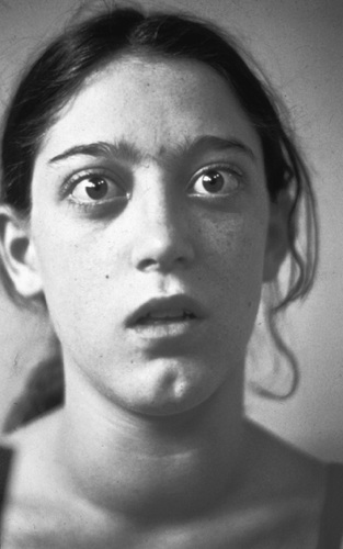
Many of the signs and symptoms of Graves disease in children are similar to those in adults (Fig. 584.1 and Table 584.3 ). However, the earliest signs and most pronounced differences in children may be related to growth and neuropsychologic systems. Tremulousness, headaches, mood disturbances, behavioral swings, difficulties with sleep, decrease in attention span, hyperactivity, and a decline in school performance are all common findings in childhood, and many hyperthyroid children are referred for evaluation of attention-deficit/hyperactivity disorder (ADHD).

Table 584.3
From De Leo S, Lee SY, Braverman LE: Hyperthyroidism, Lancet 388:906–916, 2016, Table 2.
Children with hyperthyroidism may show acceleration in growth velocity and advanced skeletal maturation. The effect on growth may be more pronounced if hyperthyroidism presents earlier in childhood. With antithyroid drug (ATD) treatment, growth velocity, and bone age approach a more normal pattern. There may also be an increase in appetite with either failure to gain weight or overt weight loss. Polyuria and more frequent defecation (although not usually frank diarrhea) contribute to changes in weight. Due to the increased risk of comorbid autoimmune disorders, screening for type 1 diabetes, celiac disease, and inflammatory bowel disease should be considered in patients who present with these symptoms.
The age of the onset of puberty does not appear to be altered by hyperthyroidism; however, menarchal females can develop secondary amenorrhea. Hyperthyroidism is also associated with increased aromatization of androgens to estrogens, but gynecomastia does not occur in males.
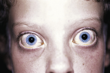
Most children with Graves disease have a diffuse goiter, but the size of the thyroid is variable. It is typically smooth and without nodules. A bruit can occasionally be auscultated over a markedly enlarged gland. If exophthalmos is present, it is typically mild. Ocular manifestations can produce pain, eyelid erythema, chemosis, decreased extraocular muscle function, and decreased visual acuity (corneal or optic nerve involvement) (Table 584.4 ). In children with thyrotoxicosis, the identification of these signs of ophthalmopathy on physical examination in the setting of diffuse goiter is highly suggestive of the diagnosis of Graves disease. Stare and lid lag are common eye findings caused by increased sympathetic activity and can be seen in thyrotoxicosis of any cause, not only Graves disease (Fig. 584.2 ). In general, ocular symptoms in children with Graves disease tend to be milder than in adults, and they improve with the restoration of euthyroidism.
Table 584.4
Clinical Assessment of the Patient With Graves Ophthalmopathy
| ACTIVITY MEASURES * |
| SEVERITY MEASURES |
|
• Lid aperture (distance between the lid margins in mm with the patient looking in the primary position, sitting relaxed, and with distant fixation) • Swelling of the eyelids (absent/equivocal, moderate, severe) • Redness of the eyelids (absent/present) • Redness of the conjunctivae (absent/present) • Conjunctival edema (absent, present) • Inflammation of the caruncle or plica (absent, present) • Exophthalmos (measured in millimeters using the same Hertel exophthalmometer and the same intercanthal distance for an individual patient) • Subjective diplopia score † • Eye muscle involvement (ductions in degrees) • Corneal involvement (absent/punctate keratopathy/ulcer) • Optic nerve involvement (best corrected visual acuity, color vision, optic disk, relative afferent pupillary defect (absent/present), plus visual fields if optic nerve compression is suspected |
* Based on the classic features of inflammation in Graves ophthalmopathy.
† Subjective diplopia score: 0, no diplopia; 1, intermittent (i.e., diplopia in primary position of gaze, when tired or when first awakening); 2, inconstant (i.e., diplopia at extremes of gaze); 3, constant (i.e., continuous diplopia in primary or reading position).
The clinical activity score (CAS) is the sum (1 point each) of all items present; a CAS ≥ 3/7 indicates active ophthalmopathy.
From Davies TF, Laurberg P, Bahn RS: Hyperthyroid disorders. In Melmed S, Polonsky KS, Larsen PR, Kronenberg HM, editors: Williams textbook of endocrinology , ed 13, Philadelphia, 2016, Elsevier, Table 12.4.

Children with hyperthyroidism have an increase in cardiac output. Tachycardia, palpitations, increased systolic blood pressure, and a widened pulse pressure are common cardiac manifestations, whereas cardiac enlargement and insufficiency, and atrial fibrillation are rare complications.
The skin is smooth and flushed, with excessive sweating. Occasionally, vitiligo or psoriasis can be present. Graves dermopathy is rare in children and typically responsive to steroids. Proximal muscular weakness is common. Thyroid hormone stimulates bone resorption, leading to low bone density and an increased fracture risk in patients with chronic hyperthyroidism. Bone density returns to normal with treatment.
Thyroid storm is an extreme form of hyperthyroidism manifested by a severe biochemical derangement, hyperthermia, tachycardia, heart failure, and restlessness (Table 584.5 ). There may be rapid progression to delirium, coma, and death. Precipitating events include trauma, infection, radioactive iodine (RAI) treatment, or surgery.
Table 584.5
Diagnostic Criteria for Thyroid Storm
| POINTS | |
|---|---|
| TEMPERATURE°F (°C) | |
| 99-99⋅9 (37⋅2-37⋅7) | 5 |
| 100-100⋅9 (37⋅8-38⋅2) | 10 |
| 101-101⋅9 (38⋅3-38⋅8) | 15 |
| 102-102⋅9 (38⋅9-39⋅4) | 20 |
| 103-103⋅9 (39⋅4-39⋅9) | 25 |
| ≥104⋅0 (>40⋅0) | 30 |
| CENTRAL NERVOUS SYSTEM EFFECTS | |
| Absent | 0 |
| Mild (agitation) | 10 |
| Moderate (delirium, psychosis, extreme lethargy) | 20 |
| Severe (seizure, coma) | 30 |
| GASTROINTESTINAL–HEPATIC DYSFUNCTION | |
| Absent | 0 |
| Moderate (diarrhea, nausea/vomiting, abdominal pain) | 10 |
| Severe (unexplained jaundice) | 20 |
| CARDIOVASCULAR DYSFUNCTION | |
| Tachycardia | |
| 90-109 | 5 |
| 110-119 | 10 |
| 120-129 | 15 |
| 130-139 | 20 |
| ≥140 | 25 |
| Congestive Heart Failure | |
| Absent | 0 |
| Mild (pedal edema) | 5 |
| Moderate (bibasilar rales) | 10 |
| Severe (pulmonary edema) | 15 |
| Atrial Fibrillation | |
| Absent | 0 |
| Present | 10 |
| Precipitating History | |
| Absent | 0 |
| Present | 10 |
In adults, a score ≥45 is highly suggestive of thyroid storm; a score of 25-44 is suggestive of impending thyroid storm; a score of <25 is unlikely to represent thyroid storm. Data are from Burch and Wartofsky.
From De Leo S, Lee SY, Braverman LE: Hyperthyroidism, Lancet 388:906–916, 2016, Table 3.
Laboratory Findings
In Graves disease, serum TSH is suppressed and free T4 and T3 are elevated. Most patients with newly diagnosed Graves disease have measurable thyrotropin receptor antibodies (TSHR-Abs). TSHR-Abs can be measured by either of 2 methods. Thyroid-stimulating immunoglobulin (TSI) is a functional assay measuring the presence of antibodies capable of stimulating TSHR-mediated cAMP generation (TRSAb). Thyrotropin-binding inhibitory immunoglobulin (TBII) is a binding assay measuring antibody binding to the TSHR, regardless of receptor-stimulating (TRSAb) or receptor-blocking (TRBAb) activity. In a patient with thyrotoxicosis, both assays are 96–97% sensitive and 99% specific for Graves disease.
When the diagnosis cannot be established by history, physical examination, and laboratory evaluation, RAI uptake can be measured. 123 I is the radionucleotide of choice for thyroid uptake and scintigraphy. The RAI uptake (typically assessed at 4 and 24 hr after isotope administration) is elevated in Graves disease, whereas it is low in other causes of thyrotoxicosis like thyroiditis or exogenous thyroid hormone ingestion. If scintigraphy is also performed, the increased RAI uptake in Graves disease is present diffusely throughout the gland, whereas focally increased uptake is observed in hyperfunctioning thyroid nodules.
Differential Diagnosis
Diagnosis of thyrotoxicosis is straightforward once the diagnosis is considered. Elevated serum levels of T4 or free T4 and T3 in association with suppressed levels of TSH are diagnostic (see Table 584.1 ). The combination of diffuse goiter and prolonged thyrotoxicosis (>8 wk) is nearly always caused by Graves disease, and the presence of circulating TSHR-Abs or characteristic eye or skin changes is diagnostic.
In cases of thyrotoxicosis in which the etiology is not clear, 123 I radioiodine uptake can be used to distinguish hyperthyroidism (increased uptake) from other causes of thyrotoxicosis, which will determine the appropriateness of antithyroid medication. If a discrete thyroid nodule is palpated, 123 I scintigraphy should be performed to assess the possibility of a hyperfunctioning nodule. Some children with toxic multinodular goiter may have either a TSHR–activating mutation or McCune-Albright syndrome. If precocious puberty, polyostotic fibrous dysplasia, or café-au-lait macules are present, then McCune-Albright syndrome is likely.
Patients with thyroid hormone resistance (see later) have elevated levels of free T4 and T3 , but levels of TSH are inappropriately elevated or normal. They must be differentiated from patients with TSH-secreting pituitary tumors who have elevated serum levels of the common α-subunit shared by TSH, luteinizing hormone (LH), follicle-stimulating hormone (FSH), and human chorionic gonadotropin (hCG). Most other causes of elevated serum T4 are uncommon but can result in erroneous diagnosis. Patients with elevated thyroxine-binding globulin levels or familial dysalbuminemic hyperthyroxinemia have high serum T4 but normal levels of free T4 and TSH and are clinically euthyroid. Rare patients with mutations in SLC16A2 (encoding the MCT8 thyroid hormone transporter) or THRA (encoding thyroid hormone receptor α) can present with high serum T3 , inappropriately normal or high TSH, and low or low-normal serum T4 concentrations.
When thyrotoxicosis is caused by exogenous thyroid hormone (thyrotoxicosis factitia), levels of free T4 and TSH are the same as those seen in Graves disease but, in contrast to Graves disease, thyroid size is small, serum thyroglobulin is very low, and 123 I radioiodine uptake is suppressed.
Treatment
Antithyroid Drugs
Most pediatric endocrinologists recommend initial medical therapy of Graves disease using antithyroid drugs (ATDs) rather than radioiodine ablation or near-total thyroidectomy, although radioiodine is gaining acceptance as initial treatment in children older than 10 yr of age. Each therapeutic option has advantages and disadvantages (Table 584.6 ). Methimazole is the first line ATD for children with Graves disease and functions by blocking the organification of iodide necessary to synthesize thyroid hormone. Methimazole has a long serum half-life (6-8 hr) that allows once- or twice-daily dosing. Propylthiouracil is a similar ATD that is effective in hyperthyroidism, but its use is not recommended in children due to its potential to cause liver failure.
Table 584.6
| TREATMENT | ADVANTAGE | DISADVANTAGE | COMMENT |
|---|---|---|---|
| Antithyroid drugs |
Noninvasive Less initial cost No risk of permanent hypothyroidism Possible remission |
Remission rate 30–50% (with long-term treatment) Adverse drug reactions Drug compliance required |
First line treatment in children and adolescents and in pregnancy Initial treatment in severe cases or preoperative preparation |
| Radioactive iodine (131 I) |
Cure of hyperthyroidism Most cost-effective |
Permanent hypothyroidism Might worsen ophthalmopathy Pregnancy must be deferred for 6-12 mo, mother cannot breastfeed; small potential risk of exacerbation of hyperthyroidism |
No evidence for infertility, birth defects, or secondary cancers with currently recommended doses |
| Surgery | Rapid, effective (especially in patients with large goiter) |
Most invasive therapy Potential complications (recurrent laryngeal nerve damage, hypoparathyroidism) Permanent hypothyroidism Most costly therapy Pain, surgical scar |
Potential use in pregnancy if major side effect from antithyroid drugs Useful when coexisting suspicious nodule is present or thyromegaly is massive Option for patients who do not desire radioiodine |
From Cooper DS: Hyperthyroidism, Lancet 362:459–468, 2003.
Adverse reactions can occur with ATDs, and although most are mild, some are life threatening. Minor adverse effects occur in approximately 10–20% of patients, and severe adverse effects occur in 2–5%. Reactions most commonly occur in the first 3 mo of therapy but can occur at any time during therapy. Transient urticarial rashes are common and may be managed with antihistamines or by a short period off therapy followed by restarting ATD. Agranulocytosis is a severe adverse reaction that occurs in 0.1–0.5% of patients and can lead to fatal infections. Therefore patients on methimazole should stop this medication and have a white blood count checked during any episode of significant fever, pharyngitis, or oral ulcers. On the other hand, transient, asymptomatic granulocytopenia (<2,000/mm3 ) is a common finding in Graves disease; it is not a harbinger of agranulocytosis and is not a reason to discontinue treatment. Other severe reactions include hepatitis (0.2–1.0%), a lupus-like polyarthritis syndrome, glomerulonephritis, and antineutrophilic cytoplasmic antibody–positive vasculitis. Severe liver disease, including liver failure requiring transplantation, has been reported with propylthiouracil. The most common liver disease associated with methimazole is cholestatic jaundice, which is reversible when the drug is discontinued. Patients with severe adverse effects should be treated with radioiodine or thyroidectomy. In rare instances in which hyperthyroidism is severe and methimazole cannot be used, a short course of propylthiouracil may be offered to restore euthyroidism prior to definitive therapy. Both methimazole and propylthiouracil have been associated with congenital malformations in infants exposed to these drugs in utero. Methimazole exposure may be associated with aplasia cutis, omphalocele, choanal atresia, and urinary system malformations, whereas propylthiouracil may be associated with malformations of the head, neck, and urinary system.
The initial dosage of methimazole is 0.5-1.0 mg/kg/24 hr (max 40 mg/day) administered once or twice daily. Smaller initial dosages should be used in early childhood. Careful surveillance is required after treatment is initiated. Rising serum levels of TSH of greater than normal indicates overtreatment and warrants a dose reduction. Clinical response becomes apparent in 3-4 wk, and adequate control is typically evident within 3-4 mo. The dose is decreased to the minimal level required to maintain a euthyroid state.
Most studies report a remission rate of approximately 25% after 2 yr of ATD treatment in children. Some studies find that longer treatment is associated with higher remission rates of 30–50% after 4-10 yr of drug treatment. Relapses usually occur within 6-12 mo after therapy has been discontinued. In case of relapse, ATD therapy may be resumed or definitive therapy with either radioiodine or thyroidectomy can be pursued. In general, adolescents, males, those with a higher body mass index, and those with small goiters and modestly elevated T3 levels are thought to have earlier remissions; however, this has not been proven in large studies, because TRSAbs tend to persist for a longer period of time in children than in adults with Graves disease.
Thyroid hormones potentiate the actions of catecholamines, including tachycardia, tremor, excessive sweating, lid lag, and stare. To help control cardiovascular symptoms, a β-adrenergic blocking agent such as propranolol or atenolol is a useful supplement to ATDs. However, these agents do not alter thyroid function or exophthalmos. Table 584.7 lists additional therapies for thyroid storm .
Table 584.7
From Goldman L, Ausiello D, editors: Cecil textbook of medicine , ed 22, Philadelphia, 2004, WB Saunders, p 1401.
Definitive Therapy
Radioiodine ablation or thyroidectomy is indicated when medical management is not possible due to patient nonadherence or severe side effects of ATDs, when an adequate trial of medical management has failed to result in remission, or if the patient prefers definitive therapy.
Radioiodine ablation with 131 I is an effective therapy for Graves disease in children. In patients with severe thyrotoxicosis, euthyroidism should be restored with methimazole prior to radioiodine ablation to deplete the gland of preformed hormone and reduce the risk of a thyrotoxic flare from radiation thyroiditis. If a patient is taking an ATD, it must be stopped 3-5 days before radioiodine administration to avoid inhibition of uptake. The goal of radioiodine ablation is to administer a sufficient dose of radioiodine to ensure complete ablation of thyroid tissue. Many centers measure radioiodine uptake before treatment and use this to calculate an 131 I dose that delivers an absorbed thyroid dose of >150 µCi/g thyroid tissue (based on thyroid gland mass estimated by clinical examination or ultrasound). Alternatively, an empiric fixed 131 I dose (usually 10-15 mCi) can be offered. The theoretical advantage of calculated doses is that they define for each individual patient the lowest administered dose that achieves the therapeutic target. This benefit is most important in small children, because the absorbed radiation dose to the bone marrow and other normal tissues is inversely proportional to body size. Based upon this concept and theoretical modeling of radiation exposure, current consensus guidelines recommend that 131 I therapy be avoided in children younger than 5 yr of age and used in children between 5 and 10 yr of age if the administered dose is <10 mCi. As with other Graves therapies, radioiodine ablation has a low failure rate (5–20%). Patients with persistent hyperthyroidism more than 6 mo after their first 131 I therapy can be offered retreatment.
Thyroidectomy is a safe procedure when performed by an experienced surgeon. Thyroid surgery should be done only after the patient has been rendered euthyroid with methimazole over 2-3 mo. A saturated solution of potassium iodide (SSKI; 1-3 drops, 2-3 times per day) may be added for 7-14 days before surgery to decrease the vascularity of the gland. In expert hands, complications of surgical treatment are rare and include hypoparathyroidism (transient or permanent) and paralysis of the vocal cords. When surgery is elected, total or near-total thyroidectomy should be performed rather than a less extensive subtotal resection. The incidence of recurrence is low, and patients become hypothyroid postoperatively. Referral to a surgeon with extensive experience in thyroidectomy and a low personal complication rate is paramount.
Graves ophthalmopathy usually remits gradually and independently of the hyperthyroidism, but control of ophthalmopathy is facilitated by maintaining consistent euthyroidism. Severe ophthalmopathy can require treatment with high-dose glucocorticoids, orbital radiotherapy, or orbital decompression surgery. Teprotumumab, a human monoclonal antibody against the insulin-like growth factor 1 receptor IGF-1R has been effective in adults with ophthalmopathy. Cigarette smoking is a risk factor for thyroid eye disease and should be avoided or discontinued to avoid progression of eye involvement.
Bibliography
Abbasi B, Sharif Z. Sprabery LR: hypokalemic thyrotoxic periodic paralysis with thyrotoxic psychosis and hypercapnic respiratory failure. Am J Med Sci . 2010;340(2):147–153.
Andersen SL, Olsen J, Wu CS, Laurberg P. Birth defects after early pregnancy use of antithyroid drugs: a Danish nationwide study. J Clin Endocrinol Metab . 2013;98(11):4373–4381.
Bahn RS. Graves ophthalmopathy. N Engl J Med . 2010;362(8):726–736.
Bahn RS, Burch HB, Cooper DS, et al. Hyperthyroidism and other causes of thyrotoxicosis: management guidelines of the American thyroid Association and American Association of Clinical Endocrinologists. Thyroid . 2011;21(6):593–646.
Barrio R, Lopez-Capape M, Martinez-Badas I, et al. Graves disease in children and adolescents: response to long-term treatment. Acta Paediatr . 1583;94(11):2005.
Bauer AJ. Approach to the pediatric patient with Graves disease: when is definitive therapy warranted? J Clin Endocrinol Metab . 2011;96(3):580–588.
Brent GA. Graves disease. N Engl J Med . 2008;358:2594–2605.
Cassio A, Corrias A, Gualandi S, et al. Influence of gender and pubertal stage at diagnosis on growth outcome in childhood thyrotoxicosis: results of a collaborative study. Clin Endocrinol (Oxf) . 2006;64(1):53–57.
Chu X, Pan CM, Zhao SX, et al. A genome-wide association study identifies two new risk loci for Graves disease. Nat Genet . 2011;43(9):897–901.
De Leo S, Lee SY, Braverman LE. Hyperthryroidism. Lancet . 2016;388:906–916.
Food and Drug Administration. FDA Drug Safety Communication: New boxed warning on severe liver injury with propylthiouracil . http://www.fda.gov/Drugs/DrugSafety/PostmarketDrugSafetyInformationforPatientsandProviders/ucm209023.htm .
Franklyn JA, Boelaert K. Thyrotoxicosis. Lancet . 2012;379(9821):1155–1164.
Gastaldi R, Poggi E, Mussa A, et al. Graves disease in children: thyroid-stimulating hormone receptor antibodies as remission markers. J Pediatr . 2014;164(5):1189–1194.
Glaser NS, Styne DM. Predicting the likelihood of remission in children with Graves disease: a prospective, multicenter study. Pediatrics . 2008;12(3):e481–e488.
Goldstein SM, Katoqitz WR, Moshang T, et al. Pediatric thyroid-associated orbitopathy: the Children's Hospital of Philadelphia experience and literature review. Thyroid . 2008;18(9):997–999.
Kaguelidou F, Alberti C, Castanet M, et al. Predictors of autoimmune hyperthyroidism relapse in children after discontinuation of antithyroid drug treatment. J Clin Endocrinol Metab . 2008;93(10):3817.
Lee HJ, Li CW, Hammerstad SS, et al. Immunogenetics of autoimmune thyroid diseases: a comprehensive review. J Autoimmun . 2015;64:82–90.
Leger J, Gelwane G, Kaguelidou F, et al. Positive impact of long-term antithyroid drug treatment on the outcome of children with Graves disease: national long-term cohort study. J Clin Endocrinol Metab . 2012;97(1):110–119.
Lytton SD, Li Y, Olivo PD, et al. Novel chimeric thyroid-stimulating hormone-receptor bioassay for thyroid-stimulating immunoglobulins. Clin Exp Immunol . 2010;162(3):436–438.
Laurberg P, Wallin G, Tallstedt L, et al. TSH-receptor autoimmunity in Graves disease after therapy with anti-thyroid drugs, surgery, or radioiodine: a 5-year prospective randomized study. Eur J Endocrinol . 2008;158(1):69–75.
Pinto T, Cummings EA, Barnes D, et al. Clinical course of pediatric and adolescent Graves disease treated with radioactive iodine. J Pediatr Endocrinol Metab . 2007;20(9):973–980.
Okawa ER, Grant FD, Smith JR. Pediatric Graves disease: decisions regarding therapy. Curr Opin Pediatr . 2015;27(4):442–447.
Regina A, Majlesi N. Thyrotoxicosis after consumption of dietary supplements purchased through the internet—Staten Island, New York, 2015. MMWR Morb Mortal Wkly Rep . 2016;65(13):353–354.
Rivkees SA, Mattison DR. Ending propylthiouracil-induced liver failure in children. N Engl J Med . 2009;360(15):1574–1575.
Rivkees SA. Pediatric Graves disease: controversies in management. Horm Res Paediatr . 2010;74(5):305–331.
Ross DS, Burch HB, Cooper DS, et al. American Thyroid Association guidelines for the diagnosis and management of hyperthyroidism and other causes of thyrotoxicosis. Thyroid . 2016;26(10):1343–1421.
Smith TJ, Hegedus L. Graves disease. N Engl J Med . 2016;375(16):1552–1565.
Smith TJ, Kahaly GJ, Ezra DG. Teprotumumab for thyroid-associated ophthalmopathy. N Engl J Med . 2017;376:1748–1761.
Congenital Hyperthyroidism
Jessica R. Smith, Ari J. Wassner
Etiology and Pathogenesis
Neonatal Graves disease is caused by transplacental passage of TRSAbs from mothers with a history of Graves disease. These mothers can have active Graves disease, Graves disease in remission, or a prior history of Graves disease treated with radioiodine ablation or thyroidectomy. Occasionally, there is a maternal history of chronic lymphocytic thyroiditis with hypothyroidism. High levels of TRSAb typically result in classic neonatal hyperthyroidism, but if the mother has been treated with ATDs, the onset of hyperthyroid symptoms may be delayed by 3-7 days until the ATD is metabolized by the neonate. If TRBAbs are also present, the onset of hyperthyroidism may also be delayed for several weeks, or neonatal hypothyroidism may even develop.
Neonatal hyperthyroidism occurs in approximately 2% of infants born to mothers with a history of Graves disease. In utero, fetal tachycardia and goiter may suggest the diagnosis, and close ultrasound surveillance is recommended in mothers with uncontrolled hyperthyroidism, particularly in the 3rd trimester. Elevated serum titers of TRSAb (more than 3 times the upper limit of normal) or a history of a prior child with neonatal thyroid dysfunction increases the likelihood of neonatal Graves disease.
Neonatal Graves disease typically remits spontaneously within 6-12 wk but can persist longer, depending on the titer and rate of clearance of the TRSAbs (and TRBAbs, if present). Rarely, classic neonatal Graves disease may not remit but persist for several yr or longer. These children typically have a family history of Graves disease. In these infants, the transfer of maternal TRSAbs exacerbates the infantile onset of autonomous Graves disease.
Clinical Manifestations
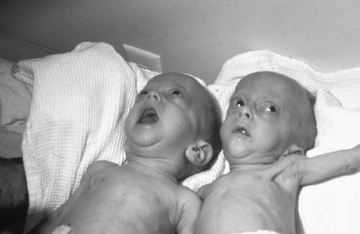
Many infants born with neonatal Graves disease are premature and have intrauterine growth restriction. Many infants also have goiter, and occasionally tracheal compression can occur if the goiter is very large. Other signs and symptoms of neonatal Graves disease include low birth weight, stare, periorbital edema, retraction of the eyelids, hyperthermia, irritability, diarrhea, feeding difficulties, poor weight gain, tachycardia, heart failure, hypertension, hepatomegaly, splenomegaly, cholestasis, jaundice, thrombocytopenia, and hyperviscosity (Fig. 584.3 ). Laboratory evaluation shows suppressed serum TSH and elevated serum levels of T4 , free T4, and T3 . TRSAbs are markedly elevated at birth and typically resolve within 3 mo of life. If symptoms and signs are not immediately recognized and treated, cardiac failure and death can occur. Craniosynostosis and developmental delay can be permanent sequelae of the hyperthyroidism.

Treatment
Treatment should be initiated at the onset of symptoms to avoid short-term and long-term complications. Therapy consists of methimazole (0.5-1.0 mg/kg/24 hr given every 12 hr) and oral or intravenous administration of a nonselective β-adrenergic blocker such as propranolol to decreases sympathetic hyperactivity. In refractory cases, Lugol solution or potassium iodide (1-2 drops per day) can be added. The first dose of iodide should be given at least 1 hr after the 1st dose of ATD to prevent the iodide from being used for further thyroid hormone synthesis. If thyrotoxicosis is severe and progresses to heart failure, parenteral fluid therapy, corticosteroids, and digitalization may be indicated. Once serum thyroid hormone levels begin to decrease, antithyroid medications should be gradually tapered to keep the infant euthyroid. Occasionally, a block-and-replace method with concurrent ATD and thyroid hormone replacement therapy may be required to ensure euthyroidism.
Most cases of neonatal Graves disease remit by 3 mo of age, but occasionally neonatal hyperthyroidism persists into childhood. Typically, there is a family history of hyperthyroidism. Neonatal hyperthyroidism without evidence for autoimmune disease in mother or infant may be caused by a mutation in the TSHR gene that results in constitutive activation of the receptor. This can be transmitted in an autosomal dominant manner or can occur sporadically. Neonatal hyperthyroidism has also been reported in patients with McCune-Albright syndrome because of an activating mutation of the stimulatory α-subunit of the G-protein. Under these circumstances, hyperthyroidism recurs when ATDs are discontinued, and these children eventually must be treated with radioiodine or surgery.
Prognosis
Advanced osseous maturation, microcephaly, and cognitive impairment occur when treatment is delayed. Intellectual development is normal in most treated infants with neonatal Graves disease, although some manifest neurocognitive problems from in utero hyperthyroidism. In some infants, in utero hyperthyroidism appears to suppress the hypothalamic-pituitary-thyroid feedback mechanism and they develop transient or permanent central hypothyroidism that requires thyroid hormone replacement. Neurocognitive development should be monitored throughout childhood.
Resistance to Thyroid Hormone
This autosomal dominant disorder is caused by mutations in the THRB gene. Because this receptor mediates the normal feedback of thyroid hormone on the hypothalamus and pituitary, patients have elevated serum levels of T4 and T3 but serum TSH levels are inappropriately normal or elevated. Goiter is almost always present, but symptoms of thyroid dysfunction are highly variable among individuals. There may be clinical features of hypothyroidism such as developmental delay, growth retardation, and delayed skeletal maturation, as well as certain clinical features of hyperthyroidism like tachycardia and hyperreflexia. Affected children also have an increased association of learning disabilities and ADHD. The clinical symptoms, goiter, and elevated thyroid hormone levels may be mistaken for Graves disease, but resistance to thyroid hormone (RTH) is confirmed by the presence of normal or elevated (not suppressed) TSH levels. This condition must also be differentiated from a pituitary TSH-secreting tumor, which is not familial and in which serum levels of the TSH α-subunit are elevated. Elevated levels of T4 on newborn screening should suggest the possibility of RTH.
More than 100 distinct mutations in THRB have been identified in patients with RTH, and genotype-phenotype correlation is poor even amongst affected members of the same family. Nearly all mutations have a dominant-negative effect in which the mutant receptor interferes with normal receptor action, leading to disease even in heterozygotes. Individuals carrying 2 mutant alleles are severely affected. A very rare autosomal recessive form of this disorder has been reported in individuals homozygous for a deletion of the THRB gene.
Treatment is usually not required unless growth and skeletal retardation are present. Different therapies, including levothyroxine and triiodothyroacetic acid, have been successful in some patients. Symptoms of hyperthyroidism can be treated with β -blockers, but ATDs or radioiodine ablation are generally not used because they increase TSH levels and goiter size.
Although RTH due to THRB mutations has been recognized for decades, the first patients with mutations in the THRA gene have been reported only recently. These mutations, too, are dominant negative and cause disease in heterozygous carriers. Clinical symptoms suggest those of untreated primary hypothyroidism, including skeletal dysplasia with short stature and macrocephaly, developmental delay, constipation, bradycardia, and macrocytic anemia. Serum thyroid function tests show subtle abnormalities of low or low-normal T4 , high or high-normal T3 (with elevated T3 /T4 ratio), and normal TSH, as well as the unique finding of markedly low reverse T3 . Treatment has not been clearly established for this condition.
Bibliography
Alexander AK, Pearce EN, Brent GA, et al. 2017 guidelines of the American Thyroid Association for the Diagnosis and Management of Thyroid Disease During Pregnancy and the Postpartum. Thyroid . 2017;27(3):315–389.
Chan W, Wong GW, Fan DS, et al. Ophthalmopathy in childhood Graves disease. Br J Ophthalmol . 2002;86(7):740.
Chester J, Rotenstein D, Ringkananont U, et al. Congenital neonatal hyperthyroidism caused by germline mutations in the TSH receptor gene. J Pediatr Endocrinol Metab . 2008;21(5):479–486.
Kempers MJ, van Tijn DA, van Trotsenburg AS, et al. Central congenital hypothyroidism due to gestational hyperthyroidism: detection where prevention failed. J Clin Endocrinol Metab . 2003;88(12):5851–5857.
Leger J. Management of fetal and neonatal Graves disease. Horm Res Paediatr . 2017;87(1):1–6.
Lewis KA, Engle W, Hainline BE, et al. Neonatal Graves disease associated with severe metabolic abnormalities. Pediatrics . 2011;128(1):e232–e236.
Mastorakos G, Mitsiades NS, Doufas AG, et al. Hyperthyroidism in McCune-Albright syndrome with a review of thyroid abnormalities sixty years after the first report. Thyroid . 1997;7(7):433–439.
van der Kaay DCM, Wasserman J, Palmert M. Management of neonates born to mothers with Graves disease. Pediatrics . 2016;137(4).