Arthrogryposis
Helen M. Horstmann, Richard S. Davidson
Arthrogryposis multiplex congenita refers to a heterogeneous group of muscular, neurologic, and connective tissue anomalies that present with two or more joint contractures at birth as well as muscle weakness. It is associated with abnormal contraction of muscle fibers, causing reduced mobility with a decreased active and passive arc of motion. Arthrogryposis is not a specific diagnosis but a descriptive term with various etiologies and complex clinical features, including multiple congenital contractures of various limb joints. It is associated with over 300 different disorders encompassing malformation, malfunction, and neurologic deficiency.
Approximately 1% of all births show some form of contractures of the joints ranging from unilateral clubfoot to the most severe amyoplasia, a condition characterized by pervasive, crippling contractures involving many joints. The overall incidence of arthrogryposis is 1 in 5,000-10,000 live births with equal gender ratios.
Although children with arthrogryposis have many other problems, such as micrognathia and feeding issues, focus is on the orthopedic problems frequently seen in this group of children. In the absence of central nervous system lesions, many children have normal intelligence.
Etiology
The main cause of arthrogryposis is fetal akinesia or decreased fetal movement. The associated pattern of abnormalities is often referred to as the fetal akinesia deformation sequence . This sequence manifests as multiple joint contractures, polyhydramnios, craniofacial anomalies (e.g., micrognathia), and pulmonary hypoplasia due to lack of movement of the diaphragm and intercostal muscles. Intrinsic and extrinsic causes of fetal akinesia are categorized into six groups (Fig. 702.1 ) and include a multitude of disorders (Table 702.1 ).

Neurologic Abnormalities
As one of the most common causes of arthrogryposis, neurologic abnormalities are present in 70–80% of cases. Patchy damage to the anterior horn cells of the spinal cord can lead to characteristic limb posturing of arthrogryposis. Neurologic disorders, such as spinal muscular atrophy and anterior horn disease, are associated with arthrogryposis; however, the type of anterior horn cell involvement is usually not from spinal muscular atrophy syndrome. Other, less-common neurologic disorders include neonatal myasthenia, myotonic dystrophy, olivo-ponto-cerebellar disorders, and neuronal migration anomalies.
Muscular Abnormalities
These rare abnormalities affect the function and structure of the muscles. Some muscular diseases associated with arthrogryposis are muscular dystrophies, congenital myopathies (central core, nemaline, centronuclear), intrauterine myositis, and mitochondrial diseases.
Limited Intrauterine Spacing
With a less than 0.1% occurrence rate, uterine constraint is rarely the primary cause of arthrogryposis. Maternal uterine anomalies will occasionally increase contractures of fetal limbs with arthrogryposis already existing. Other known causes are lack of amniotic fluid within the uterus and tumors, such as fibroids, that can prevent movement by impinging on uterine space.
Connective Tissue Abnormalities
When the tendons, bones, joints, and joint lining develop atypically, decrease in fetal movement causes congenital contractures. Diseases such as diastrophic dysplasia, campomelic dysplasia, and metatropic dysplasia result from connective tissue not developing properly. These are specific diagnoses resulting in limited joint motion and not true distal arthrogryposis. Other cases show that individuals who lack normal joint movement have distal joint involvement because the connective tissue develops normally but does not attach to the proper location around a joint bone or joint.
Maternal Diseases
Maternal diseases, such as multiple sclerosis, diabetes mellitus, myasthenia gravis, maternal hyperthermia, infection, drugs, and trauma, are associated with an increased incidence of arthrogryposis. In approximately 10% of neonates born to mothers with myasthenia gravis, maternal antibodies enter the fetal circulation through the placenta, causing transient myasthenia gravis; this inhibits fetal acetylcholine receptors, which leads to damaged fetal muscles.
Intrauterine Vascular Compromise
Inadequate vascular supply to the fetus causes fetal hypoxia resulting in anterior horn cell death, which decreases neurologic and myopathic function, resulting in fetal akinesia and secondary joint contractures. Multiple congenital contractures have been reported in individuals after bleeding throughout pregnancy or after a failed attempt at terminating the pregnancy.
Classification
Arthrogryposis multiplex congenita is divided into subgroups with different signs, symptoms, and causes as a practical way to make a differential diagnosis. Disorders involving primarily limbs such as amyoplasia and distal arthrogryposis are the most common subgroups (Table 702.2 ). Disorders involving limbs and other body parts typically represent a form of multiple pterygium, which is characterized by a weblike membrane that forms across joints affecting a child's ability to extend and causing fixed flexion. Disorders with limb involvement and abnormal neurologic function are caused by atypical central nervous system, peripheral nervous system, and damaged or absent anterior horn cells.
Table 702.2
From Kowalczyk B, Feluś J: Arthrogryposis: an update on clinical aspects, etiology, and treatment strategies. Arch Med Sci 12(1):10–24, 2016 (Table 1, p 16).
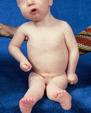
Amyoplasia, also known as classic arthrogryposis, is a sporadic symmetric disorder that causes fibrotic replacement of the muscles. Symptoms include internally rotated and adducted shoulders, extended elbows, pronated forearms, flexed fingers and wrists, dislocated hips, feet with severe equinovarus contractures, and extended knees. Involved muscles are hypoplastic and fibrotic. Often patients have midfacial hemangioma. Intelligence is usually normal (Figs. 702.2 and 702.3 ).
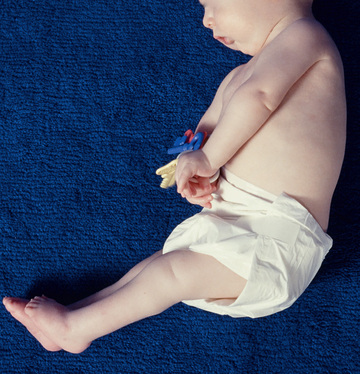
Distal arthrogryposis is an autosomal dominant disorder that primarily affects the distal joints of the limbs. Characteristics of the upper limbs are medially overlapping fingers, clenched fists, ulnar deviation of fingers, camptodactyly, and hypoplasia. Lower limbs show talipes equinovarus, calcaneovalgus, vertical talus, or metatarsus varus (Fig. 702.4 ).
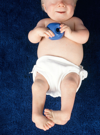
Ten different types of distal arthrogryposis have been categorized based on specific traits they share with each other.
Management of Orthopedic Problems of Arthrogryposis
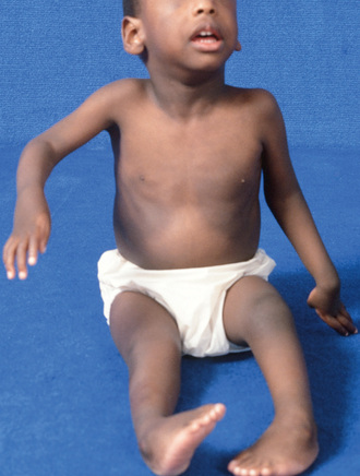
When a child is born with arthrogryposis, the many stiff or dislocated joints pose issues of timing and best practices of management. Typically, a child can have stiff elbows, dislocated hips, dislocated, hyperextended, or contracted knees, and clubfeet (Fig. 702.5 ). The stiffness and deformity need to be aggressively addressed through a combination of modalities. A team of clinicians, including therapists for the upper and lower extremities, orthotists, and orthopedic surgeons, will be involved.
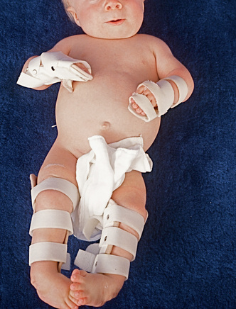
Initially, passive range-of-motion exercises and judicious splinting directed and assisted by physical and occupational therapy will help to address the various deformities. Splinting and casting can be augmented by a taping program which can be taught to the family so that the taping can be redone frequently to take advantage of improved range of motion. The ingenuity of the therapists and/or orthotists to create the right splints and braces using appropriate thermoplastics, neoprene, Velcro, and other materials can be simple yet effective (Fig. 702.6 ).
The therapeutic and orthopedic goal for the child with arthrogrypotic limb deformities is to achieve maximal joint motion and to optimize joint position for function. In the lower extremities, the foot needs to be plantigrade. The knees need to have optimal motion for sitting and standing. Hips need to be stabilized especially if the child has walking potential. In the upper extremities, the goals should include positioning of 1 arm for feeding and the other for toileting in cases where there is extreme stiffness. Two-handed activities require some symmetry, which can be a challenging goal with extreme contractures and limited muscle strength.
Although scoliosis is common, it usually does not become a problem until adolescence.
Foot Problems
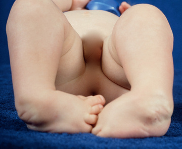
Clubfoot deformities are the most commonly seen deformities with arthrogryposis (Fig. 702.7 ). A clubfoot has components of hindfoot equinus, midfoot varus, and forefoot adduction. Feet in arthrogryposis tend to be resistant to improvement but the traditional methods of treatment are nevertheless employed. Casting is begun shortly after birth in a method known as the Ponseti method. Casts are changed weekly until a plateau is reached and heel cord lengthening is needed. Other deformities such as vertical talus are also seen and are addressed in a similar approach although with appropriately differing techniques.
Persistent stiffness often leads to more comprehensive soft tissue releases. This is typically done around age 6-12 mo and is followed by 3 mo of further casting and additional bracing as needed, especially as the foot is growing. When deformities are not corrected in early childhood, additional bony surgery may be needed later. Some of the approaches to this involve bony wedge osteotomies, lateral column lengthening, bone decancellation, or talectomy. Ilizarov ring or multiaxial monolateral external fixation with or without osteotomies are used in late correction of residual deformities.
Children with significant deformities are often in ankle foot orthoses through much of their lives to avoid deformity recurrence and to augment the standing base due to weak leg muscles. A plantigrade, pain free, stable foot is the goal of foot management. Foot stiffness is anticipated and unavoidable in arthrogryposis involving the foot.
Knee Problems
Knee issues, including knee extension or flexion, subluxation, and stiffness, respond well to therapy and splinting. Knee flexion is more common in arthrogryposis. Infrequently it can structurally be complex and associated with skin webbing known as pterygiums. Pterygiums are resistant to nonsurgical intervention and require plastic Z lengthenings. In the case of a flexion contracture, the quadriceps musculature is often deficient and weak. Sometimes the casting and splinting of the knee contractures is insufficient. Hamstring lengthenings with additional posterior knee capsular releases are often needed.
In the case of knee hyperextension, the quadriceps are sometimes fibrotic and weak in spite of seeming to overpower the hamstrings. Casting and splinting should begin shortly after birth, which can be done in conjunction with clubfoot casting following the principles of Ponseti. If splinting and therapy fail, lengthening of the quadriceps can be achieved through release of the lateral medial quadriceps, with proximal detachment of the rectus femoris, lengthening of the quadriceps either percutaneously or through a mini open procedure which may minimize scarring.
Long-standing stiffness may lead to joint surface flattening permanently reducing the arc of motion. Repositioning the arc of motion may improve sitting or standing, a choice to be made by the patient, family, and physician. Follow-up bracing can help to compensate for weak, fibrotic muscles of the legs.
Hip Problems
Teratologic hip dislocations are common within the spectrum of arthrogryposis and usually require open reduction of the hip. Hips in a child with less upper-extremity involvement and more supple hips that are not pathologically stiff may respond to early treatment with a Pavlik harness. Knee hyperextension can often be treated with physical therapy and serial casting. Careful observation of the hip during knee flexion as tightening of the quadriceps and hip flexors can push the hip into posterior dislocation. Once some knee flexion has been achieved, the Pavlik harness can be useful in further flexing the knee and maintaining hip stability in the infant. Most often, the hips are stiff and not reducible closed. For these, open reduction with pelvic reconstruction and femoral osteotomy are commonly required, typically at 1 yr of age. There is some controversy about reducing bilateral hip dislocations as a high failure rate can result in asymmetry of the pelvis, pain, leg length inequality, and stiffness. If a child has little ambulation potential, he may do as well retaining the bilateral hip dislocations and positioning the hips for sitting. Management judgment should be made in conjunction with the family guided by a pediatric hip surgeon.
Ambulation
As would be expected, walking is more difficult for children with arthrogryposis due to the muscle weakness and limited joint motion. Children with arthrogryposis who walk have lower activity levels and take fewer steps than their peers. Not surprisingly, muscle fatigue and pain on exertion were noted in a study that included adults with distal arthrogryposis.
Upper-Extremity Problems
If splinting and a movement exercise program do not result in optimally functional upper extremities, surgical management may improve use of the arms of the child with arthrogryposis. A typical child with arthrogrypotic involvement of the upper extremities has internally rotated arms, extended elbows, flexed wrists, and thumb-in-palm or clasp-thumb deformities (see Figs. 702.2 and 702.3 ).
Treatment is geared toward optimizing use of the arms and hands particularly for critical activities of daily living, such as feeding and toileting. Therapy to improve motion of the joints is started immediately after birth. Pediatric hand therapists are the optimal leaders of the mobility treatment program. Therapy is augmented by use of splints so that less-extensive surgery will be required. The elbow is the critical length adjuster of the arm, allowing the arm to reach out as is necessary for toileting or to approach the mouth for feeding. If necessary, lack of these motions can be compensated by modified silverware and other adaptive equipment, including arm extenders for grabbing.
Surgery of the Upper Extremity
Surgical correction of arthrogrypotic upper extremity contractures should be started after 1-3 mo and completed by age 12 mo so that the child can optimize his or her motor development. This allows for improved results optimizing the joint growth remodeling plasticity. One-stage procedures yield the best results. Delays in surgery result in more problems of intraarticular adhesions as well as fixed joint incongruity.
Shoulder
Because of the rotational capacity of the shoulder, derotation osteotomy of the humerus is only occasionally needed. This is usually done in later childhood.
Elbow
A stiff elbow that does not respond to therapy requires surgical intervention starting with soft tissue and capsular release. Capsulotomy of the posterior elbow combined with a V-Y or Z reconstructive lengthening of the triceps allows improved elbow flexion. The triceps may need to be lengthened. Muscle transfer to the forearm can permit active elbow flexion. Each child needs individual assessment as to available flexor source. Most commonly available is the triceps. An elbow with some flexion is extremely important for arm function. Use of the triceps can create elbow flexion overpowering and contracture.
Wrist
Wrist flexion deformity is improved with soft tissue balancing as well as partial carpectomies. The carpectomies need to be trapezoidal with more removed from the dorsum and the radial side to balance the wrist flexion contracture as well as the tendency for ulnar deviation. Thumb adduction may require an adductor release with an opponensplasty. Tendon transfers such as transfer of the extensor indicis pollicis to the extensor pollicis longus is helpful for improved function of the thumb in clasp thumb deformity.
Finger stiffness and wrist contractures often respond to therapy and bracing without need for surgery.
Scoliosis
Scoliosis is frequent in arthrogrypotic children, although the reported incidence of between 28% and 66% is probably skewed upward in reports as they reflect the experience of scoliosis surgeons. Scoliosis can be congenital or paralytic. The scoliosis is often accompanied by hip contractures associated with hip dislocation and compensatory lumbar lordosis. Curves <30 degrees can be treated initially with bracing in a thoracolumbar spinal orthosis (TLSO brace). After 40 degrees, spinal fusion is warranted.
Surgical Staging
Surgical treatment of the lower limbs usually begins distally and works proximally. The feet are corrected around 6 mo of age, the knees around 8 mo of age, and the hips around 12 mo of age as pelvic osteotomy is often needed to stabilize the hips properly.
The upper extremities are corrected during infancy when the child is seen early. Hand, physical, and occupational therapy are a critical part of the team to optimize function prior to and after surgery. Further surgery during childhood may be needed to tweak and optimize functional use of the upper and lower extremities.