13
Neonatal resuscitation and post-resuscitation care
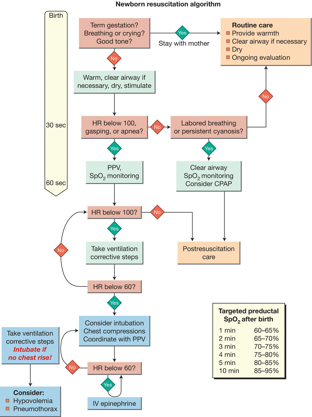
Relatively few infants require resuscitation at birth. The aim of resuscitation is to optimize the airway, breathing and circulation as quickly as possible. Most infants respond to lung inflation. Very few require chest compressions or medications. As the need for resuscitation has become uncommon, there is increasing emphasis on simulation and teamwork training to ensure proficient resuscitation when it is required.
Preparation
The presence of antenatal or intrapartum risk factors often allows anticipation that resuscitation may be required. This enables those skilled in neonatal resuscitation to be present. However, the need for resuscitation cannot always be predicted. There should always be at least one health professional skilled in basic resuscitation readily available at every delivery. Staff skilled in advanced resuscitation should attend high risk deliveries and be readily available at all times (see video: Basic newborn resuscitation).
Before delivery
- Introduce yourself to parents and explain why you are present.
- Review maternal history and obstetric records.
- Turn on the radiant warmer.
- Wash hands and put on gloves.
- Check equipment is present and functional:
- – several warm towels (and plastic wrap or bag for preterm infants)
- – clock
- – stethoscope
- – suction equipment
- – gas supply and pressure-limited T-piece circuit (e.g. Neopuff®) or self-inflating bag and mask
- – oropharyngeal (Guedel) airway
- – laryngoscope, tracheal tubes and introducers
- – exhaled CO2 detector
- – pulse oximeter
- – venous access equipment and drugs.
Specific questions to consider
- Will you need help? If so, call for it early.
- Is neonatal transport going to be needed?
Cord clamping
Delay in cord clamping of at least 1 minute from complete delivery of the infant is recommended for uncompromised infants. For the compromised infant, resuscitation is the priority and a plan of action should be agreed with the obstetric team. Resuscitation tables that can be placed directly adjacent to the mother to allow resuscitation with intact cord have been developed; studies are currently determining their impact on outcomes.
Temperature control
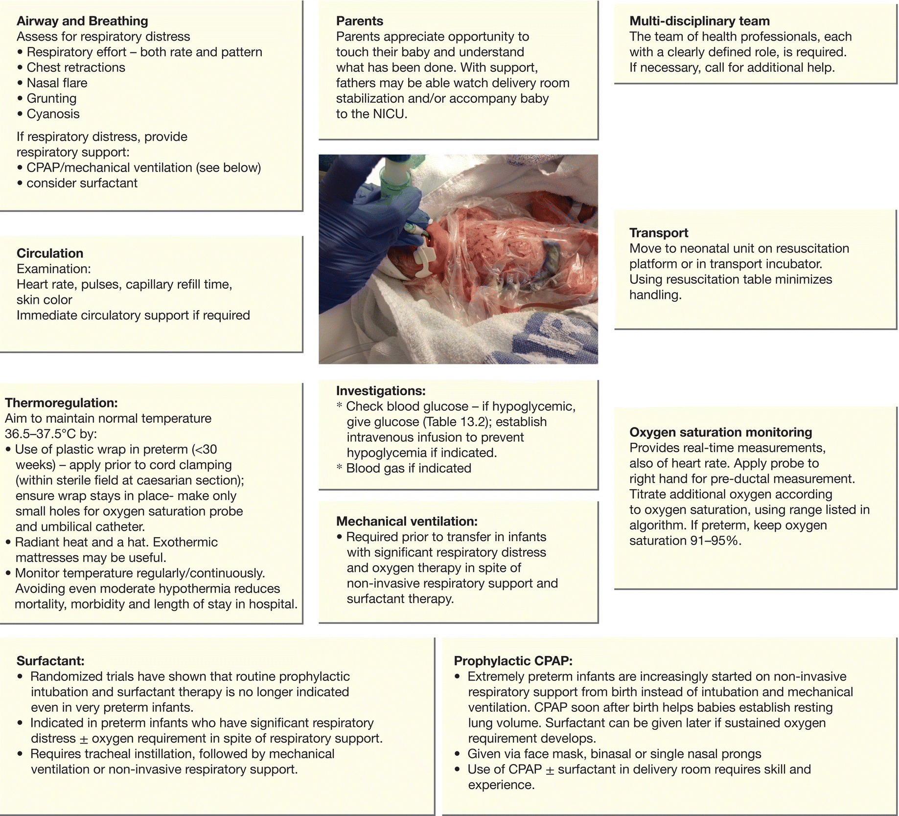
Thermoregulation is critical as hypothermia contributes to hypoglycemia, acidosis and even mortality, especially in extremely preterm infants (see video: Resuscitation of preterm).
Action
- Resuscitation area should be warm and draft free.
- Perform resuscitation under radiant warmer.
- Dry infant, remove wet towel, then wrap in dry towel.
- For preterm infants (<30 weeks), place wet infant directly in plastic wrap (bag) with only face exposed. Cover head with hat. An exothermic mattress may also be used.
Initial assessment at birth
Start the clock or note the time.
Is the baby:
- term gestation?
- crying or breathing vigorously?
- good muscle tone?
If this is the case, the baby should dried, placed skin-to-skin with the mother if desired and covered with dry linen to maintain temperature.
If not, assess breathing (rate, depth and symmetry), heart rate (stethoscope on apex), color and tone and initiate resuscitation as required. Drying often produces enough stimulation to induce breathing.
A – Airway
If gasping or not breathing – open airway:
- Position – optimize head position, avoiding head overextension or flexion, using a towel beneath the shoulders if desired.
- Suction – only if obvious obstruction (e.g. blood, meconium) and unable to achieve chest rise. Suction should be under direct vision with a wide-bore suction device (bulb syringe or Yankauer). Tracheal suctioning of meconium-stained fluid is only indicated for depressed, hypotonic babies. Care is required as pharyngeal suctioning may cause laryngeal spasm and vagal bradycardia. Suctioning meconium from the oropharynx at the perineum, routinely practiced for many years, does not improve outcome.
- For congenital airway abnormalities – choanal atresia, severe micrognathia (small jaw) or macroglossia (large tongue) – consider using an oral airway or laryngeal mask airway.
B – Breathing
Assessment
- Look for chest movement – check if inadequate or absent or if heart rate is <100 beats/min.
- Attach oxygen saturation monitor to right hand if resuscitation is anticipated, if baby remains cyanotic or if positive-pressure ventilation or supplementary oxygen required. Oxygen saturation monitors provide continuous reading of heart rate and oxygen saturation but take time to apply and may not function if poor perfusion. In healthy term infants following vaginal birth, it takes about 10 minutes for preductal oxygen saturation to reach levels >90%.
Action
If inadequate respiratory effort, gasping or not breathing or heart rate is <100 beats/min, the priority is to inflate the lungs with mask ventilation (Fig. 13.1), using a T-piece pressure device or self-inflating bag (Fig. 13.2).
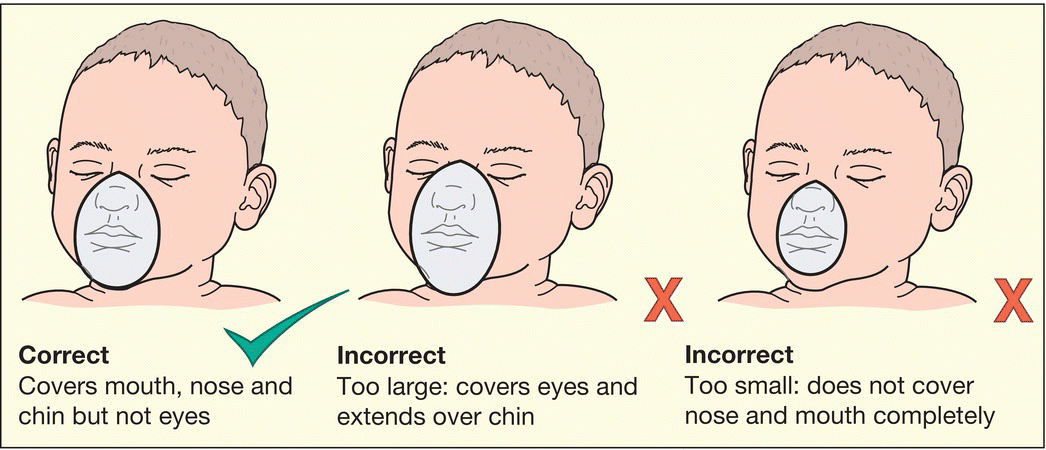
Fig. 13.1 Correct size and position of face mask. It should cover the mouth, nose and chin.
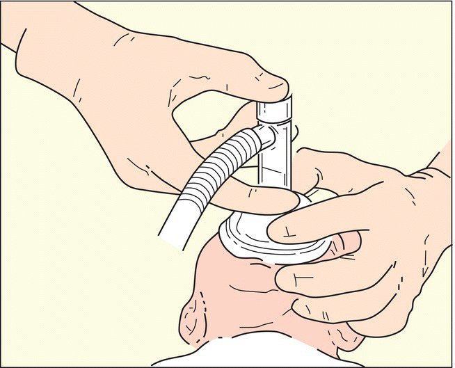
Fig. 13.2 Mask ventilation via a T-piece connected to air/oxygen blender from a pressure-limited circuit.
Mask ventilation
Most infants will respond to lung inflation.
- Term infants – start with air. Titrate additional oxygen according to oxygen saturation level.
- Preterm infants – as above but use air/oxygen blender and pulse oximetry.
Use minimum oxygen required, according to preductal saturation targets in first 10 minutes of life. Avoid oxygen saturation above target range 91–95% as hyperoxemia can be damaging to eyes, lungs and brain.
Achieving lung inflation
The aim is good chest rise using the lowest pressure.
Assess regularly, observing for:
- Increasing heart rate to >100 beats/min.
- Improving color (or saturations).
- Spontaneous breathing.
- If lung inflation is not achieved and the heart rate is not responding, further airway maneuvers may be required. Recheck the airway position and consider suction (if obstruction seen), increased peak pressure and inspiratory time, airway adjuncts (Guedel airway or laryngeal mask airway) or endotracheal intubation.
Endotracheal intubation (see Chapter 74)
Indications
- If effective mask ventilation is not sustaining adequate ventilation (but only when all the basic measures described above have been checked).
- Consider in all babies who require cardiac compressions to maintain the airway.
- If prolonged ventilation is needed.
- Congenital diaphragmatic hernia and some congenital upper airway abnormalities.
- If direct tracheal suction is needed for thick meconium or other obstruction in depressed, hypotonic infant.
However:
- Should only be attempted by those with advanced airway skills.
- Limit each attempt to <30 seconds.
- Confirm tube in trachea with symmetrical air entry and end-tidal CO2 detector.
- Provide effective mask ventilation between attempts if necessary.If no response to intubation consider DOPE:
- Displaced tube:
- – Is it in oesophagus? (no chest movement, air entry over stomach greater than chest, no CO2 detected)
- – Is it in one (usually right) main bronchus?- asymmetrical chest rise or air entry, CO2 may still be detected.
- Obstructed tracheal tube (especially meconium) – aspirate or replace tube, or tube too small for baby
- Patient:
- – Lung disorders i.e. lung immaturity, surfactant deficiency, pneumothorax, diaphragmatic hernia, lung hypoplasia, pleural effusion
- – Shock from blood loss, anemia
- – Neonatal encephalopathy – perinatal asphyxia, head trauma, CNS hemorrhage or abnormality
- Equipment failure-exhausted or disconnected gas supply.
C – Circulation
- Assess heart rate regularly with stethoscope or feel pulse at base of the umbilical cord (or observe heart rate on saturation monitor, if connected).
- Start chest compressions (Fig. 13.3) if heart rate <60 beats/min despite adequate ventilation.
- Call for help – if not already available.
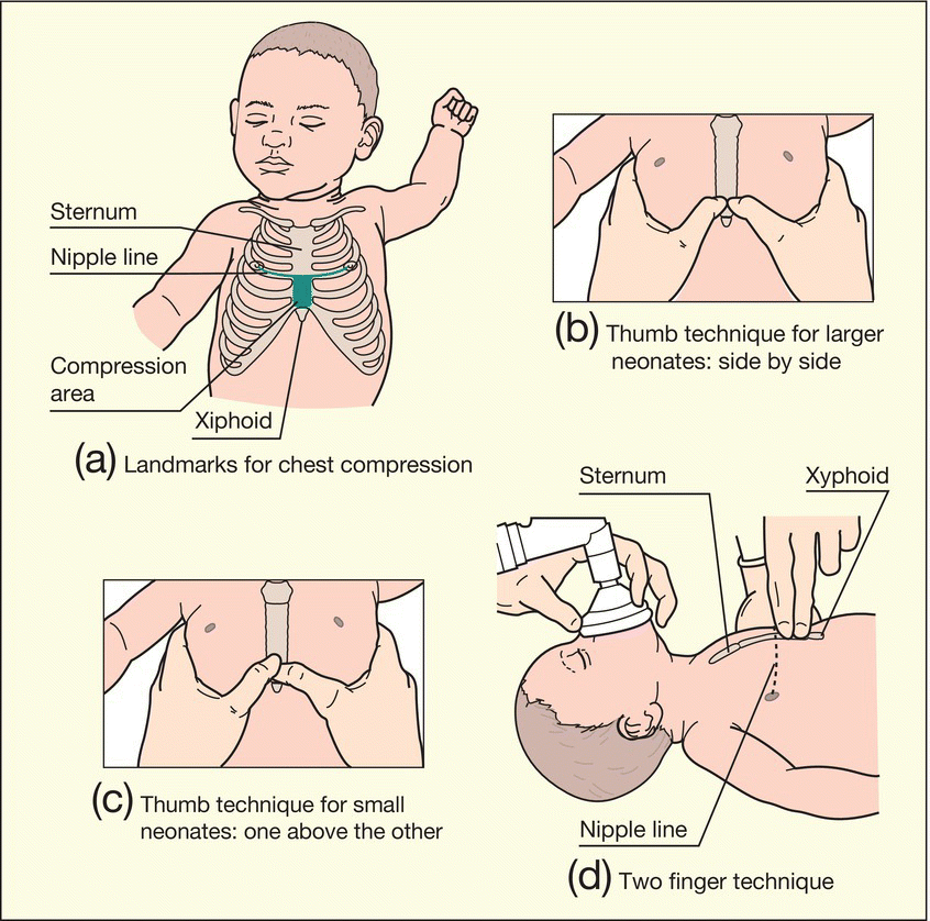
Fig. 13.3 (a) Apply pressure to lower third of sternum, just below an imaginary line joining the nipples. Avoid the xiphoid. Depress to reduce anteroposterior diameter of the chest by one-third with no bounce. The thumb technique (b and c) is more effective than the two-finger technique and is recommended, but the two-finger technique (d) is easier if you are alone.
There should be a 3:1 ratio of compressions to breaths (approximately 90 compressions and 30 breaths/minute) to maximize ventilation at an achievable rate. Compressions and ventilations should be coordinated to avoid simultaneous delivery.
Drugs (Table 13.1)
Table 13.1 Resuscitation drugs
| Medication | Concentration | Dosage/route | Indications |
| Epinephrine (adrenaline) | 1 in 10 000 (0.1 mg/mL) |
IV: 0.1 to 0.3 ml/kg Endotracheal: 0.5–1 ml/kg |
No response in spite of visible lung inflation and cardiac compressions |
| Volume expander |
Normal saline Whole blood |
10 mL/kg IV over 5–10 min | Suspected acute blood loss and/or signs of hypovolemia |
| Sodium bicarbonate | 0.5 mEq/mL (0.5 mmol/mL) |
1–2 mEq/kg (1–2 mmol/kg) (2–4 mL/kg of 4.2% solution) |
Consider only after prolonged arrest in spite of effective ventilation |
| Glucose | 10% | 2–2.5 mL/kg (250 mg/kg) IV | For documented hypoglycemia |
Drugs are rarely indicated; only use if no response in spite of:
- visible lung inflation
- cardiac compressions for at least 30 seconds.
Epinephrine (adrenaline) administered intravenously via umbilical venous catheter is recommended. Whilst intravenous access is obtained, administration in higher dose via endotracheal tube may be considered, but its efficacy has not been established.
Volume expansion with saline or blood may be indicated when blood loss is suspected e.g. pale infant, history of hemorrhage.
The role of sodium bicarbonate is controversial. It may be considered in prolonged resuscitation to correct metabolic acidosis, but may potentially be harmful as it may exacerbate intracellular acidosis if breathing is inadequate.
Prevention of hypoglycemia should be considered following prolonged resuscitation.
Drugs should be flushed with a bolus of saline.
Withholding and discontinuing resuscitation
Difficult decisions arise at the margins of viability and for infants with conditions that have unacceptably high morbidity or almost certain early mortality. Attitudes and practices vary widely between practitioners, institutions and countries. Whenever possible, decisions should be made with parental agreement.
Discontinuation of resuscitation should be considered if the heart rate is undetectable at birth and remains so for 10 minutes despite full resuscitation. If there is doubt, rather than hasty decisions in the delivery room, it is often best to transfer to the neonatal unit for detailed assessment.
