55
Neutrophil and thrombotic disorders
Thrombotic disorders (thrombophilia)
These are a group of disorders characterized by an increased tendency for abnormal clot formation. Thrombosis occurs in approximately 5 per 100 000 births; 50% of episodes are arterial and 50% are venous.
Predisposing factors
These are:
- indwelling catheters (80–90% of episodes)
- acute bacterial and viral infection
- asphyxia (ischemia), shock
- cardiac abnormality
- polycythemia
- prematurity
- twin–twin transfusion
- genetic.
Maternal and familial conditions associated with thrombophilia
These include:
- multiple fetal losses
- anticardiolipin antibodies
- SLE (systemic lupus erythematosus)
- maternal diabetes
- placental abruption
- myocardial infarction
- deep venous thrombosis
- pulmonary embolism.
Inherited causes of thrombosis
Gene mutations have been identified for some of the most common thrombotic disorders:
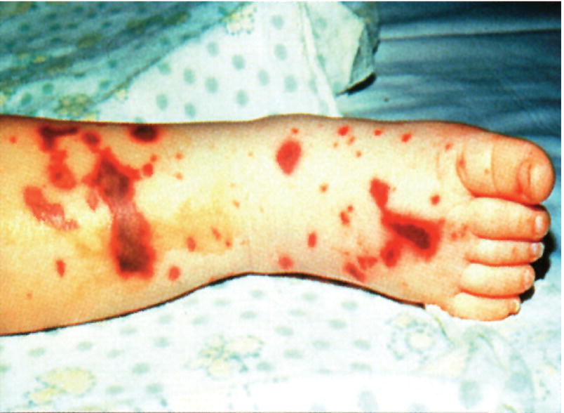
- protein C deficiency (Fig. 55.3)
- protein S deficiency
- antithrombin deficiency
- factor V Leiden mutation (APC resistance)
- prothrombin gene mutation.

Fig. 55.3 Infant with microthrombi in the skin from protein C deficiency.
Diagnosis
Most thrombi are asymptomatic.
Clinical signs of thrombosis depend on location of the clot, which may embolize:
- Arterial – limb may become mottled in color, cool and discolored with reduced pulses. In time may become gangrenous with zone of demarcation (see Fig. 66.5). Thrombus in aorta may lead to heart failure or stroke.
- Venous – portal vein, renal vein thrombosis causing abdominal mass, hematuria, oliguria and hypertension. Thrombus in right atrium may lead to stroke.
Imaging
Depends on site:
- Ultrasound, echocardiography, MRI for diagnosis and follow-up.
- Angiography is the gold standard but may become difficult or impossible to perform or not justified, e.g. for stroke. MR angiography is now available.
Management
Options include:
- If catheter-related, may be due to arterial spasm or too large a catheter or hypovolemia. If does not respond promptly to partial withdrawal of the catheter or correction of hypovolemia, the catheter should be removed.
- Observe and follow up for increase in clot size and functional compromise.
- Anticoagulation with unfractionated or low molecular weight heparin (e.g. enoxaparin).
- Clot lysis with fibrinolytic agents (tissue plasminogen activator), but contraindicated if there has been a recent intraventricular hemorrhage.
- Surgical thrombectomy – rarely required or possible.
- Factor concentrate if thrombosis and inherited deficiency (e.g. protein C, antithrombin).