Figure 14.1 The excavation site at the churchyard in Longyearbyen, Spitsbergen, an island off the coast of Norway where seven young men who had died of Spanish influenza were buried in the permafrost.
What was so special about the 1918 Spanish influenza virus, which killed more people than the two world wars combined? This was a hugely important unanswered question in the 1980s. We needed to get hold of that virus so we could find out its secrets. Unfortunately, since the influenza virus was not isolated until the 1930s, no samples were saved during the 1918 pandemic. Our only hope lay in tissue samples that had been saved from soldiers or influenza patients and were preserved in formalin. These display jars were located in various departments of pathology and in pathological museums. Another possible source of samples was from sufferers who had died in the Arctic regions and were buried in the permafrost. Sixty years later, could we find samples of the 1918 influenza virus in either of these unlikely places?
At scientific meetings in the 1980s, the team at St Jude Children’s Research Hospital asked colleagues if they knew of any pathology departments that might have formalin-preserved lung or other tissue from people who had been diagnosed with Spanish influenza. We heard that the US Armed Forces Institute of Pathology in Washington, DC, had a large collection of such tissues from young soldiers who had died in military camps during the peak of the 1918 pandemic. I immediately wrote to Douglas Weir at that institute and proposed a joint study to try to find out why the 1918 influenza virus had been so severe. We knew we would not obtain live virus from the samples, because formalin is also used to kill influenza viruses during the preparation of vaccines. However, we wanted to determine the virus’s genetic code, and we were hopeful that the chemicals making up the genetic material of viruses would be sufficiently well preserved for analysis.
We were overjoyed with Weir’s immediate and affirmative reply. On 2 February 1990 we received a shipment of formalin-fixed lung samples from nine known victims of 1918 Spanish influenza. Since this was precious material, we developed and then fine-tuned our methods on formalin-fixed lung and respiratory tract tissues of mice and ferrets that had been infected with influenza viruses known to cause severe and lethal disease. Once we were satisfied with our methods, we turned eagerly to the nine human lung samples provided by Weir.
But although the animal tissues had yielded tiny portions of influenza virus genetic code, results from the human tissues were disappointing: we found very little genetic code that we could attribute to influenza. It seemed as if the molecules making up the genetic code of influenza virus had broken apart during their almost 70 years in formalin.
Although our initial foray into this area was frustrating, we were delighted several years later to learn that we had not been barking up the wrong tree. Jeffery Taubenberger, from the Armed Forces Institute of Pathology, had investigated lung tissues from 1918 influenza victims that had been fixed in formalin and then embedded in paraffin blocks for preparation of histological sections. These tissues, which had not been exposed to formalin for as long as the tissues we had analysed, provided the first bits of the genetic code of the 1918 Spanish influenza virus. This groundbreaking work was published in the prestigious journal Science.82
Now Taubenberger’s group had another problem. While the tissue blocks yielded information about the shorter pieces of the influenza virus’s genome, our knowledge of the larger pieces of the genome was still incomplete – and they were running out of the lung tissue in the paraffin blocks.
Meanwhile, a team of scientists led by Kirsty Duncan from the University of Windsor, Canada, was attempting to obtain tissue samples of young men who had died of 1918 Spanish influenza en route to the coal mines on Spitsbergen, a Norwegian island approximately a thousand kilometres north of mainland Norway. Each summer the coal mine owners would recruit strong young men in Tromsø, a town on the mainland. The men could earn enough money in one year of mining to buy a small farm, so the competition for positions was fierce.
Duncan found records indicating that seven young men aged 19 to 28 years had been infected while on the ship to Spitsbergen and died of severe influenza shortly after they arrived. They were buried in the churchyard in Longyearbyen, Spitsbergen, on 27 October 1918.83 Duncan obtained all the necessary permissions to exhume the bodies and collect tissue samples. The international team she assembled comprised geologists, archaeologists, forensic pathologists, physicians and influenza scientists, including myself and other senior influenza virologists from the WHO network.84 It took a very long time to organise such a complex expedition – six years from inception in 1992 to beginning the exhumation in 1998.
The initial question was whether the crosses in the churchyard cemetery marked the actual graves of the miners, for during World War II the town had suffered extensive bombardment, and most of its structures had been destroyed. To search for burial sites, Duncan’s team used ground-penetrating radar that would detect any ground disturbances and their depth. Ground disturbances down to two metres were found for all seven marked graves. Since the active layer in permafrost melts each summer and re-freezes, to a depth of 0.8–1 metre, these findings suggested that the bodies had been frozen in situ for 79 years.
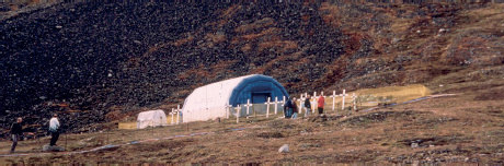
Figure 14.1 The excavation site at the churchyard in Longyearbyen, Spitsbergen, an island off the coast of Norway where seven young men who had died of Spanish influenza were buried in the permafrost.
This raised the serious issue of whether live 1918 influenza virus might be released if we indeed found permanently frozen tissues containing the virus. While the scientists agreed that it would be extremely unlikely, none of them could say it was impossible. An already complicated expedition now had to factor in biosafety and biosecurity measures to protect all personnel and the environment.
The work area in the churchyard was entirely covered by an inflatable mobile surgical unit equipped with chemical and decontamination showers, and everyone wore masks and protective clothing. The Longyearbyen locals undoubtedly thought we were a group of mad scientists as they watched several freezers and a shipping container full of assorted equipment being dragged up the hillside (Figure 14.1).85
As an additional precaution, we had a supply of the anti-influenza drug Tamiflu on hand in case anyone was exposed during tissue collection. Excavations began, and almost immediately the person digging broke into the first coffin. In keeping with the planned safety measures, he was given Tamiflu right away, but the next morning he complained of severe stomach pain and nausea. Had he released something from the coffin? It seemed highly unlikely, and sure enough he was soon back to normal.
In the event, all our careful planning and safety precautions turned out to be quite unnecessary. The seven coffins were in fact buried in shallow graves, in the active layer of the permafrost, and had been repeatedly frozen and thawed for 79 years. Only the men’s skeletons, brain tissue and bone marrow remained, and later analysis of the latter two failed to provide the genetic information we sought. Some of the bodies were wrapped in newspaper, and the dates on the pages coincided with the dates of burial. It seems that the ground-penetrating radar had failed to detect the wooden coffins and the remains, and the ground disturbance two metres down had probably been caused by blasting powder used by the gravediggers to loosen the permafrost.
Although the immaculately planned and executed Spitsbergen expedition came up empty, Taubenberger soon received his much-needed samples – from a completely unexpected source. After publishing his paper on the partial genetic sequence of the 1918 Spanish influenza virus, he received a letter from Johan Hultin, a retired San Francisco physician, asking him if he would like to receive additional tissue from Alaskan victims of the 1918 influenza that were buried in permafrost. Taubenberger could not believe his good fortune, and was delighted when Hultin offered to go to Alaska the following week to obtain the material.
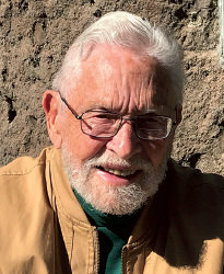
Figure 14.2 Johan Hultin obtained the samples from Brevig Mission that permitted Jeffery Taubenberger to complete the sequence of the 1918 influenza virus.
Hultin, it transpired, was driven by a passion similar to Taubenberger’s own: to understand why the 1918 influenza had killed so many young people so quickly. Forty-six years earlier Hultin, while a graduate student at Iowa State University, had been part of an expedition to Alaska to isolate the 1918 influenza virus. Back in June 1951 Hultin, Robert McKee and Jack Layton, all scientists from Iowa State, had flown to Alaska, where they were joined by Otto Geist, a palaeontologist from the University of Alaska, to exhume bodies at Brevig Mission on Seward Peninsula. Samples of lungs were recovered from 1918 victims buried deep in the permafrost. These samples were taken frozen to the university, and Hultin had been sure they would succeed in isolating the influenza virus. But it was not to be. The team completely failed in their attempts to grow the virus in chicken embryos (Figure 14.2).86
The 1918 influenza outbreak in this Inuit fishing village is a classic example of just how deadly the 1918 Spanish influenza could be in an isolated community. Hultin told me that in November 1918 a quarantine was in effect in Alaska for all ships with influenza cases on board. The mail ship had stopped at Nome without any known cases of influenza and dropped off mail. But someone on board must have been in the early stages of developing influenza, because the dogsled driver on his 100km drive to the Brevig Mission region was found comatose and later died of severe influenza. He brought the monster virus to the region and at Brevig Mission 72 of the 80 people died. The survivors were mostly children; there were reports of children being found alive in homes where the starving dogs had begun eating the dead parents (Figure 14.3).87
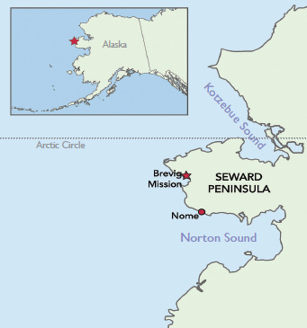
Figure 14.3 A map showing the location of Brevig Mission and Nome in Alaska. On 10 November 1918 the sled-dog musher brought Spanish influenza to the region. Five days later, 72 of the 80 inhabitants of Brevig Mission were dead; remarkably, children survived.
In 1997 Hultin still knew exactly where to find the bodies buried in a mass grave at Brevig Mission. One week after agreeing to obtain samples for Taubenberger, he packed his wife’s pruning shears (his only equipment) and flew to Alaska. He met with the village matriarch, who knew that Hultin had previously exhumed bodies, then met the mayor and obtained permission to reopen the mass grave. The village provided four young men to help with the digging.
They dug over two metres deep in the permafrost and located the bodies as expected. From an obese woman in her mid-20s dubbed Lucy, they obtained samples from the optimally frozen lungs, which were filled with blood. They also collected lung samples from less well-preserved bodies. All of the samples were placed in a solution that would kill influenza virus but preserve genetic material (guanidinium thiocyanate). After closing the graves, Hultin built two crosses – one 3.3 metres tall and the other 2.1 metres – to mark the graves and honour the people interred. After returning to San Francisco he divided the samples into several insulated boxes and sent them separately for safety: one by Federal Express, one by the United States Postal Service and one by United Parcel Service. Taubenberger received them all, and the samples from Lucy provided all the material needed for his group to complete the genetic sequence of the entire 1918 Spanish influenza virus.88
It was inevitable that comparisons would be made between the successful expedition to Brevig Mission and the unsuccessful expedition to Spitsbergen. In any research project, there is a certain amount of luck involved. At their outset, both expeditions held equal promise. At Brevig Mission the bodies were buried deep in the permafrost, while the Spitsbergen bodies were not. Perhaps the most useful information from the Spitsbergen expedition was that giving Tamiflu on an empty stomach can cause severe stomach ache!
So did finding the entire genetic sequence of the 1918 Spanish influenza virus provide all the answers as to why it was so deadly? Unfortunately the answer is no. The genetic code of the virus on its own was not enough.