When is an ultrasound indicated in prenatal care?
A 22-year-old woman who has never been pregnant before presents to you after having a positive home pregnancy test. She has no significant medical history. Upon further questioning, she states that she is unsure of the date of her last menstrual period. She denies any symptoms and is worried as she has not felt the baby move thus far. She is also concerned as she recently had dental x-rays taken prior to discovering that she was pregnant. Patient denies the use of any drugs, alcohol, or tobacco. She inquires about when she can get an ultrasound and a genetic test to rule out Down syndrome.
When is an ultrasound indicated in prenatal care?
What laboratory studies are routinely indicated at an initial prenatal visit?
What is the risk to the pregnancy based on the radiation exposure that the patient has encountered?
When is the optimal time for screening with a trisomy screen test?
Summary: A 22-year-old primigravida woman with no significant past medical history presents for initial prenatal care visit. She has numerous questions regarding her care and recently has had dental x-rays taken.
• Indications for an ultrasound in pregnancy: According to the American College of Obstetricians and Gynecologists (ACOG), an ultrasound is not mandatory in routine, low-risk prenatal care. An ultrasound is indicated for the evaluation of uncertain gestational age, size/date discrepancies, vaginal bleeding, multiple gestations, or other high-risk situations.
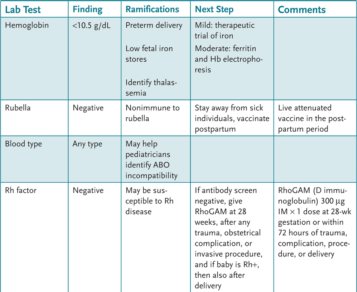
• Laboratory studies recommended at the initial prenatal visit: Complete blood count (CBC), hepatitis B surface antigen (HBsAg), HIV testing, syphilis screening with a rapid plasma reagin (RPR), urinalysis and urine culture, rubella antibody, blood type and Rh status with antibody screen, Papanicolaou (Pap) smear, and cervical swab for gonorrhea and Chlamydia.
• Risk to the pregnancy based on the radiation exposure from dental x-rays: Risk for the baby is increased once the radiation exposure is greater than 5 rad; the radiation exposure from routine dental x-rays is 0.00017 rad.
• The optimal time for the trisomy screen: The options for trisomy screening include: first trimester testing for nuchal translucency (NT) via ultrasound or a combined test of NT and serum markers hCG and pregnancy-associated plasma protein A (PAPP-A) testing between 10 and 13 weeks and second trimester triple (AFP, hCG, estriol) or quadruple (triple screen and inhibin-A) screen between 16- and 18-week gestation; however, it may be performed between 15-and 20-week gestation, if necessary. Emerging evidence shows that combining results of first- and second-trimester screening tests improves trisomy detection rate; consequently, the optimal time for screening should be discussed at initial prenatal visit.
1. Learn the components of the preconception counseling and the initial prenatal visit.
2. Know the recommended screening tests and visit intervals in routine prenatal care.
3. Learn the relevant psychosocial aspects of providing prenatal care, including important counseling issues.
Prenatal, or antenatal, care affords the opportunity to both perform appropriate medical testing and provide counseling and anticipatory guidance. Pregnancy can be a time of anxiety and patients frequently have many questions. One of the goals of prenatal care is to provide appropriate education in order to help reduce anxiety and help women to be active participants in their own care.
ADVANCED MATERNAL AGE: Pregnant woman who will be 35 years or beyond at the estimated date of delivery (EDD).
ISOIMMUNIZATION: The development of specific antibodies as a result of antigenic stimulation by material from the red blood cells of another individual. For example, Rh isoimmunization means a Rh-negative woman who develops anti-D (Rh factor) antibodies in response to exposure to Rh (D) antigen.
ASYMPTOMATIC BACTERIURIA (ASB): 100,000 cfu/mL or more of a pure pathogen of a mid-stream voided specimen without clinical symptoms. ASB in pregnant women increase risk of acute pyelonephritis, preterm delivery, and low birth weight; therefore, early detection is paramount and treatment is mandated.
GENETIC COUNSELING: An educational process provided by a health-care professional for individuals and families who have a genetic disease or who are at risk for such a disease. It is designed to provide patients and their families with information about their condition or potential condition and help them make informed decisions.
VERTICAL TRANSMISSION: Infectious passage of infection from mother to fetus, whether in utero, during labor and delivery, or postpartum.
ANTENATAL TESTING: A procedure that attempts to identify whether the fetus is at risk for uteroplacental insufficiency and perinatal death. Some of these tests include nonstress test and biophysical profile.
In the United States, the first visit for prenatal care frequently is at 8 weeks of gestation or later, and yet it is the time preceding this that poses the greatest risk to fetal development. A preconception visit is an ideal opportunity for the patient to discuss with her physician any issue related to possible pregnancy or contraception occurring within 1 year of pregnancy. The preconception visit can be included during visits for many reasons, including fertility problems, contraception, periodic health assessment, recent amenorrhea, or specifically for preconception counseling. Roughly one-half of patients with a negative pregnancy test may have some risk that could adversely affect a future pregnancy. Because roughly 50% of pregnancies are unplanned or unintended, physicians should consider the potential of pregnancy when writing each prescription.
Women who intend to become pregnant should be advised to avoid, whenever possible, potentially harmful agents such as radiation, drugs, alcohol, tobacco, over-the-counter (OTC) medications, herbs, and other environmental agents. Radiation exposure greater than 5 rad is associated with fetal harm. Most commonly performed x-ray procedures, including dental, chest, and extremity x-rays, expose a fetus to only very small fractions of this amount of radiation. Fetuses are particularly sensitive to radiation during the early stages of development, between 2 and 15 weeks after conception. Whenever possible, the abdomen and pelvis should be shielded and x-rays performed only when the benefit outweighs the potential risk. Magnetic resonance imaging studies have not been proven to cause harm, but are not recommended in pregnancy, if avoidable. Ultrasound has not been shown to be harmful.
Women should refrain from OTC medicines, herbs, vitamins, minerals, and nutritional products until cleared by their obstetric provider. They should also be instructed to start a folic acid supplement at least 1 month prior to attempting to conceive. For low-risk women, 400 to 800 lg of folic acid daily is recommended to reduce the risk of neural tube defects. Higher doses are recommended in the presence of certain risk factors. For women with diabetes mellitus or epilepsy, 1 mg of folic acid a day is recommended. A woman who has had a child with a neural tube defect should take 4 mg of folic acid daily.
Women from certain ethnic backgrounds may be offered specific genetic screening. African and African-American women may be offered sickle cell trait screening. A French-Canadian or Ashkenazi Jewish background is an indication to consider screening for a Tay-Sachs carrier state. Southeast Asian and Middle Eastern women may be offered screening for thalassemia. Ashkenazi Jews and Caucasian women may be offered screening for cystic fibrosis.
Women who will be 35 years old or older at the anticipated time of delivery should be educated about age-related risk, particularly the increased risk of Down syndrome. They should be counseled about the available screening and diagnostic testing available, along with the appropriate time frame in which each test may be performed.
Women with medical conditions such as diabetes, asthma, thyroid disease, hypertension, lupus, thromboembolism, and seizures should be referred to providers with experience in managing high-risk pregnancies. Women with psychiatric disorders should be comanaged with a psychiatrist and counselor/therapist so that the patient can benefit from pharmacologic and behavioral therapy. These patients may require more frequent visits. Patients who have drug, tobacco, or alcohol dependence should be educated about the risks and referred to rehab/treatment centers to quit the drug prior to conception. Women should also be educated about proper nutrition and exercise during pregnancy. Preconception counseling may also address issues such as financial readiness, social support during pregnancy and the postpartum period, and issues of domestic violence.
The initial visit should address all the concepts in the preconception visit, if no preconception counseling was done. Ideally, the initial visit should be in the first trimester. A detailed history and physical examination, initial obstetric laboratory tests, and counseling regarding the logistics for prenatal care should be done at this visit. The history should begin with an assessment of the last menstrual period (LMP) and its reliability. One of the most crucial pieces of information is the accuracy of the dating. The first day of the LMP is used to obtain the estimated delivery date (EDD) using Naegele’s rule (from the first day of the LMP subtract 3 months and add 7 days). The LMP is considered reliable if the following criteria are met: the date is certain, the last menstrual period was normal, there has been no contraceptive use in the past 1 year, the patient has had no bleeding since the LMP, and her menses are regular. If these criteria are not met, an ultrasound should be performed. ACOG has established further criteria that can be used to ensure that a fetus is mature at the time of delivery, which include criteria such as early sonography and the timing of the positive pregnancy test.
History should also be obtained with particular attention to medical history, prior pregnancies, delivery outcomes, pregnancy complications, neonatal complications, and birth weights. Gynecologic history should focus on the menstrual history, contraceptive use, and history of sexually transmitted diseases (STDs). Allergies, current medications—both prescription and OTC—and substance use should also be investigated. Social history should consider whether the pregnancy was planned, unplanned, or unintentional. A discussion of social supports for the patient during the prenatal and postpartum period is also warranted. Genetic history should be obtained for the patient and partner’s family, if known.
The initial examination should be thorough and should assess height, weight, blood pressure, thyroid, breast, and general physical and pelvic examinations. Pregnancy-specific examinations, including an estimation of gestational age by uterine size or fundal height measurement and an attempt to hear fetal heart tones by Doppler fetoscope should be performed. Heart tones should be obtainable by 10-week gestation using a handheld Doppler fetoscope. Pelvimetry has been removed as a recommended required intervention, but it may be useful to have a subjective assessment for risks of problems during delivery.
The initial laboratory screen (Table 4–1) should include blood type and Rh status antibody screen, rubella status, HIV, hepatitis B surface antigen, rapid plasma reagin (RPR), urinalysis, urine culture, Pap smear, cervical swab for gonorrhea and Chlamydia, and a CBC.

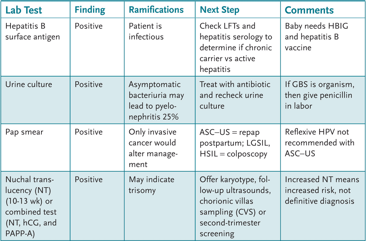
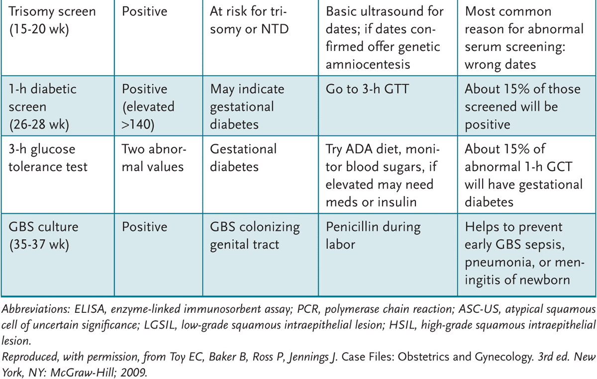
Table 4–1 • SUMMARY OF PRENATAL LABORATORIES, RAMIFICATIONS, AND EVALUATION
The logistics of the prenatal visits should be addressed. A typical protocol includes follow-up visits every 4 weeks until 28-week gestation, every 2 weeks from 28- to 36-week gestation, and every week from 36-week gestation until delivery. More frequent visits should be performed if any problems arise or if all issues are not addressed in the scheduled visits.
The ACOG does not stipulate routine ultrasonography in patients without complications. Ultrasound is considered accurate for establishing gestational age, fetal number, viability, and placental location. Therefore, ultrasonography should be performed in patients without reliable dating criteria, with a discrepancy between the measured and expected uterine growth, and in case of a postdated pregnancy, suspicion for twin gestation, suspicion for placental issues, chromosomal abnormalities, or other problems. For gestational-age estimations, ultrasonography is accurate to within 1 week if performed in the first trimester, 2 weeks in the second trimester, and 3 weeks in the third trimester. If the ultrasound dates and LMP are off by more than the aforementioned intervals, the due date should be recalculated based on the ultrasound findings.
The visit should end with an adequate explanation of all patient/partner concerns. Women should be counseled that sexual activity is not associated with any harm during an uncomplicated pregnancy, although there may be conditions that arise during the course of a pregnancy that would make sexual activity inadvisable. A follow-up visit should be scheduled prior to her leaving the office. She should also be educated about preterm labor precautions, signs of ectopic pregnancy, and situations in which to call the physician or go to the obstetrics triage unit for evaluation.
At follow-up prenatal visits, concerns or questions brought up by the patient should be addressed. The examiner should ask questions specifically targeted at symptoms suggestive of complications that include gestational hypertension, preeclampsia, infections (urinary tract, vaginal, etc), fetal compromise, placenta previa/abruption, and preterm labor or premature rupture of membranes. At each visit, the patient should be asked about vaginal bleeding, loss of fluid, headaches, visual changes, abdominal pain, dysuria, facial or upper-extremity edema, vaginal discharge, and subjective sensation of fetal movements.
The examination on each subsequent visit should include weight, blood pressure, fundal height measurement, and fetal heart tones by handheld Doppler. In addition, a urinalysis should be performed at every visit to assess for protein, glucose, or infection.
At 15 to 20 weeks’ gestation (preferably between 16 and 18 weeks’ gestation), a multiple marker test, which screens for Trisomy 21, Trisomy 18, and neural tube defects, should be offered to patients. The two most common modalities of screening the fetus for these anomalies are the triple screen and the quad screen. The triple screen tests for serum human chorionic gonadotropin (hCG), unconjugated estriol, and α-fetoprotein; the quad screen tests for those three-plus inhibin-A. The triple screen has a sensitivity of approximately 65% to 69% and specificity of 95% for detecting aneuploidy. The quad screen increases sensitivity to approximately 80% without reducing specificity. The most common cause for a false-positive serum screen is incorrect gestational age dating. During the first trimester, fetal nuchal translucency can be measured by ultrasonography combined with maternal serum analyte levels (ie, free hCG and pregnancy-associated plasma protein A [PAPP-A]). This testing can be performed at 10 to 14 weeks’ gestation. Sensitivity and specificity of these tests is determined by the risk cutoff used (eg, for Trisomy 21, sensitivity is 85.2% when specificity is 90.6%; at 95% specificity, the sensitivity is 78.7%). Women should be counseled about the limited sensitivity and specificity of the tests, the psychological implications of a positive test, the potential impact of delivering a child with Down syndrome, the risks associated with prenatal diagnosis and second-trimester abortion, and delays inherent in the process.
Women at increased risk of aneuploidy should be offered prenatal diagnosis by amniocentesis or chorionic villus sampling (CVS). Persons at increased risk include women who will be older than 35 years at delivery and who have a singleton pregnancy (older than 32 years at delivery for women pregnant with twins); women carrying a fetus with a major structural anomaly identified by ultrasonography; women with ultrasound markers of aneuploidy (including increased nuchal thickness); women with a previously affected pregnancy; couples with a known translocation, chromosome inversion, or aneuploidy; and women with a positive maternal serum screen. Amniocentesis may be performed after 15-week gestation and is associated with a 0.5% risk of spontaneous abortion. CVS is performed at 10- to 12-week gestation and has a 1% to 1.5% risk of spontaneous abortion. CVS may be associated with transverse limb defects (1 per 3000 to 1 per 1000 fetuses). Women undergoing CVS also should be offered maternal serum α-fetoprotein testing for neural tube defects. Women older than age 35 years at time of delivery may opt for serum screening and ultrasonography before deciding whether to proceed with amniocentesis. Although the risk for trisomy 21 increases with maternal age, an estimated 75% of affected fetuses are born to mothers younger than age 35 years at time of delivery.
The ACOG and the American Diabetes Association recommend that all pregnant women be screened for gestational diabetes at 24 to 28 weeks’ gestation, except women who are at low risk (eg, younger than age 25 years, belonging to a low-risk ethnic group, normal prepregnancy weight, no history of abnormal glucose metabolism, no previous poor obstetric outcomes, and no first-degree relatives with diabetes). Screening is standard in the United States, with 94% of physicians reporting universal screening.
At 24 to 28 weeks’ gestation, patients should be screened for gestational diabetes with a 1-hour 50-g glucose challenge test. Most guidelines consider a value above 140 mg/dL as abnormal, whereas new studies advocate using a value of 135 mg/dL. A value of 200 mg/dL or greater is generally diagnostic of gestational diabetes. When the screening test is positive, a 3-hour glucose tolerance test (GTT) should be performed (after an overnight fast) by giving the patient a 100-g glucose load and obtaining fasting, 1-hour, 2-hour, and 3-hour postload serum glucose samples; two out of four positive values generally establish the diagnosis of gestational diabetes. A diagnosis of gestational diabetes impacts the pregnancy, but also increases the risk of type II diabetes in the patient throughout her life.
At 28 weeks’ gestation, a repeat RPR and hemoglobin/hematocrit should be obtained in those at risk for syphilis and anemia, respectively. In addition, a patient who is Rh-negative should receive Rho(D) immune globulin (RhoGAM) at this time. An Rh-negative patient should also receive Rho(D) immune globulin at delivery and in any instance of trauma. Nonsensitized, Rh-negative women also should be offered a dose of Rho(D) immune globulin after spontaneous or induced abortion, ectopic pregnancy termination, CVS, amniocentesis, cordocentesis, external cephalic version, abdominal trauma, and second- or third-trimester bleeding. Administration of Rho(D) immune globulin can be considered before 12-week gestation in women with a threatened abortion and live embryo, but Rh alloimmunization is rare.
The Centers for Disease Control and Prevention and ACOG recommend that all women be offered group B Streptococcus (GBS) screening by vaginorectal culture at 35 to 37 weeks’ gestation and that colonized women be treated with intravenous antibiotics at the time of labor or rupture of membranes in order to reduce the risk of neonatal GBS infection. The proper method of collection is to swab the lower vagina, perineal area, and rectum. Of tested women, 10% to 30% will test positive for GBS colonization. Because GBS bacteriuria indicates heavy maternal colonization, women with GBS bacteriuria at any time during their pregnancy should be offered intrapartum antibiotics and do not require a vaginorectal culture. Similarly, women with a previous infant who was diagnosed with a GBS infection should be offered intrapartum antibiotics.
If a patient does not go into labor spontaneously by 42 weeks’ gestation, induction of labor should be considered to reduce the risk of neonatal mortality and morbidity. Several studies have shown reduced risks with induction at 41 weeks’ gestation. ACOG recommends twice-weekly testing for fetal well-being in prolonged pregnancies, starting in the 42nd week of gestation.
Women who will be in their third trimester during flu season should be offered the trivalent IM influenza vaccine. Influenza vaccine is safe in any stage of pregnancy provided there is no allergy to any of its components. Tetanus toxoid vaccination can also be given safely during pregnancy. Varicella, rubella, and the live attenuated intranasal influenza vaccinations are not advised during pregnancy. For pregnant mothers with a rubella nonimmune status, a rubella vaccination should be given after delivery of the infant.
4.1 A 24-year-old woman presents for an initial prenatal visit. She is at 9 weeks’ gestation based on her LMP but, on further questioning, she is not certain of the first day of her LMP. Which of the following would be the most accurate estimate of her gestational age?
A. Using her LMP if her uterine size is consistent
B. A first-trimester ultrasound
C. A second-trimester ultrasound
D. A quantitative serum hCG level
4.2 A 38-year-old pregnant woman presents for initial visit at 12 weeks’ gestation. She requests a “genetic screen” because she is concerned about her advanced maternal age. She does not want any invasive testing that may cause a potential miscarriage. Which of the following is most appropriate to offer this patient?
A. If no prior personal or family history of genetic defects, no screen is needed.
B. Draw and send blood for the triple or quad screen, as patient has advanced maternal age.
C. Nuchal translucency screening and hCG and pregnancy-associated plasma protein A (PAPP-A) testing.
D. Offer the patient chorionic villus sampling.
4.3 A 28-year-old woman with a history of epilepsy presents for a preconception consultation visit. Which of the following is the most important advice to give to this patient?
A. Diabetes screening prior to pregnancy.
B. EEG reading that is normal prior to conception.
C. Preconception folate supplementation.
D. Stop epilepsy medication prior to pregnancy and through the first trimester.
4.4 A 28-year-old G1P0 woman at 16 weeks’ gestation is noted to be Rh negative. Which of the following is the most appropriate next step for this patient?
A. Administer RhoGAM at this time.
B. Check the patient’s antibody screen (indirect Coombs).
C. Schedule the patient for amniocentesis to assess for isoimmunization.
D. Counsel the patient to terminate the pregnancy.
4.1 B. A first-trimester ultrasound is accurate to within ±1 week for gestational dating and would be the most accurate assessment of gestational age of the options listed.
4.2 C. In the 10 to 13 weeks’ gestational age, first-trimester trisomy screening may be performed by ultrasound looking at a echolucent area behind the fetal neck called the nuchal translucency. That measurement together with serum hCG and PAPP-A can give a risk for trisomy.
4.3 C. Women with a history of epilepsy should receive 1 mg of folic acid supplementation daily to help prevent neural tube defects. In general epilepsy medications should be continued, although the type of medication may be changed. For instance, valproic acid has a relatively high rate of neural tube defects associated with its use, and if possible, another medication should be used.
4.4 B. For women who are Rh negative, the next step is to assess the antibody screen or indirect Coombs test. If the antibody screen is negative, there is no isoimmunization, and RhoGAM is given at 28 weeks’ gestation and again at delivery if the baby is confirmed as Rh positive. The RhoGAM is given to prevent isoimmunization. If the antibody screen is positive and the identity of the antibody is confirmed as Rh (anti-D), then assessment of its titer will assist in knowing the probability of fetal effect. A low titer can be observed, whereas a high titer should initiate further testing such as ultrasound and possibly amniocentesis.
The initial prenatal visit often is scheduled after fetal organogenesis has occurred. For this reason, a preconception visit can be very beneficial. Furthermore, when prescribing medications, physicians must consider the possibility that any woman of reproductive age may become pregnant.
Genetic counseling should be offered to any woman who will be 35 years old or older at her estimated date of confinement (EDC).
Folic acid supplementation is important for every woman, and the recommended daily dose is based on individual risk factors such as anticonvulsant therapy or a previous pregnancy with a neural tube defect.
If all criteria are met, Naegele’s rule can be used to determine the EDC (subtract 3 months, add 7 days). If there is any uncertainty, the dating should be confirmed by ultrasound, preferably in the first trimester.
Briscoe D, Nguyen H, Mencer M, et al. Management of pregnancy beyond 40 weeks’ gestation. Am Fam Physician. 2005;71(10):1935-1941.
Brundage SC. Preconception health care. Am Fam Physician. 2002;65(12):2507-2514.
Graves JC, Miller KE, Sellers AD. Maternal serum triple analytic screening in pregnancy. Am Fam Physician. 2002;65(5):915-920.
Institute for Clinical Systems Improvement (ICSI). Health Care Guideline: Routine Prenatal Care. Bloomington, MN: Institute for Clinical Systems Improvement (ICSI); July 2010.
Kirkham C, Harris S, Grzybowski S. Evidence-based prenatal care: part I. General prenatal care and counseling issues. Am Fam Physician. 2005;71(7):1307-1316.
Kirkham C, Harris S, Grzybowski S. Evidence-based prenatal care: part II. Third-trimester care and prevention of infectious diseases. Am Fam Physician. 2005;71(8):1555-1560.
National Collaborating Centre for Women’s and Children’s Health. Antenatal Care: Routine Care for the Healthy Pregnant Woman. London: RCOG Press; June 2008.
Veterans Health Administration, Department of Defense. DoD/VA Clinical Practice Guidelines for the Management of Uncomplicated Pregnancy, version 2.0. Washington, DC: Department of Veteran Affairs; 2009.
Wapner R, Thom E, Simpson JL, et al. First-trimester screening for trisomies 21 and 18. N Engl J Med. 2003;349:1405-1413.