What is your diagnosis?
A 58-year-old woman presents to your office for follow-up of an emergency department visit. She was seen 1 week earlier in the emergency department for abdominal pain and was diagnosed with nephrolithiasis. Ultimately, she was sent home with pain medications and given instructions to strain her urine for stones and to follow up with her primary care physician. Today, she is asymptomatic. She takes no medications on a regular basis. Her family history is significant only for a father with high blood pressure. She had several routine laboratory tests drawn in the emergency department, copies of which she brings with her. Upon your review of the laboratory values, you note the following (normal values are in parenthesis): sodium 142 mEq/L (135-145); potassium 4.0 mEq/L (3.5-5.0); chloride 104 mg/dL (95-105); bicarbonate 28 mEq/L (20-29); blood urea nitrogen (BUN) 20 mg/dL (7-20); creatinine 0.9 mg/dL (0.8-1.4); calcium 12.5 mg/dL (8.5-10.2); albumin 4.2 g/dL (3.4-5.4). The complete blood count (CBC) was within normal limits.
The renal calculus was detected by helical CT scanning without contrast and was located in the right midureter.
Your patient has brought with her the stone that she has strained from the urine. Upon questioning, you learn that she has had multiple episodes of “kidney stones” in the past 2 years. You send the stone to the lab for analysis and order a repeat serum calcium level. The results show that the stone is made of calcium oxalate the serum calcium is still elevated at 11.9 mg/dL.
What is your diagnosis?
What is the most likely cause?
What is the next step?
Summary: This is a 58-year-old woman with a history of recurrent nephrolithiasis, presenting for follow-up and found to have calcium oxalate stones. She had an initial serum calcium level that was elevated, as was the repeat serum calcium 1 week later. At the time of her follow up, she was completely asymptomatic. She takes no medications, and has a family history only significant for hypertension.
• Diagnosis: Hypercalcemia and recurrent nephrolithiasis
• Most likely cause: Hyperparathyroidism
• Next step: Further laboratory workup, including serum parathyroid hormone (PTH) level
1. Be familiar with the differential diagnosis of hypercalcemia, especially the most common etiologies.
2. Understand the workup of hypercalcemia.
3. Learn the basics of calcium regulation.
4. Learn about management options of hyperparathyroidism.
This patient illustrates one common presentation of hypercalcemia. Many times, patients with hypercalcemia are asymptomatic and an elevated calcium level is found unexpectedly on routine laboratory studies. The diagnostic workup begins with a careful review of the patient’s history, as clues to its etiology may often be elicited here. The diagnostic workup is designed to distinguish parathyroid dysfunction from other etiologies so that optimal treatment and management can be pursued.
HYPERPARATHYROIDISM: Condition of elevated parathyroid hormone usually due to excessive production by the parathyroid glands, leading to hypercalcemia.
SECONDARY HYPERPARATHYROIDISM: Condition as the parathyroid glands overproduce PTH to respond to low serum calcium levels. This may occur as a response to low dietary calcium intake or a deficiency of vitamin D.
TERTIARY HYPERPARATHYROIDISM: Elevated PTH in patients who have renal failure.
Before discussing the differential diagnosis of hypercalcemia, it is essential to review the basic mechanism by which normal calcium levels are maintained in the body. Most of the calcium in the body is found in the skeleton (approximately 98%). The remaining calcium is found in circulation. Of this remaining 2%, about half is bound to albumin and other proteins, and half is “free,” or ionized. It is the ionized calcium that has physiologic effects. Because the serum calcium is partially bound to albumin, abnormally low serum albumin levels will affect the measurement of calcium, thus causing a misinterpretation of an abnormal calcium level. With patients found to have a concomitant hypoalbuminemia, the ionized calcium can be measured directly. However, there is a useful formula that can correct for this error. A “corrected” serum calcium is provided by the formula:
“Corrected” serum calcium = [0.8 × (Normal albumin)–(Patient’s albumin level)] + (Serum calcium)
PTH, calcitonin, and 1,25-dihydroxyvitamin D3 (calcitriol) are responsible for regulating calcium levels and maintaining calcium homeostasis. Causes of hyper-calcemia include an increase in calcium resorption from bone, decreased renal excretion of calcium, or an increase in calcium absorption from the gastrointestinal tract. When calcium levels increase, calcitonin, produced by the thyroid parafollicular cells, attempts to lower calcium levels through renal excretion of calcium and by opposing osteoclast activation. When calcium is excreted through this pathway, phosphate is also excreted. Conversely, low levels of circulating calcium normally result in PTH secretion. This promotes osteoclast activation, which mobilizes calcium from bone and effects calcium resorption at the kidneys, thereby retaining circulating calcium. While PTH will increase the calcium in the blood, it has the opposite effect on serum phosphate levels. PTH also increases calcitriol levels, which act at the gastrointestinal tract to promote both calcium and phosphate absorption.
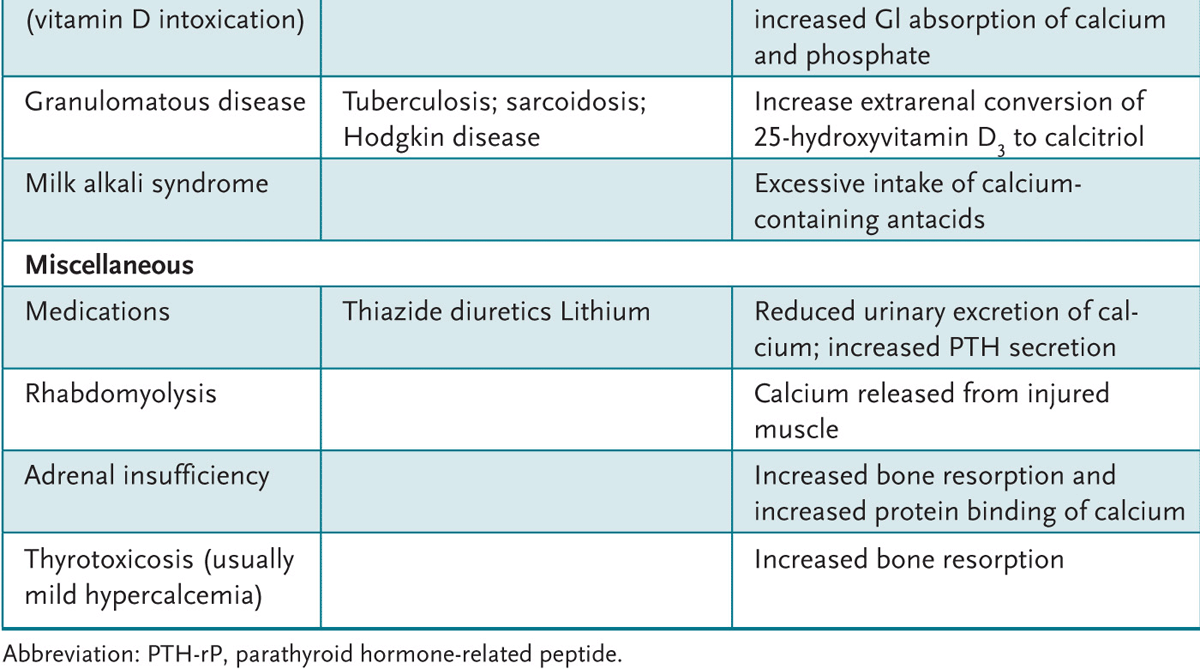
Any process that increases gastrointestinal calcium absorption, decreases renal excretion, or activates osteoclastic activity will raise serum calcium levels. If this occurs beyond the normal bounds of maintaining calcium homeostasis, hypercalcemia will occur. The most common cause of hypercalcemia in the ambulatory patient is hyperparathyroidism. Cancer ranks as the second leading cause, as hypercalcemia is often an early manifestation of malignancy. Hyperparathyroidism and cancer combined account for 90% of hypercalcemia cases. It is useful to categorize the etiologies of hypercalcemia into five main areas: parathyroid hormone-related, malignancy, renal failure, high bone turnover, and those related to vitamin D (Table 17–1).
Table 17–1 • COMMON CAUSES OF HYPERCALCEMIA
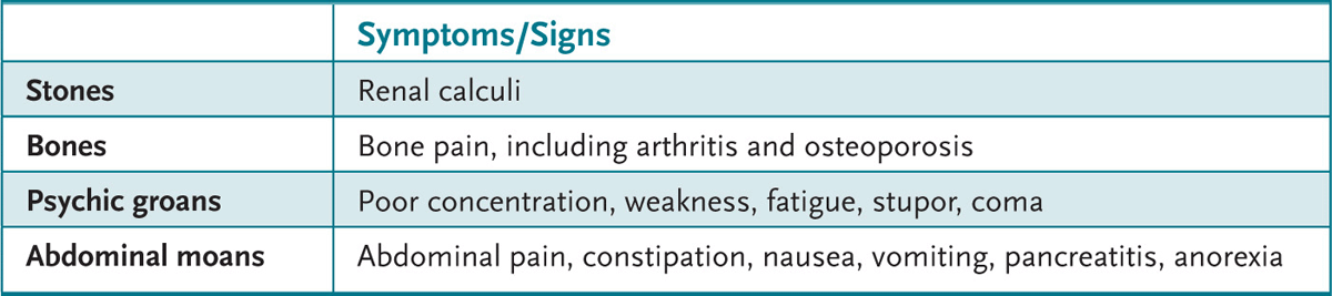
Normal values of serum calcium range from 8 to 10 mg/dL, which correspond to an ionized calcium level of 4 to 5.6 mg/dL. Levels of serum calcium between 10.5 and 12 mg/dL are classified as mild hypercalcemia; patients are frequently asymptomatic at these levels. As calcium levels increase, physical manifestations may become apparent. The classic mnemonic “stones, bones, psychic groans, and abdominal moans” is useful to categorize the constellation of physical symptoms associated with hypercalcemia (Table 17–2). Other clinical manifestations include the cardiac sequelae of shortening QT interval and arrhythmias.
Table 17–2 • PHYSICAL MANIFESTATIONS OF HYPERCALCEMIA
The diagnostic approach to hypercalcemia begins with a careful history, including the manifestations of elevated calcium levels. When the aforementioned etiologies are taken into consideration, it becomes clear that the history should include family history of calcium disorders, such as renal stones or malignancy. The patient’s risk factors for malignancy, such as smoking, should be investigated. A careful review of medications should also take place, to include not only prescription medications but also over-the-counter supplements. Dietary history is also an important component. At this point, if the hypercalcemia is mild and the patient asymptomatic, it is acceptable to stop the suspect medication and repeat the serum calcium level.
The next step, if a causative medication is not found, is to measure a serum intact PTH level. This level will either be suppressed, normal, or elevated. As with many endocrine disorders, it is useful not to think of normal or abnormal values; rather, one should understand what is appropriate for a given situation. For example, in normal subjects, an increased calcium load will normally depress the PTH hormone level, thus a low PTH level in this situation is normal, or appropriately suppressed. If a patient has an elevated calcium level and the PTH is “normal,” it is said to be inappropriately normal, because in the face of hypercalcemia it should be low, or suppressed.
If our patient with hypercalcemia has a normal or elevated PTH level, then the normal feedback loop is not responding. In this situation, the pituitary is producing PTH without check, which, in turn, is elevating the calcium level. This is hyper-parathyroidism. Primary hyperparathyroidism occurs when the parathyroid gland overproduces PTH and does not respond to the negative feedback of elevated calcium levels. The vast majority of primary hyperparathyroidism is caused by an adenoma (benign tumor) of one of the four parathyroid glands.
Secondary hyperparathyroidism occurs as the parathyroid glands overproduce PTH to respond to low serum calcium levels. This may occur as a response to low dietary calcium intake or a deficiency of vitamin D. Tertiary hyperparathyroidism occurs in patients who have renal failure. Patients in renal failure usually present with hypocalcemia, hyperphosphatemia, and low vitamin D levels. If this is untreated, it leads to hyperplasia of the parathyroid glands, an increased PTH secretion, and subsequent hypercalcemia.
There is a condition that can produce inappropriately high PTH levels unrelated to the parathyroid production. This is familial hypocalciuric hypercalcemia (FHH), a genetic disorder related to a defect in a gene that codes for a calcium-sensing receptor. Consequently, simply measuring PTH alone may confound this diagnosis, which may be mistaken for primary hyperparathyroidism. To distinguish these entities, a 24-hour urinary calcium level is obtained. In hyperparathyroidism, the kidneys spill calcium into the urine at a normal or elevated level. With FHH, the urinary calcium level is low.
A PTH level that is low with elevated serum calcium suggests that the parathyroid gland is responding appropriately to the high calcium environment. The etiology in this scenario must be some process that causes calcium to be released from bone or calcium to be absorbed from the gut despite the suppressed PTH. This is seen when tumors produce a hormone that mimics the active site of the PTH molecule, particularly in respect to the bone and renal effects, but that have no counter regulatory mechanism for suppression when calcium levels rise. This molecule is called parathyroid hormone-related peptide (PTH-rP). PTH-rP is produced by lung cancers, squamous cell cancers of the head and neck, and renal cell cancer. PTH-rP effects osteoclastic bone resorption, increases calcitriol, and promotes calcium resorption from the kidneys, resulting in increased levels of serum calcium. The continued production of PTH-rP effectively takes the parathyroid gland out of the loop in calcium homeostasis. Because cancer is a common etiology for hyper-calcemia, the search for malignancy is paramount at this step in diagnosis, before other, less common, disorders are considered.
If a malignancy is not found, other etiologies must be considered. These fall into the category of endocrine disorders other than parathyroid and include hyperthyroidism, adrenal insufficiency, and acromegaly. The workup thus includes thyroid-stimulating hormone (TSH), a cortisol level, and a pituitary imaging study, respectively.
The treatment of hypercalcemia is directed at the underlying disorder. Patients with mild hypercalcemia may be treated with preventative measures aimed at avoiding aggravating factors. These measures include adequate hydration (dehydration aggravates nephrolithiasis), avoiding thiazide diuretics or other offending medications, encouraging physical activity, and avoiding prolonged inactivity. Other interventions for mild hypercalcemia are disease specific.
For the treatment of primary hyperparathyroidism, surgical parathyroidectomy is the definitive treatment. Surgery is appropriate for patients with symptomatic hyperparathyroidism. Surgery may be an option for selected asymptomatic patients, including those who have developed osteoporosis or renal insufficiency, who have markedly elevated calcium levels, or who are younger than age 50 years.
17.1 A 60-year-old man comes into your office with the complaint of fatigue and constipation. He has had no dietary changes recently. A history reveals that he has hypertension, treated with medications, and an inguinal hernia that was repaired 10 years earlier without complications. The examination was nonspecific. You decide to obtain an electrolyte panel and find that the calcium level is elevated at 11.5 mg/dL (normal 8.5-10.2). Other laboratory values were normal. Which of the following is the next step?
A. Consult vascular surgery for placement of a dialysis catheter and schedule for dialysis.
B. Advise the patient to drink plenty of fluids and repeat the laboratory tests in 1 month.
C. Explore the patient’s hypertension, including what medications he takes.
D. Obtain a chest x-ray, looking for possible malignancy.
17.2 A 48-year-old man presents for follow-up of an elevated calcium level of 12.3 mg/dL found on routine screening laboratory tests at his last well-man visit. He takes no medications other than an occasional antihistamine for allergies. He recently started smoking a half-pack of cigarettes per day. He was prompted to attend to his well-man visit by his wife who claims that he has become forgetful, has a decreased appetite, and has had a 10-lb weight loss over the past 2 months. As part of his follow-up laboratory tests, you obtain a serum PTH, which comes back within the normal range. Which of the following is the next step in diagnosis?
A. Chest x-ray
B. Repeat calcium after hydration
C. Measurement of PTH-rP levels
D. Measurement of urinary calcium excretion
17.3 You obtain follow-up laboratory tests for a hypercalcemic patient and find that the PTH level is suppressed. There are no suspect medications. You suspect lung cancer based on a 30 pack-year smoking history, but the chest x-ray is normal. Which of the following is the next most appropriate step?
A. Continue a malignancy workup.
B. Check TSH, as a thyroid disorder may be the cause.
C. Refer the patient to an endocrinologist, as hypercalciuric hypercalcemia is an exceedingly rare genetic cause of an elevated calcium that requires specialist care.
D. Measure urinary calcium excretion.
17.1 C. When presented with a patient who has elevated calcium levels, the first step is to determine if there are any causative medications. Hydrochlorothiazide is a commonly used antihypertensive medication that may contribute to elevated calcium levels (thiazide diuretic).
17.2 D. This patient has symptomatic hypercalcemia. He has an inappropriately normal PTH level, which should be suppressed with this degree of hypercalcemia. The next step is to measure a 24-hour urinary calcium excretion to determine if this condition represents primary hyperparathyroidism (most common) or familial hypocalciuric hypercalcemia (rare).
17.3 A. In a hypercalcemic patient, a suppressed PTH first should be considered a sign of malignancy until proven otherwise. A chest x-ray is insufficient to rule out malignancy, as there are other malignancies that can cause hypercalcemia, mediated either by way of PTH-rP or through direct osteoclastic bone resorption. Multiple myeloma, granulomatous disease such as tuberculosis, sarcoidosis, and Hodgkin lymphoma, breast cancer, and squamous cell cancers of the head and neck can cause an elevated calcium with an appropriately suppressed PTH.
Be sure to question any patient with hypercalcemia regarding all medications—both prescription and over-the-counter—as both megadose vitamins (A and D) and excessive use of calcium carbonate antacids may play a role.
Most cases of primary hyperparathyroidism occur in postmenopausal women, who are often already at increased risk of osteoporosis. Be sure to check their bone density with a dual-energy x-ray absorptiometry (DEXA) scan.
Hypercalcemia with a suppressed PTH should be considered malignancy until you can prove otherwise.
Agus Z. Etiology of hypercalcemia. 2003. Available at: http://www.Uptodate.com. Accessed May 2009.
Agus Z, Fuleihan G. Management of asymptomatic primary hyperparathyroidism. 2005. Available at: http://www.Uptodate.com. Accessed May 2009.
Al Zarani A, Levine MA. Primary hyperparathyroidism. Lancet. 1997;349:1233-1238.
Carroll M, Schade D. A practical approach to hypercalcemia. Am Fam Physician. 2003;67:1959-1966.
Potts JT. Diseases of the parathyroid gland and other hyper- and hypocalcemic disorders. In: Fauci AS, Braunwald E, Kasper DL, et al, eds. Harrison’s Principles of Internal Medicine. 17th ed. New York, NY: McGraw-Hill; 2008:2377-2397.
Taniegra ET. Hyperparathyroidism. Am Fam Physician. 2004;69:333-340.