What is the most likely diagnosis?
A 52-year-old man presents to the office with approximately 2 weeks of upper abdominal pain. His symptoms are difficult for him to describe, but include some “discomfort” in the epigastric region that comes and goes. He has had some “heartburn” and nausea, but no vomiting or diarrhea. He has noticed that his stool looks darker than it used to, but he has not seen any blood. He feels full quickly after eating. He tried taking some over-the-counter antacid, which helps a little bit. His only other medication is an over-the-counter nonsteroidal anti-inflammatory drug (NSAID) that he takes “once or twice” a day because of arthritis in his knees. He does not smoke cigarettes or drink alcohol. On examination, he is pale appearing, but in no acute discomfort. He is afebrile, his blood pressure is 120/80 mm Hg, his pulse is 95 beats/min, and his respiratory rate is 14 breaths/min. Head, ears, eyes, nose, and throat (HEENT) examination is notable only for pale conjunctiva. Cardiac and pulmonary examinations are normal. His abdomen has normoactive bowel sounds and tenderness in the epigastrium. There is no mass, rebound, or guarding. Rectal examination reveals normal tone, no masses, and dark black stool that is strongly fecal occult blood test (FOBT) positive. The remainder of his examination is unremarkable.
What is the most likely diagnosis?
What evaluation and treatment is indicated at this point?
What can be done to reduce the risk of recurrence of this problem?
Summary: A 52-year-old man presents with vague upper abdominal discomfort, nausea, and early satiety. He is a daily NSAID user. He appears pale on examination, suggesting that he may be anemic. He has mild abdominal tenderness and melanotic stool on examination.
• Most likely diagnosis: Bleeding peptic ulcer.
• Evaluation and treatment at this point: A stat complete blood count (CBC), discontinuation of his NSAID, upper GI endoscopy, and testing for Helicobacter pylori. He should be treated with a proton pump inhibitor (PPI) and antibiotics for H pylori, if tests confirm its presence. He may need a blood transfusion (dependent on the result of his CBC). He will also require evaluation with a colonoscopy.
• Reduce risk of recurrence by: Discontinuation and avoidance of NSAID or, if unable to completely discontinue, use of PPI or misoprostol with the NSAID; eradication of H pylori.
1. Learn management of dyspepsia.
2. Learn the risk factors for the development of peptic ulcer disease (PUD).
3. Know how to diagnose and treat peptic ulcers.
4. Understand the role of H pylori in PUD, including methods for testing for and treatment of PUD.
5. Know the “alarm symptoms” for which endoscopy is indicated.
The Rome III Committee defines dyspepsia as one or more of the following symptoms: postprandial fullness, early satiation, epigastric pain or burning. Approximately 20% of dyspepsia is caused by peptic ulcer disease. Other common causes include gastroesophageal reflux disease (GERD) and functional dyspepsia. The diagnostic workup and treatment of patients with dyspepsia varies and is dependent on the age of the patient, the presenting symptoms and signs, and the response to the initial management offered.
Peptic ulcer disease is a problem of the gastrointestinal tract characterized by mucosal damage secondary to pepsin and gastric acid secretion. It usually occurs in the stomach and proximal duodenum; less commonly, it occurs in the lower esophagus, the distal duodenum, or the jejunum, as in unopposed hypersecretory states such as Zollinger-Ellison syndrome, in hiatal hernias (Cameron ulcers), or in ectopic gastric mucosa (eg, in Meckel diverticulum).
Early diagnostic endoscopy should be considered for patients with new-onset dyspepsia who are older than age 55 years or who have symptoms that may be associated with upper GI malignancy (Table 47–1). The cutoff age may be more appropriate at 45 to 50 for patients of Asian, Hispanic, or Afro-Caribbean descent. For those younger than age 55 years and without alarm symptoms, testing for H pylori, by IgG serology rather than the 13-C urea breath test or stool antigen testing, is recommended due its low cost and ease of collection. In this demographic population it is recommended to confirm a positive serological result with urea or stool antigen testing. For those who test positive, treating the H pylori followed by acid-suppression therapy is indicated. For persons who test negative, empiric therapy with a PPI for 4 to 8 weeks is a cost-effective intervention. Endoscopy or reconsideration of the diagnosis should be considered for those who continue to be symptomatic following these interventions.
Table 47–1 • “ALARM” SYMPTOMS FOR WHICH EARLY UPPER GI ENDOSCOPY IS RECOMMENDED
H2 BLOCKER: Class of medications that are competitive antagonists of histamine binding to gastric parietal cell H2 receptors, which prevent activation of the pathway that mediates release of acid into the gastric lumen.
PROTON PUMP INHIBITOR (PPI): Class of medications that suppress gastric acid production by irreversibly inhibiting the H+K+ ATPase proton pump in gastric parietal cells.
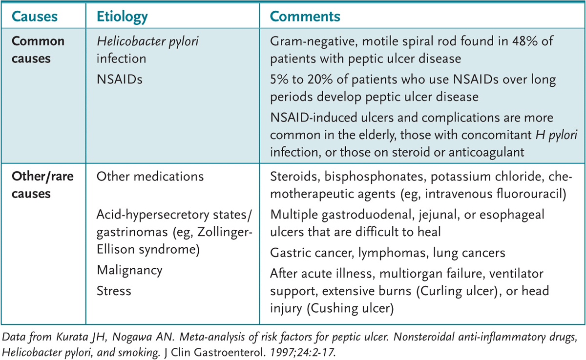
PUD is a term generally used to describe both duodenal and gastric ulcers. Duodenal ulcers are more prevalent overall, whereas gastric ulcers are more common in NSAID users. Risk factors for the development of PUD include H pylori infection, the use of an NSAID, cigarette smoking, and personal or family history of PUD. Black and Hispanic populations have a higher likelihood of developing PUD as well. The lifetime risk of developing PUD in the United States is approximately 10%. Table 47–2 summarizes other causes of PUD.
Table 47–2 • CAUSES OF PEPTIC ULCERS Causes Etiology Comments Common causes
History and Examination
Dyspepsia symptoms are common and there is significant overlap between the symptoms of PUD, GERD, and functional dyspepsia. Patients with symptoms primarily of heartburn or acid regurgitation are more likely to have GERD. Classic symptoms associated with PUD include epigastric abdominal pain that is improved with the ingestion of food, or pain that develops a few hours after eating. Nocturnal symptoms are also common with PUD, usually between 11 PM and 2 AM, when the circadian stimulation of acid secretion is maximal. The symptoms are often gradual in onset and present for weeks or months. Patients often self-medicate with over-the-counter antacid medications, which usually provide some relief, prior to presenting to the physician.
The examination should both attempt to confirm your suspicion of PUD and rule out other diagnoses that may present with abdominal pain. PUD often will only have the examination finding of epigastric tenderness. The presence of GI bleeding may be documented by stool occult blood testing; however, the bleeding from PUD may be episodic and a negative single office occult blood test does not completely rule out bleeding. Signs of anemia (pale conjunctiva or skin, tachycardia, hypotension, orthostasis) should be evaluated and managed as needed.
Many potential diagnoses must be considered in your differential. Finding right upper quadrant tenderness may suggest gallbladder or biliary disease. Appendicitis, while classically causing right lower quadrant pain, may present with only vague abdominal symptoms (especially with a retrocecal appendix). Epigastric pain radiating to the back and associated with nausea and vomiting may be pancreatitis. Pelvic infections, pelvic pathology, and even ectopic pregnancy must be considered as possibilities in women. Myocardial ischemia should be considered in those at risk.
Helicobacter pylori is a corkscrew-shaped gram-negative bacillus that is the causative agent of most non–NSAID-related ulcers. H pylori is also associated with the development of gastric cancer. The presence of the organism is associated with five to seven times increased risk of the development of PUD. H pylori infection is usually maternally acquired as a child and is more common in developing countries. Several tests are available to diagnose infections with H pylori. Stool antigen testing is now the preferred non-invasive office test for H pylori, due to its superior positive predictive value and ability to be used posttreatment to test for eradication. However, for this test to be accurate, patients must not have been treated with PPIs for at least 2 weeks prior to testing. Serologic testing for anti–H pylori antibodies is widely available, inexpensive, and noninvasive. It is highly sensitive for the presence of a history of infection but cannot distinguish an active infection from a treated infection.
Active infection can be confirmed by urea breath testing. This test is performed by having the patient ingest a carbon-labeled urea compound, which is then metabolized by urease from the H pylori organism. The labeled CO2 released by this process is measured in exhaled breath. This test is highly sensitive and specific, but is limited by availability and expense.
The gold standard for diagnosis is endoscopy with biopsy testing for H pylori. The bacterium can either be visualized microscopically using a variety of staining methods, cultured, or detected by rapid testing of the specimen. Endoscopy also allows for direct visualization of ulcers and evaluation for the presence of malignancy or other pathology in the esophagus, stomach, or duodenum. Endoscopy is invasive and expensive, limiting its utility to certain clinical situations.
About 25% of patients with peptic ulcer disease have a serious complication such as hemorrhage, perforation, or gastric outlet obstruction. Silent ulcers and complications are more common in older patients and in patients taking NSAIDs.
Upper gastrointestinal bleeding occurs in 15% to 20% of patients with peptic ulcer disease. It is the most common cause of death and most common indication for surgery in the disease. The risk of rebleeding and death is increased based on age, comorbidities, and hemodynamic status.
After the initial history and physical examination, focused testing appropriate to evaluate the suspected clinical syndromes should be ordered. A CBC should be drawn to evaluate for anemia, even when stool studies are negative for occult blood. A patient who has been vomiting or not eating should have basic chemistry studies performed. Liver enzymes, amylase, and lipase tests may be ordered when biliary or pancreatic disease is suspected. An ECG can be done if cardiac disease is a consideration, and an upright chest x-ray is the test of choice for possible abdominal visceral perforation. Abdominal ultrasonography is indicated when gallstones are suspected. A pregnancy test should be ordered on reproductive-age women, and cervical cultures performed if infection is suspected.
Patients with significant anemia, hemodynamic instability (hypotension, tachycardia, orthostasis), or suspected acute abdomen should be hospitalized. IV rehydration and blood transfusion should be performed when necessary. Urgent surgical evaluation should be obtained if an acute abdomen is present.
Dyspepsia in patients younger than age 55 years with no alarm symptoms can be managed with a noninvasive H pylori “test-and-treat” protocol followed by acid suppression preferably using a PPI especially if symptoms remain. A test for an active H pylori infection (stool antigen or serum IgA ELISA [enzyme-linked immunosorbent assay] antibodies) should be performed. A negative test rules out ulcer in dyspeptic patients. If positive, treatment to eradicate the infection, along with a PPI to suppress acid production, should be prescribed (Table 47–3 lists H pylori treatment regimens).
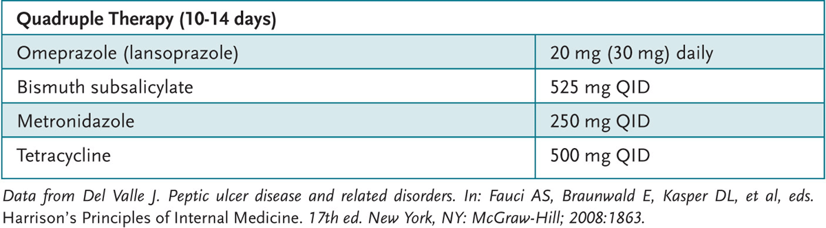
Table 47–3 • HELICOBACTER PYLORI TREATMENT REGIMENS
Generally PPI have greater efficacy in suppressing acid production and hastening ulcer healing than H2 blockers. Those with no evidence of active infection can be treated with acid suppression alone for 4 to 8 weeks. If symptoms resolve, no further testing is indicated. Along with treatment, offending agents, such as NSAIDs and tobacco, should be discontinued.
Patients older than age 55 years or with alarm symptoms should be referred for upper GI endoscopy to exclude the possibility of malignancy. Endoscopy is preferred over radiologic procedures because of better visualization and because of the ability to perform biopsy. Endoscopy also can be therapeutic, if a source of bleeding can be identified and cauterized. A patient who is older than age 50 years and who has blood in the stool should also undergo a colonoscopy regardless of the upper endoscopic findings, to ensure that there is not a colon cancer also contributing to the GI blood loss.
Surgical treatment for PUD is rarely needed. However it may be warranted for cases of hemorrhage that cannot be controlled, perforated, or obstructed.
47.1 A 30-year-old woman with no known medical problems comes to you for advice. She attended a health fair where she tested positive for H pylori on a blood test. She has had no abdominal discomfort, nausea, vomiting, or diarrhea. Her stool has been negative for blood. She occasionally has to use over-the-counter antacids after eating spicy foods. Which of the following would you tell her about the result of this test?
A. She may or may not have an H pylori infection.
B. She probably has a peptic ulcer.
C. She has an H pylori infection, but may or may not have an ulcer.
D. She should be treated immediately for H pylori.
47.2 A 62-year-old man presents to clinic with increasing shortness of breath and fatigue. Cardiac examination is negative and lungs are clear to auscultation bilaterally. No jaundice, jugular venous distention (JVD), or peripheral edema is noted. Mucous membranes are pink with no evidence of cyanosis and capillary refill is good. CBC reveals a microcytic anemia and a gastric ulcer is diagnosed on upper GI endoscopy. A biopsy and testing confirm an H pylori infection. His last colonoscopy was 2 years ago and was normal. Which of the following further testing is indicated at this time?
A. Upper GI radiographic series with small bowel follow through
B. Abdominal ultrasound
C. Colonoscopy
D. Urea breath test
47.3 A 41-year-old man presents for evaluation of upper GI discomfort that he has had for about 2 months. He says that he has a “full” sensation in the epigastric region. He recently began smoking after increased stress at work. He has had no blood in his stool, no vomiting, and no dysphagia. He has lost about 10 lb, but does not exercise. His mother has hemorrhoids, but no family member has ever had colon cancer. He has never had a colonoscopy. Which of the following is most appropriate?
A. H pylori “test-and-treat”
B. Empiric therapy for H pylori C. FOBT with reassurance if negative
D. Referral for endoscopy
47.4 A 19-year-old woman arrives at the emergency room with a 15-hour history of abdominal pain, nausea, and vomiting. She was awoken early in the morning by severe abdominal pain. She does admit to drinking heavily the prior evening that is not unusual during the weekends. She does not use NSAIDs regularly. Her blood pressure is 100/60 mm Hg, pulse rate is 130 beats/min, respirations are 14 breaths/min, and her temperature is 39°C. Acute abdominal series upon admission displayed substantial amount of free air under the right hemidiaphragm. Which of the following is the most likely diagnosis?
A. Perforated peptic ulcer
B. Alcohol-related gastritis
C. Appendicitis
D. Gastroenteritis
E. Kidney stones
47.1 A. H pylori blood tests are testing for anti–H pylori antibodies. They cannot distinguish active infections from old infections nor can they diagnose the presence of ulcers. Treating a positive serum test in an asymptomatic person is not indicated.
47.2 C. The presence of blood in the stool or anemia in a patient older than age 50 years, even when an ulcer is found, is an indication for colonoscopy, as this may also represent a presentation of a concomitant colon cancer. An urea breath test may be beneficial after completion of treatment to confirm eradication of the infection.
47.3 D. This patient presents with the alarm symptom of weight loss. He should be referred for early endoscopy.
47.4 A. The acute abdomen and free air under the diaphragm indicates a perforated viscus. This patient has perforated ulcer with hemodynamic instability. Additional workup includes a chemistry panel, CBC, and urgent laparotomy.
Persons who require long-term NSAID therapy may benefit from testing for active H pylori infection, followed by eradication, if positive, as this may lower their risk of developing an ulcer. PPI therapy, along with the NSAID, can also lower the risk.
Commonly held beliefs, such as ulcers being caused by stress or spicy foods, are incorrect. The vast majority of ulcers are caused by H pylori and NSAIDs.
Del Valle J. Peptic ulcer diseases and related disorders. In: Fauci AS, Braunwald E, Kasper DL, et al, eds. Harrison’s Principles of Internal Medicine. 17th ed. New York, NY: McGraw-Hill; 2008: 1855-1872.
Kurata JH, Nogawa AN. Meta-analysis of risk factors for peptic ulcer. Nonsteroidal anti-inflammatory drugs, Helicobacter pylori, and smoking. J Clin Gastroenterol. 1997;24:2-17.
McColl KE. Clinical practice. Helicobacter pylori infection. N Engl J Med. 2010:362:1597.
Ramakrishnan K, Salinas RC. Peptic ulcer disease. Am Fam Physician. 2007;76(7):1005-1012.
Talley NJ. American Gastroenterological Association medical position statement: Evaluation of dyspepsia. Gastroenterology. 2005;129(5):1753-1755.
The Medical Letter, Inc. Treatment of peptic ulcer and GERD. Treatment guidelines from The Medical Letter. 2008:55-57.
Townsend CM, Beauchamp RD, Evers BM, et al. Sabiston Textbook of Surgery. 17th ed. Philadelphia, PA: WB Saunders; 2004:1289.