9
The Brain and the Nervous System
THE BRAIN
Evolutionarily, the vast majority of organisms above the level of the primitive invertebrate phyla have evolved sense organs in the anterior portion (front end) of their bodies that respond to the high concentration of incoming information there. The most anterior ganglion, or enlarged, organized, integrative mass of nervous tissue, is called the brain. It is the brain that is responsible for processing most of the incoming information.
In many species of invertebrates, the brain is not much larger than the other ganglia located along the rest of the longitudinal nerve cords (see Figure 9.1). The brain of an invertebrate usually has considerably less dominance over the rest of the nervous system, and therefore the body, than is true for a vertebrate brain in the same size category. The brain in most lower vertebrates is not capable of significantly more complex tasks than most invertebrate brains. But the early vertebrate brains reflect evolutionary trends that led to many of the brain developments that have helped distinguish the vertebrates from other groups of organisms.
In the higher invertebrates as well as in the vertebrates, the brain functions in coordination with, or in place of, the many localized, segmented ganglia that are usually little more than a stimulus-and-response apparatus. This large accumulation of nervous tissue receives and transmits sufficiently large amounts of data to give it considerable control over the rest of the organism. The brain also makes it possible for many of these organisms to learn.
MEMBRANES COVERING THE BRAIN
The brain is protected by a system of membranes, the meninges. Outside the meninges is the hard, bony covering protecting the head, called the skull. The cranium is the part of the skull covering the brain. One of the three layers of meninges, the dura mater, lies just under the skull; it is tough and fibrous. Just under the dura mater is the meninge that resembles a cobweb, the arachnoid. And just over the brain is the pia mater, which is tightly molded around it. Between the pia mater and the arachnoid is cerebrospinal fluid, which acts as a protective cushion, protecting the brain from mechanical injury. Cerebrospinal fluid also fills the central canal that penetrates the entire length of the spinal cord and extends all the way into the brain, where it forms a series of cerebrospinal fluid-filled compartments, known as ventricles.

Figure 9.1 Grasshopper's nervous system (brain and ganglia are shaded).
THE VERTEBRATE BRAIN AND ITS EVOLUTION
Even the most primitive vertebrate brain has three principal divisions; each is also found in the advanced vertebrate brain. The most anterior of these three divisions is called the forebrain. It is composed of the olfactory lobes, cerebrum, thalamus, hypothalamus, and pituitary. The midbrain connects the forebrain with the hindbrain. It is composed largely of the optic lobes. The hindbrain consists of the cerebellum and the medulla oblongata. These parts of the brain are described below, and they are illustrated both on page 147 (Figure 8.3) and on page 161 (Figure 9.2).
Forebrain
The forebrain includes two olfactory lobes (or bulbs) that are always associated with the sense of smell. The posterior part of the forebrain consists of three other main structures. The thalamus is the major sensory integrative area in the forebrain of lower vertebrates. In higher vertebrates, it also integrates some of this information, but these functions have largely, through evolutionary history, become relegated to the cerebrum. The cerebrum, a major part of the forebrain, is the main center for controlling sensory and motor responses, as well as memory, speech, and most factors associated with intelligence. Underneath the cerebrum, near the thalamus, is the hypothalamus, which controls visceral functions such as blood pressure, body temperature, hostility, hunger, pain, pleasure, reproductive behavior, thirst, and water balance.
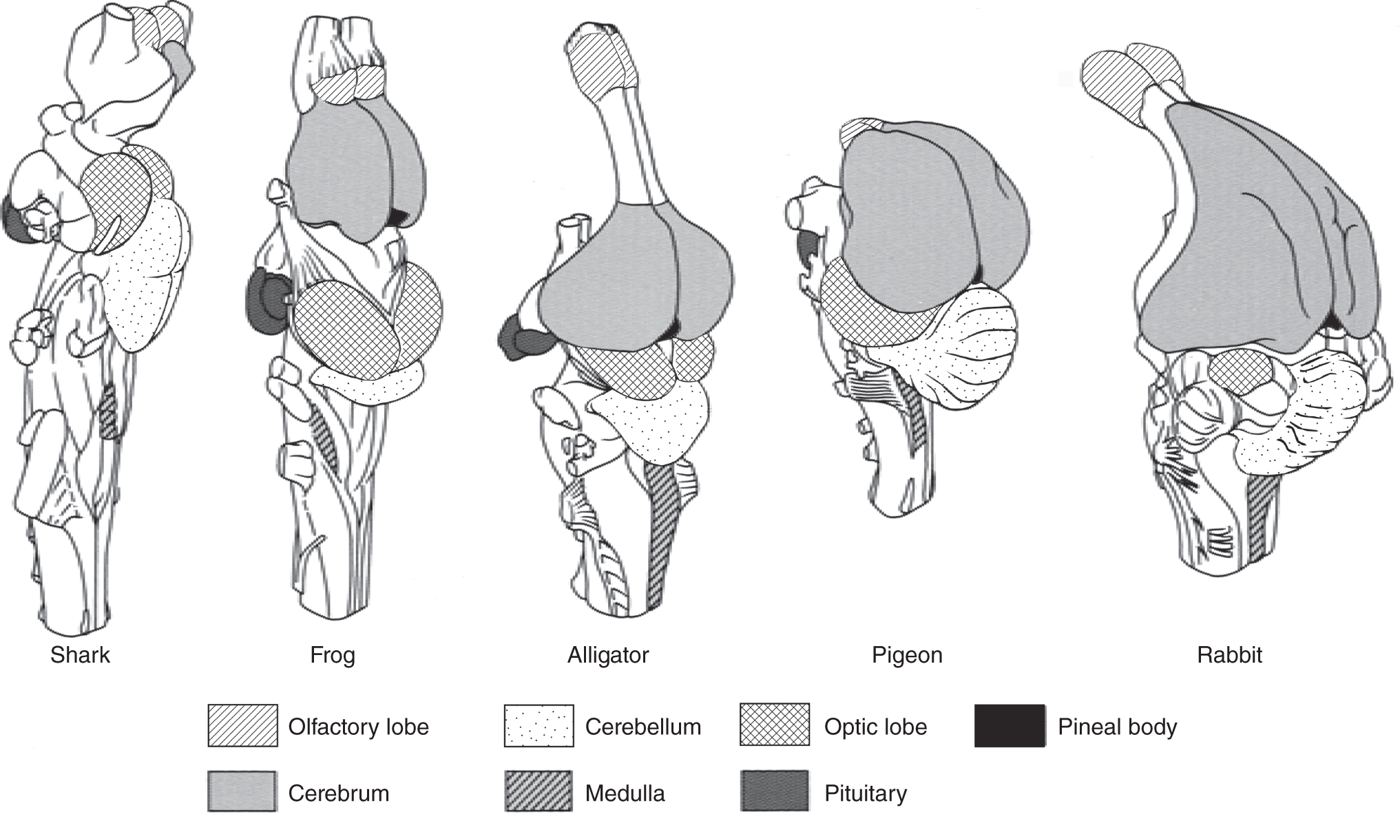
Figure 9.2 The brains of five vertebrates (not drawn to scale) illustrate varying sizes of different parts.
Midbrain
During evolutionary history, in several of the more advanced lineages, the forebrain increased in relative size and importance. Accordingly, the midbrain decreased in relative size and importance (see Figure 9.2). The most important parts of the midbrain are the specialized areas known as the optic lobes. These are the visual centers connected to the eyes by the optic nerves.
Hindbrain
The anterior portion of the hindbrain became enlarged and specialized as the cerebellum, which controls balance, equilibrium, and muscular coordination. The ventral portion of the hindbrain, the medulla oblongata, became increasingly specialized as the center of control for such visceral functions as heartbeat and breathing. This is the part of the brain that connects the nerve tracts from the spinal cord to the rest of the brain.
For the most part, gray matter consists of cell bodies and synapses. In fish, gray matter is primarily involved in relaying information from the olfactory lobes to the brain. The synapses connecting these neurons act as little more than relays, moving about the neuronal impulses without much integration. Amphibians have more gray matter, indicating that their cerebrum functions less as a simple conduit and more as an integrator of incoming information.
Concomitant with this expansion of internal gray matter, the gray matter moved from the inside part of the brain to the surface, where it is called the cerebral cortex. In amphibians and many reptiles, this surface layer of the brain is involved in smell. In some cases, its role expanded to the point where it may have become involved in the control of emotions. Certain advanced reptiles developed an additional component to the cortex, the neocortex, which expanded in primitive mammals into a covering over most of the forebrain. The neocortex of more advanced mammals increased in size and became folded, or convoluted, which increased its surface area. As this occurred, the ancestral (olfactory) part of the cortex was relegated to a more internal position in the forebrain.
Both birds and mammals evolved from reptiles, but birds evolved from reptiles without a primitive neocortex. The result has been that modern birds do not have the large, convoluted cerebral cortex found in higher mammals. Since it was another part of the bird cerebrum that grew in size, it is thought that this distinctly different origin of most of the modern cortex of birds accounts for the differences in their behavior. Birds rely more on innate responses. To a greater extent, mammals appear to develop much of their behavior depending on their experiences. However, when dealing with behavior, there is considerable overlap between and among different groups of organisms.
Originally the midbrain was important as a coordinating center. Later the thalamus area of the forebrain took over much of this function. Eventually, the neocortex of higher mammals preempted much of the midbrain and thalamus control, relegating the midbrain to little more than a link between the forebrain and the hindbrain. The midbrain of modern advanced vertebrates does have some control over several reflexes and minor eye functions, and there is a degree of interaction with emotional responses.
Even though researchers have made great progress in understanding which parts of the brain are involved with specific functions, we still have very little understanding of how the brain accomplishes each of these functions. For instance, although we might know which region of the brain contains certain memories, we cannot yet say with any degree of certainty how memories are made, nor can we say what they are made of or, for that matter, how they are retrieved and integrated.
THE NERVOUS SYSTEM
Neurons are among the most excitable cells in the body. As a group, they respond to a wide range of electrical, chemical, thermal, or mechanical stimuli, transmitting messages to one another, to muscles, and to endocrine organs (hormone-secreting glands). Together, all the neurons and their supporting cells (glial cells) compose the central nervous system.
Neurons do not exist in sponges (phylum Porifera) or more primitive organisms. The first neurons appear among the coelenterates (phylum Cnidaria or Coelenterata), which include jellyfish, hydra, and anemones. Of all living organisms, coelenterates have the simplest nervous arrangement, with only two types of nerve cells: receptor-conductor cells (those with receptor sites at the tips of the sensory nerve endings that respond to the stimuli and pass it on through the long part of the neuron, called the axon) and effector cells (those that contract when the stimulus reaches them). They have none of the alternative types that allow for the increased flexibility of response typical of higher organisms.
As the neurons in a nervous system increase in number, so does the complexity of behavioral responses an animal can have. Since one neuron communicates with nearby neurons, which in turn communicate with other neurons, the total number of possible neuron connections increases exponentially as the total number of neurons increases.
A roundworm (phylum Nematoda) is an organism that moves very little. It has only about 160 neurons. The leech (phylum Annelida), slightly more mobile, has about 13,000 nerve cells. An octopus (phylum Cephalopoda), which has considerable control over its movements and behavior, has over 1 billion neurons. And humans (phylum Chordata) have more than 10 billion neurons.
EVOLUTION OF THE NERVOUS SYSTEM
Nerve Net
Although many groups of lower invertebrates lack a nervous system, cnidarians do possess a simple nervous system termed a nerve net. A connected network of neurons without any apparent central control, the nerve net depends on the strength of the original stimulus to transmit a generalized reaction throughout the other neurons. The result may be contraction of other cells that later relax, leading to varying responses that enable simple organisms to maintain rather complex life histories.
Directed Movement
The cnidarians such as jellyfish, sea anemones, and coral represent a successful group of organisms, but radial symmetry, where the organism has similar parts radiating in a regular pattern from the center, turned out to be an evolutionary dead end. The flatworms (phylum Platyhelminthes), however, represent a significant advance in neural organization. Flatworms have a distinct top and bottom, front and back, head and tail. Rather than being sessile (attached to the substrate) or drifting about at random, flatworms control the direction of their movement; they have directed movement.
Among those species groups with directed movement, natural selection has favored the clustering of neurons in the anterior region, where the incoming information may be processed before being passed on to other neurons. Such neuronal clusters are known as ganglia (singular: ganglion).
The evolutionary trend toward the construction of animals with a body axis and directed movement (and away from the basically spherical) led to the neuronal development of the anterior region. This directed and lateral arrangement is termed bilateral symmetry. The anterior region of such organisms is the head. The large ganglion in the head that maintains considerable control over much of the entire nervous system, and therefore over much of the body, is often referred to as the brain. Such organisms are said to have a centralized nervous system.
NEURONS
Neurons are the only cells that transmit signals or nervous impulses; glial cells appear to provide nutrition to the neurons. Neurons are usually only a few micrometers in diameter, and most are quite small, though some extend from the spinal cord to the fingertips. Depending on the type of neuron, the long part is usually called the axon and the thicker part is the cell body, which contains the cell's nucleus. Dendrites are the short, branching projections extending from the cell body. They conduct nervous impulses toward the cell body. Axons usually conduct impulses away from the cell body, although they can also carry impulses toward it. In this case, however, there is no effect on an effector organ or cell. Bundles of neurons are called nerves.
Cnidarian nerve fibers are more primitive than the generalized neurons described above; they are not differentiated into dendrites and axons. The impulses are conducted in either direction, moving at random throughout the nerve net.
Neuronal responses include the ability to add up many incoming signals and integrate the information. Together, this is accomplished with three basic types of neurons. Sensory neurons carry information about environmental change to such integration centers as the brain or spinal cord. Interneurons (or association neurons) are the major components of the integration centers. They relay messages from one neuron to another. Most neurons in complex animals fall into this category. Motor neurons carry the impulses away from the integration centers to muscles or glands (see Figure 9.3).
Associated with the neurons are different types of glial cells. Together, all the glial cells are known as the neuroglia, which account for at least half of the nervous system's volume. Some axons are encircled by one type of glial cell, the Schwann cell, which provides nutrition to the neuron (see Figure 9.4). Some Schwann cells' plasma membranes envelop certain axons and nerves. Such membranes are called myelin sheathing. This resembles fatty insulation and seems to be related to increasing the rate at which the nervous impulses are conducted along the axons. The nodes of Ranvier occur at intervals along such axons. These are constricted junctions where one Schwann cell ends and the next begins.
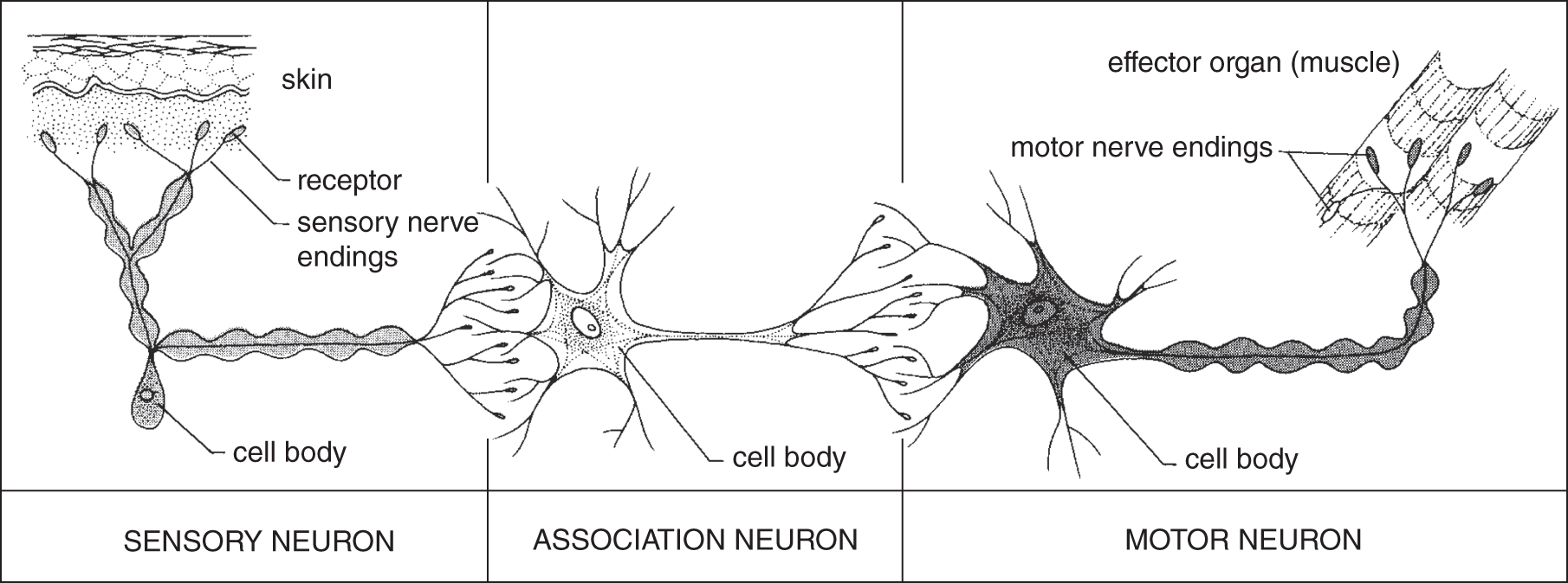
Figure 9.3 Sensory, association, and motor neurons and their relationships.

Figure 9.4 A nerve cell and associated Schwann cells with microstructural details.
In the brain and spinal cord, much of the nervous tissue consisting of myelinated axons is called white matter because the myelinated axons are whitish in appearance. Much of the brain's nervous tissue, lacking a fatty sheath surrounding the axons, is called gray matter.
NERVOUS IMPULSE
Both the endocrine system (hormone-secreting glands and their products) and the nervous system control many of the body's activities by regulating and integrating much of what an organism does throughout its life. One of the major functional differences between the nervous system and the endocrine system is the speed with which the nervous system reacts. A nervous impulse can travel through an entire organism in a fraction of a second, while hormones (which move through the blood) elicit a slower response.
The speed at which a nervous impulse can travel through myelinated nerves is about 200 km s−1. Nervous impulses traveling through nonmyelinated nerves travel about half as fast (100 km s−1).
Changes in the physical or chemical environment (i.e. due to motion, sound, light, heat, or chemicals) can be converted into nervous impulses. The environmental change is known as the stimulus, and the neuronal response is the neural impulse. When part of the nervous system receives a neural impulse, it may respond by sending another impulse to the appropriate effectors. Many effectors are muscles, which respond by contracting. However, there are many other types of effectors, such as photoreceptor cells or glandular cells. In addition, the nervous impulse may reach another neuron, which triggers it to the next neuron, and so on, although such an impulse may eventually dissipate to the point that it can no longer elicit a response from an effector.
A neural impulse is triggered by a change in the neuron's electrical charge, which is the result of rapid movement of certain ions. Like most other living cells, neurons have an asymmetric distribution of ions across their plasma membranes. The interior of a resting neuron (one that is not transmitting an impulse) contains more negatively charged ions than the outside of the cell, where there are more positively charged ions. This uneven distribution of electrical energy, which might be described as an electrical potential difference across the membrane, is usually referred to as membrane potential and is critical to the neuron's ability to transmit an impulse along its entire length.
A resting neuron has a net negative charge inside the axon, primarily from negative chloride ions (Cl−), and a higher concentration of positively charged sodium ions (Na+) outside the neuron. Inside the neuron, along with the negative chloride ions, are positively charged potassium ions (K+). Upon a neuronal stimulus of sufficient strength, a response, or a nervous impulse, is initiated. The intensity of stimulus required to activate this kind of response is called threshold.
At the site where the neuron is stimulated, the membrane initially becomes more permeable to sodium ions, which then rush across the membrane to the inside of the cell, momentarily producing a slightly more positive charge inside the cell relative to the outside. The membrane also becomes a little more positively charged relative to the outside of the cell. This change in membrane potential is called depolarization.
After the neuron becomes more permeable to sodium ions, it becomes more permeable to potassium ions. So in the fraction of a second following the influx of sodium ions, potassium ions rush out of the cell. This exit of positively charged ions restores the charge inside the cell as well as that of the membrane to its initial negative charge. Once depolarization has been initiated at one end of a neuron, it passes down the entire length of the neuron. This sequence of events is known as a nervous impulse.
This wave of depolarization, also called an action potential, rapidly passes down the entire length of the axon. This is why an action potential is termed “all or nothing.” Then, immediately following the inrush of sodium ions and the outflow of potassium ions, the sodium ions are pumped back out of the nerve cell and the potassium ions are pumped back in (against their concentration gradient, so energy is required to fuel the active transport). The pump that restores the original ionic balance is the sodium-potassium pump. It does this after a very brief refractory period, when the neuron can't conduct a neural impulse (see Figures 9.5 and 9.6).
Upon reaching the end of the neuron, which is called an axon ending or axon terminal, the action potential stimulates the release of chemicals known as neurotransmitters, which travel across the synapse (a short gap) to the next neuron (see Figure 9.7). If the neurotransmitters fit into the receptor sites (see Figure 9.7), these chemicals trigger a new depolarization, and then another action potential travels along the next neuron too (see Figure 9.8). The neurotransmitters that are short-chain polypeptides are called neuropeptides. To read about neuropeptides, please refer to the glossary.
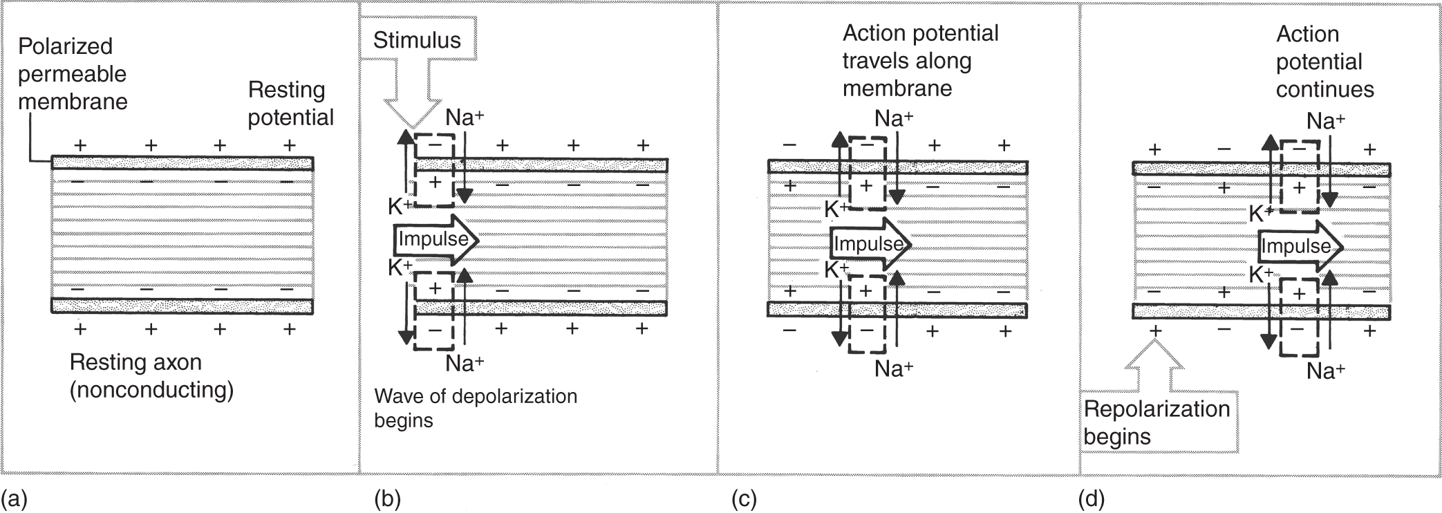
Figure 9.5 Initiation and transmission of a nerve impulse as illustrated along a small section of a neuron: (a) resting nerve fiber; (b) impulse begins with depolarization of cell membrane as sodium ions (Na+) move into the cell and potassium ions (K+) move out; (c) the wave of depolarization moves along the nerve fibers; (d) after the impulse passes, the membrane repolarizes by pumping out the sodium ions.

Figure 9.6 This graph illustrates the change in a nerve fiber's membrane potential when measured at one location along the neuron during a nerve impulse. The movement of sodium ions into the cell depolarizes it, and the movement of potassium ions out of the cell repolarizes it.
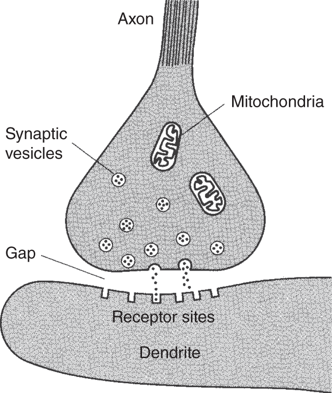
Figure 9.7 A synapse, the junction between two neurons. Neurotransmitters are released from synaptic vesicles and picked up by receptor sites.
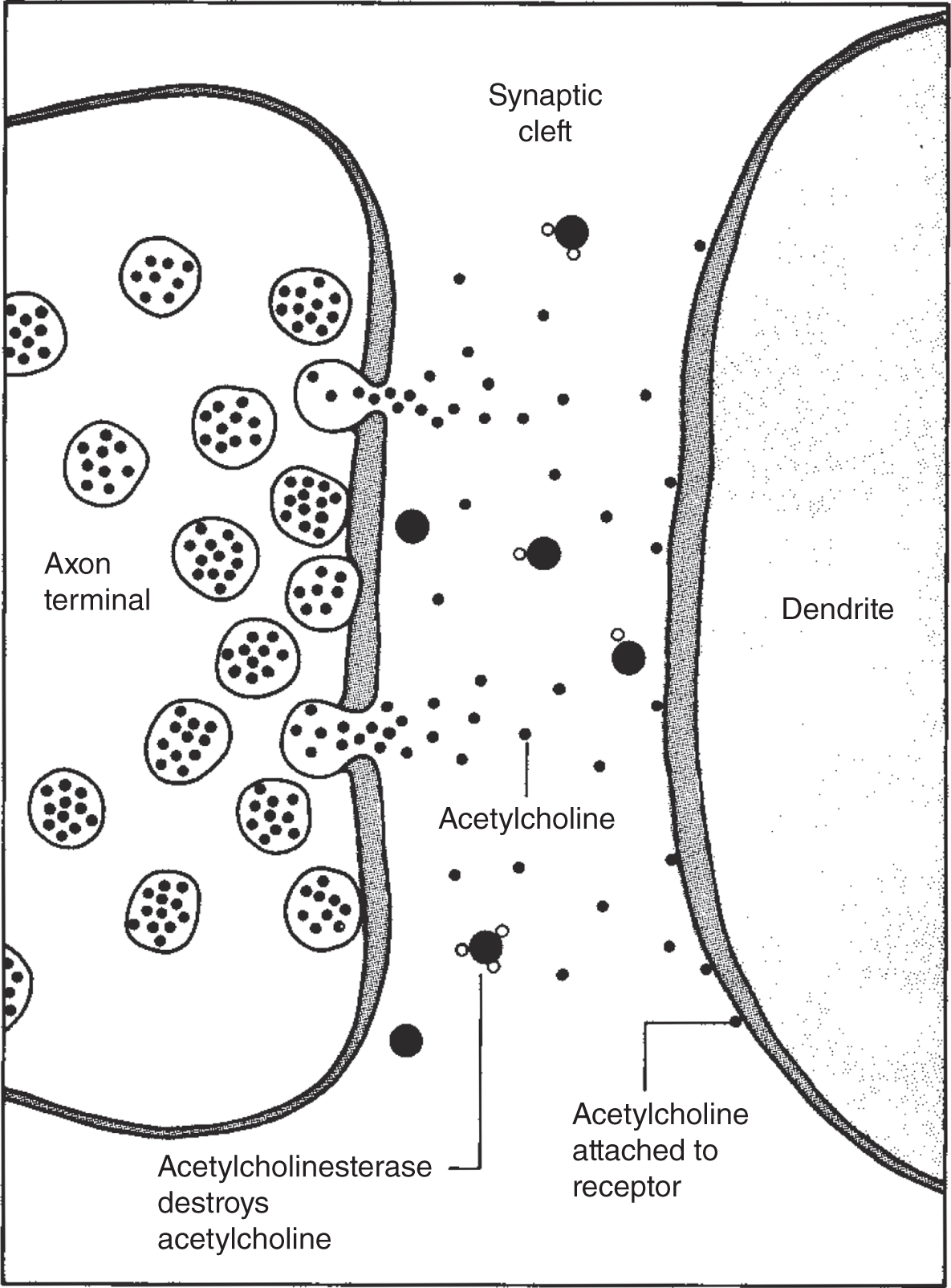
Figure 9.8 After the neurotransmitter molecules attach to the receptor sites, they are deactivated by specific enzymes.
SYNAPSE AND NEUROTRANSMITTERS
The term “synapse” is used to describe both the junction between a neuron and another neuron and that between a neuron and the cell it acts upon. The gaps between the cells at the synapse are about 20 nanometers wide (a nanometer is a unit of length equal to one billionth of a meter). There are synapses between the ending of an axon and a dendrite, between an axon and a cell body, and sometimes between two axons. Most neurons synapse (connect; synapse can also be used as a verb) with a number of neurons, although many also synapse with other cells such as muscles and glands. The axons produce neurotransmitters, which cross the synapse and are picked up at receptor sites on the dendrites of an adjoining neuron.
The difference between inhibition or stimulation depends on the amount and type of neurotransmitter as well as on the type of receptor site. Over 10 different neurotransmitters have been identified to date; these include acetylcholine, dopamine, glutamate, gamma-aminobutyric acid, histamine, nitric oxide, noradrenalin, and serotonin. Each affects the response of a neuron. Normally, neurotransmitters are rapidly broken down enzymatically following release. But tranquilizers, caffeine, nerve gas, many insecticides, and curare (the chemical used in poison arrows in South America) can interfere with neurotransmitters – by stimulating or retarding neurotransmitter production, by binding to a receptor site, or by affecting the enzymes that normally destroy the neurotransmitters.
REFLEX ARCS
A reflex arc is usually based on a small group of neurons where the entire neural impulse totally circumvents the brain. The most simple reflex arc contains a sensory neuron, an association neuron, and a motor neuron that synapses with an effector cell. Each reflex arc contains one sensory neuron, sometimes extremely long, such as those running from a large mammal's foot to its spinal cord. The cell bodies of these sensory neurons are always located in ganglia (dorsal-root ganglia) just outside the spinal cord (see Figure 9.9). The axons enter the spinal cord dorsally, where they connect (synapse) with several association neurons in the gray matter of the spinal cord. Then they synapse with a motor neuron (also in the gray matter of the spinal cord) and exit the spinal cord ventrally. Figure 9.9 uses a cross section of a spinal cord to illustrate the path of a reflex arc.
Some of the association neurons directly synapse with motor neurons that run back to where the sensation originated. The advantage of a reflex arc is speed. The signal is sent straight back, causing an immediate response, such as a knee-jerk reaction, an eye blink, or the formation of a tear drop. Reflex arcs are not the result of any conscious control. However, it is possible for some association neurons to synapse with other association neurons that pass up the spinal cord to the brain, where the information is relayed to other centers. There it may then be possible to consciously inhibit part of the reflex or add to it.

Figure 9.9 Cross section of the spinal cord showing major features and the route of a reflex arc.
Still other reflexes may be far more complex than the simple knee-jerk reaction. Two such examples are breathing and the control of one's heartbeat. Though largely involuntary, it is possible to exert a considerable amount of conscious control over these behaviors.
All the nerves connecting with the spinal cord contain sensory and motor neurons and are called mixed nerves. Not all the nerves intercept with the spinal cord. Humans have 12 pairs of nerves that directly connect with the brain. These are known as the cranial nerves; some cranial nerves contain just sensory or just motor neurons, and others are mixed.
ORGANIZATION OF THE NERVOUS SYSTEM
The brain and spinal cord compose the central nervous system. The somatic nervous system conducts nervous impulses that have already been processed away from the central nervous system to the skeletal muscle tissue.
The somatic nervous system is under voluntary control. All the parts of the nervous system, excluding the brain and spinal cord, are collectively known as the peripheral nervous system.
The autonomic nervous system consists of the nerves that carry nervous impulses from the central nervous system to the heart (cardiac muscles), to the muscles in the digestive system (smooth muscles), and to the glands (see Figure 9.10). All of these muscles and glands contract and function involuntarily. The autonomic nervous system is subdivided into two parts, the sympathetic and parasympathetic systems. These function in opposition to one another; the first inhibits organs, while the latter usually excites organs.

Figure 9.10 Autonomic nerves innervate smooth muscles found in organs and glands. Most organs receive innervation from sympathetic and parasympathetic portions of the nervous system.
KEY TERMS
| action potential | neocortex |
| arachnoid | nerve net |
| association neurons | nerves |
| autonomic nervous system | nervous impulse |
| axon | neural impulse |
| bilateral symmetry | neuroglia neurons |
| brain | neurotransmitters |
| cell body | nodes of Ranvier |
| central nervous system | olfactory lobes |
| cerebellum | optic lobes |
| cerebral cortex | optic nerves |
| cerebrospinal fluid | parasympathetic nervous system |
| cerebrum | peripheral nervous system |
| cranial nerves | pia mater |
| cranium | pituitary |
| dendrites | radial symmetry |
| depolarization | receptor sites |
| directed movement | receptor-conductor cells |
| dorsal-root ganglia | reflex arc |
| dura mater | refractory period |
| effector cells | Schwann cell |
| forebrain | sensory nerve endings |
| ganglia | sensory neurons |
| glial cells | skull |
| gray matter | sodium-potassium pump |
| hindbrain | somatic nervous system |
| hypothalamus | stimulus |
| interneurons | sympathetic nervous system |
| medulla oblongata | synapse |
| membrane potential | thalamus |
| meninges | threshold |
| midbrain | ventricles |
| motor neurons | white matter |
| myelin sheathing |
SELF-TEST
Multiple-Choice Questions
The Brain and Its Membranes
- The brain is protected by a membranous system known as the ___________.
- ventricles
- olfactory sheaths
- oblongata
- meninges
- medullas
- The ___________ is the part of the skull that covers only the brain.
- dura mater
- arachnoid
- pia mater
- cerebrospinal fluid
- cranium
- The spinal cord has a central canal that extends into the brain, becoming a series of hollow compartments called ___________, which are filled with cerebrospinal fluid.
- ventricles
- dura mater
- pia mater
- meninges
- thalamus
- The first of the meninges that lies just under the skull, and is tough and fibrous, is the ___________.
- dura mater
- pia mater
- ventricle
- thalamus
- gray matter
- Between the pia mater and the arachnoid is the ___________ that bathes the entire region, providing a cushion to protect the brain from mechanical injury.
- albumin
- ovalbumin
- lymph
- cerebrospinal fluid
- protoplasm
- The part of the brain that consists of the olfactory bulbs, cerebrum, thalamus, hypothalamus, and pituitary is the ___________.
- forebrain
- midbrain
- hindbrain
- medulla oblongata
- cerebellum
- The cerebellum and the medulla oblongata are part of the ___________.
- forebrain
- midbrain
- hindbrain
- thalamus
- hypothalamus
- The ___________ is (are) the major sensory integrative area in the forebrain of lower vertebrates.
- olfactory bulbs
- medulla oblongata
- cerebellum
- thalamus
- hypothalamus
- The ___________ control(s) visceral functions such as blood pressure, body temperature, hostility, hunger, pain, pleasure, reproductive behavior, thirst, and water balance.
- thalamus
- hypothalamus
- medulla oblongata
- olfactory bulbs
- cerebrum
- The ___________ control(s) balance, equilibrium, and muscular coordination.
- thalamus
- hypothalamus
- gray matter
- olfactory bulbs
- cerebellum
- The part of the brain that consists of cell bodies and synapses is known as the ___________.
- optic nerves
- optic lobes
- olfactory bulbs
- white matter
- gray matter
The Nervous System, Its Evolution, Neurons, Impulses, Synapse, Neurotransmitters, and Reflex Arcs
- Together, all the neurons and their supporting cells comprise the ___________.
- glial cells
- myelin sheathing
- nerve net
- nervous system
- central nervous system
- Among members of the phylum Cnidaria, the connected network of neurons that creates the nervous system lacking central control is known as a(n) ___________.
- central nervous system
- ganglion
- brain
- axon
- nerve net
- Flatworms have a top and bottom, a front and back, a head and tail; this type of body construction is known as ___________.
- radial symmetry
- lateral symmetry
- bilateral symmetry
- trilateral symmetry
- asymmetry
- All groups of animals above the evolutionary level of sponges have a ___________.
- radially symmetrical arrangement
- bilaterally symmetrical arrangement
- nerve net
- tentacle
- nervous system
- Receptor cells ___________ stimuli.
- receive
- conduct
- speed up
- slow down
- circulate
- Conductor cells are specialized for ___________ stimuli.
- conducting
- stopping
- slowing down
- speeding up
- associating
- Effector cells are usually ___________ or___________.
- muscle, bone
- bone, gland
- gland, muscle
- none of the above
- all of the above
- As the entire nervous system became more complex in terms of its increased flexibility of response, there was a trend toward ___________.
- specialization
- cephalization
- minimization
- maximization
- a and b
- The part of a neuron that contains the cell's nucleus is the ___________.
- axon
- dendrite
- cell body
- glial cell
- myelin sheath
- ___________ are usually short, branching projections extending from the cell body.
- perikaryons
- dendrites
- cell bodies
- Schwann cells
- axons
- In terms of total volume, all the glial cells account for about ___________ of the nervous system.
- 10%
- 25%
- 50%
- 75%
- 100%
- All of the ___________ are known as the neuroglia.
- glial cells
- neurons
- axons
- cell bodies
- dendrites
- Some of the axons are encircled by ___________ that provide nutrition and perhaps other forms of support.
- Schwann cells
- cell bodies
- perikaryons
- nerves
- nodes of Ranvier
- The ___________ resemble(s) a coiled, fatty insulation around certain axons.
- myelin sheath
- nodes of Ranvier
- ganglia
- sensory neurons
- association neurons
- Myelinated axons are ___________ in appearance.
- greenish
- reddish
- bluish
- grayish
- whitish
- The junction between two or more neurons is called a ___________.
- neural impulse
- neurotransmitter
- synapse
- dorsal-root ganglion
- nervous junction
- The movement of the electrical impulse across the synapse requires specific chemicals known as ___________.
- neural impulses
- neurotransmitters
- synapse jumpers
- dorsal-root ganglia
- nervous junction chemicals
- Bundles of individual axons are called ___________.
- synapses
- sensory neurons
- nerves
- gray matter
- association neurons
- Each reflex arc contains one sensory neuron that has its cell body located just outside the spinal cord in the ___________.
- motor neuron
- association neuron
- spinal cord
- dorsal-root ganglion
- gray matter
- All the parts of the nervous system, excluding the brain and spinal cord, are collectively known as the ___________.
- afferent system
- efferent system
- peripheral nervous system
- somatic nervous system
- autonomic nervous system
- The ___________ consists of nerves that carry nervous impulses from the central nervous system to the smooth muscles, the heart muscle, and to the glands.
- afferent system
- efferent system
- peripheral nervous system
- somatic nervous system
- autonomic nervous system
- The parasympathetic system usually ___________ an organ.
- inhibits
- excites
- carries lymph to
- carries lymph from
- a and b
- The sympathetic system usually ___________ the particular organ.
- inhibits
- excites
- carries lymph to
- carries lymph from
- a and b
ANSWERS
- d
- e
- a
- a
- d
- a
- c
- d
- b
- e
- e
- d
- e
- c
- e
- a
- a
- c
- e
- c
- b
- c
- a
- a
- a
- e
- c
- b
- c
- d
- c
- e
- a
- b
Questions to Think About
- What protective layers envelop the vertebrate brain?
- How are vertebrate brains similar in basic construction?
- What do the terms “forebrain,” “midbrain,” and “hindbrain” refer to?
- What is the difference between gray and white matter?
- What are three basic types of neurons?
- Define myelin sheathing and explain its function.
- What is the all-or-nothing principle?
- Shortly after being released, what normally happens to neurotransmitters?
- How does a nervous impulse pass down a neuron?