10
Bones and Muscles
BONES
Muscles and bones work together. The bones make up the skeletal system, which provides structural support, sites for muscle attachment, and organ protection. Osseous tissue, or bone, as it is more often called, consists of cells and collagen fibers interspersed in a matrix of intercellular material containing calcium phosphate and calcium carbonate, which are responsible for hardness. Together, these substances account for two-thirds of the weight of bones, while the collagen fibers, which reinforce the tissue, account for the other third.
In addition to bone, another important connective tissue in most skeletal systems is cartilage, which, unlike bone, is both firm and flexible. Bone is usually considerably harder and more brittle. Most sharks and rays have skeletal systems composed of all cartilage and no bone. Some other “primitive” groups of fish have less bone than cartilage in their skeletal systems. In most other vertebrates, however, cartilage is located only where firmness and flexibility are needed, such as in joints, nose, ears, larynx, and trachea. During the development of the skeletal system of these vertebrates, embryos begin with cartilaginous skeletons. Gradually most of the cartilage is replaced by true bone.
Depending on the construction of the particular bony tissue, it can range in consistency from being completely spongy to being very compact. The spongy bone contains many spaces filled with marrow (which is either composed of fat or involved in the production of blood cells). In the case of relatively lighter animals such as birds, the spaces may be filled with air sacs. Compact bony tissue is thicker and usually involved in support. Such bones can resist considerable weight and stress.
Compact bones are penetrated by blood vessels and nerves through small narrow openings, some of which are known as Haversian canals, whose microscopic structure is identified by the characteristic concentric rings of bony tissue surrounding them. These rings are composed of cells that were involved in producing the bony tissue. Spongy bone doesn't contain Haversian systems, nor does cartilage. Materials are exchanged through the blood vessels and bone cells that penetrate the Haversian canals. This is the only way for materials to move to and from the cells living throughout bony tissue.
The main bones in the human body, from head to toe, are as follows. The fused bones creating the cranium compose the skull; the lower teeth are located in the mandible, or jaw. The collar bone is the clavicle. The “wings” in the upper back are called scapulas. The bone connecting all the ribs in the middle of the chest is the sternum. The ribs are connected in the back to the vertebral column (backbone), which is composed of vertebrae. The vertebrae in the neck are called cervical vertebrae; thoracic vertebrae articulate with the ribs; lumbar vertebrae descend from the thoracic vertebrae to the pelvis; and together, the fused bones in the pelvis compose the sacrum. The tail is composed of caudal vertebrae. In humans, the “tail” is called the coccyx (see Figure 10.1).

Figure 10.1 The human spinal column consists of 33 bones. Within the spinal column is the spinal cord, branching from which are 33 pairs of spinal nerves that emerge from between the bones.
The bone in the upper arm is the humerus, and the two bones in each lower arm are the radius and ulna. The wrist bones are called carpals. At the base of the fingers, located within the part of the hand known as the palm, are the metacarpals, and the smaller bones extending out to the fingertips are the phalanges.
The largest bones in the body are the femurs, the thigh bones that connect the upper leg with the pelvis. The distal end of each femur is attached to the lower leg. The upper and lower legs meet at the knee, and covering that is the kneecap, or patella. Each lower leg has two long bones, the tibia and fibula. The little bones in the ankle area are the tarsals; then come the metatarsals. The rest of the bones extending to the toe's tips are, like the fingers, called phalanges (see Figure 10.2). Note similarities between the human and pigeon skeleton (Figure 10.3).
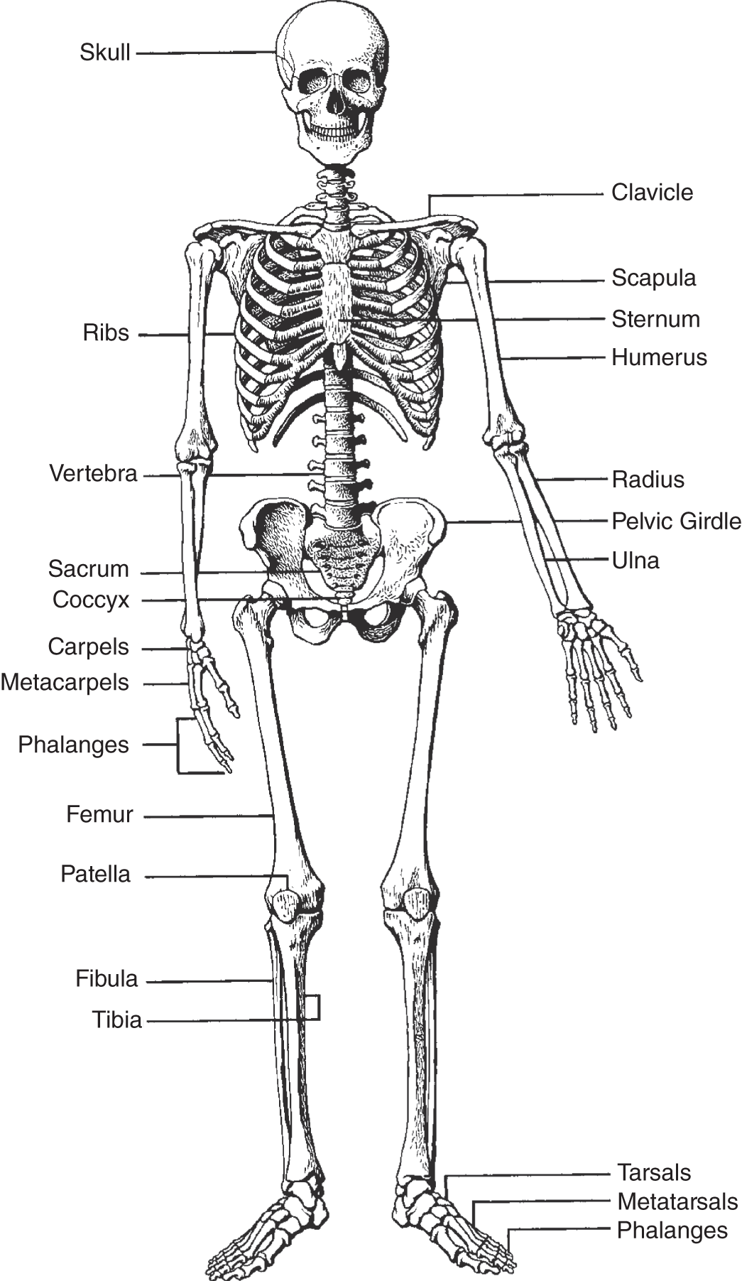
Figure 10.2 The human skeleton.
Some bones are held together with fused, immovable joints, such as the sutures located between several skull bones. Other joints are movable and are held together with ligaments, the flexible tissues that connect bones and cartilage. Similarly, tendons are flexible tissues connecting muscles to bones. The end of a muscle attached to the bone nearest to the axis of the body (proximal) is known as the origin. The muscle end attached to the farther bone (distal), such as in the hand or foot, is known as the insertion. In Figure 10.4, for example, the biceps originates on both the humerus and the scapula, and it inserts on the radius.
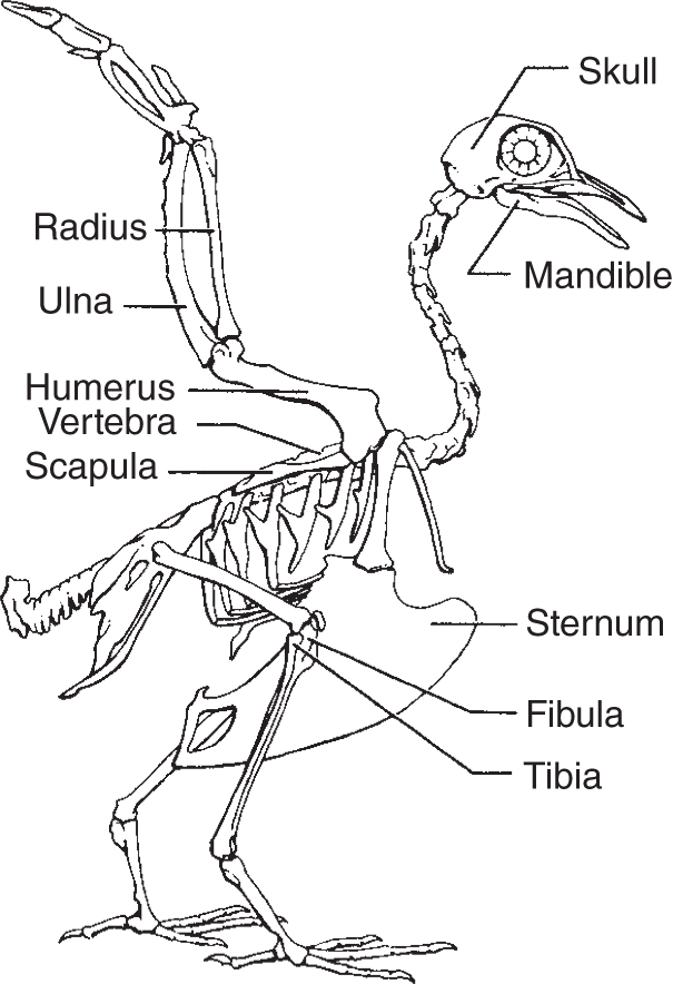
Figure 10.3 The pigeon skeleton.
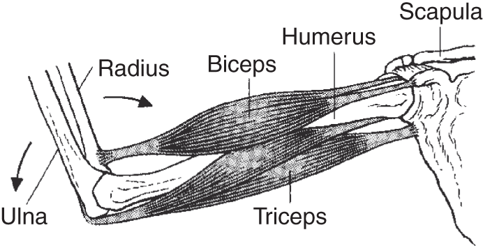
Figure 10.4 With balance sensors located in muscles and tendons, the antagonistic action of muscle pairs controls the tensing and releasing of different muscles. Together, this antagonistic action enables an organism to control its body movements.
The movements of different parts of the body all depend on muscular contraction, on the location of the origins and insertions, and on the type of joint involved. Muscles usually work antagonistically. That is, when one group of muscles contracts, it will pull part of the body one way. Alternately, when the antagonistic group of muscles contracts, it will pull the same body part in the other direction. The biceps and triceps in Figure 10.4, for example, form an antagonistic muscle pair.
MUSCLES
Muscles are composed of muscle cells, which look like long, thin fibers (often 3 cm long). They are also called muscle fibers and myofibers. Three types of muscles have been recognized in vertebrates. These are smooth, or visceral, muscle; skeletal muscle; and cardiac, or heart, muscle. Smooth muscles line internal organs such as the intestines and bladder. They also line the walls of the arteries and veins and many ducts and tubes found throughout the body. For the most part, the smooth muscles contract involuntarily (they are innervated by the autonomic nervous system). Together, these muscle fibers form thin broad sheets.
Responding to conscious control, the skeletal muscles are commonly called the voluntary muscles (they are innervated by the somatic nervous system). Skeletal muscles move the arms and legs, back, face, jaw, and eyes, as well as many other parts of the body. Skeletal muscle cells (fibers) are coenocytic (contain many nuclei) and, when viewed microscopically, are crossed by many thin dark lines, which is why they are called striated muscles. Together, many skeletal muscle fibers form bundles, which are wrapped in connective tissue to form muscles.
Like skeletal muscle cells, cardiac muscle fibers have striations. They are also multinucleate, but they are innervated by the autonomic nervous system. The heart has a pacemaker, which spontaneously begins each heartbeat. This specialized area located in the wall of the right atrium (see Figure 12.4, page 216) is called the sinoatrial node. Once the heart begins to contract, the impulse spreads to the node lying near the atrium between both ventricles, which is called the atrioventricular node; once stimulated, it initiates the ventricular contraction.
Unlike vertebrate muscle, all insect muscles are striated, even those lining the internal organs. Many other invertebrates, however, have both smooth and striated muscle, and some have only smooth muscle. One of the key differences between each of these muscle types is that striated muscle contracts very rapidly but, unlike smooth muscle, cannot be held in the contracted position for very long.
Smooth muscle cells are connected to (thus innervated by) two nerve fibers. When one fiber is stimulated, the muscle cell contracts, and, when the other is stimulated, the muscle cell relaxes. Sometimes, as is the case with involuntary muscle contraction, such as that in the throat when swallowing, the contractions constituting the wave of peristalsis may occur without direct nervous stimulation.
MUSCLE CONTRACTION
Like nerve cells, muscle cells contract either entirely or not at all. When a muscle receives a nervous stimulus, the actual response, or how strongly the muscle contracts and how much work it can do when contracting, depends on the number of muscle cells stimulated. That, in turn, depends on the strength of the initial stimulus. Not all of the individual muscle fibers are alike; some respond to stronger stimuli than others. By increasing the stimulus, more muscle fibers contract until all contract, and that is the maximal stimulus. After reaching a maximal stimulus, any increase in stimulus will not elicit a stronger muscular contraction.
When a muscle is stimulated, a certain base level of electricity is necessary to produce a simple twitch, and, after this contraction, it takes a brief interval for the muscle to contract and then relax before it can contract again. The time taken to contract is the contraction period, and the time taken to relax before the muscle can contract again is the relaxation period. The amount of time from when the initial stimulus is administered until the contraction begins is called the latent period. Together, the latent, contraction, and relaxation periods constitute a single simple muscle twitch.
If a muscle is not allowed to relax completely before being stimulated again, the next contraction stimulated by the same electrical input elicits a stronger response. If one continually stimulates the muscle, eliciting stronger and stronger contractions until the maximum contraction is reached, the period of increased contractions is called summation, and the leveling off to one sustained contraction is called tetanus. Afterward the muscle fatigues. Figure 10.5 shows a kymograph, which records the intensity of muscle contractions over time.
The energy required for muscle contraction is fueled by adenosine triphosphate (ATP), which is stored in the muscles until needed. ATP is a triple-phosphorylated organic compound that functions as “energy currency” in most organisms. The oxygen found in the muscles is stored in myoglobin, a compound quite similar to hemoglobin. The harder muscles have to work, the more oxygen they consume. Without enough available oxygen, working muscles continue to contract, deriving energy through a different biochemical pathway. This alternative, known as anaerobic respiration, causes a lactic acid buildup, viafermentation, which can be poisonous. Anaerobic respiration causes what is termed an oxygen debt, which means that after strenuous muscular activity, one breathes very deeply to acquire the needed oxygen to convert this potentially dangerous lactic acid to glycogen, which is a useful carbohydrate. (The concepts mentioned in this paragraph are explained in Chapter 5.)
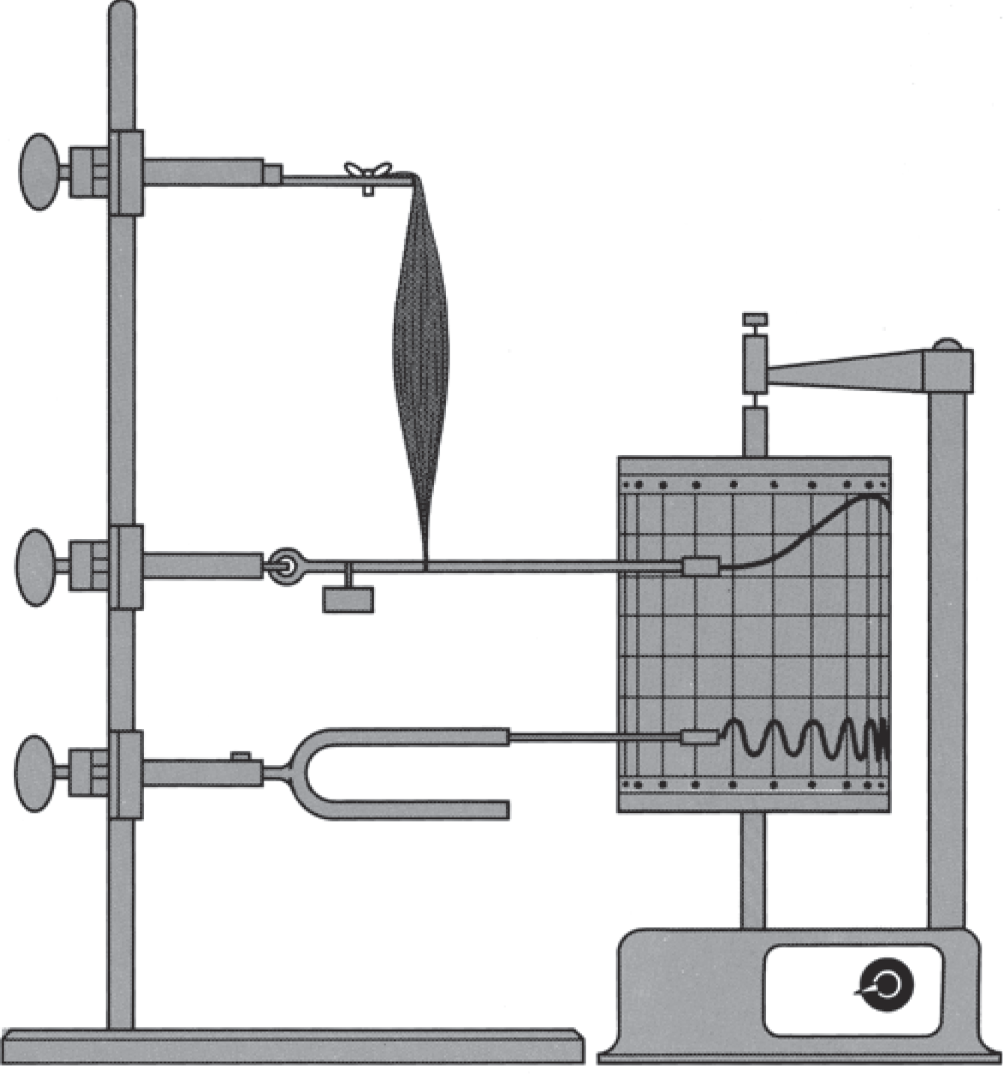
Figure 10.5 A kymograph is used in laboratory experiments to study muscle contractions. A pen, which is attached to a system of levers, records muscle contractions. The muscle can be stimulated with known amounts of electricity for known durations. Each muscle contraction is then recorded on a revolving drum. The muscle can be treated with chemical stimulants or depressants, and the muscle's reaction recorded. A timing device or a tuning fork creates a series of values for comparison and calibration.
SLIDING-FILAMENT THEORY
The contractile elements that make up most of the muscles' bulk consist of two proteins, actin and myosin. Alone, neither protein will contract, but together, they form an actomyosin complex that, in the presence of ATP, will contract. An individual muscle fiber is composed of many long thin myofibrils, each of which looks like a long ribbon with alternating light and dark bands. The wide light bands are the I-bands; they are composed of actin. In the middle of each I-band is a dark line called the Z-line. The broad dark bands, the A-bands, are composed of myosin. Each has a lighter H-zone through the middle. The unit from one Z-line to the next, along a single myofibril, is called a sarcomere (see Figure 10.6).

Figure 10.6 The muscle elements described in the text are illustrated with this breakdown of a muscle's organization.

Figure 10.7 Muscle contraction results when the protein polymers called actin and myosin slide together. Actin filaments are thinner than myosin filaments.
When the muscle contracts, the actin and myosin slide together with the light and dark areas overlapping. When the muscles relax, the proteins slide apart again (see Figure 10.7).
The contraction of a muscle fiber depends on the depolarization of a polarized, resting cell. A nerve carries an electrical signal that stimulates a transmitter at the neuromuscular junction. This stimulates a momentary reduction of polarization. The depolarized muscle cell admits an inflow of calcium ions (Ca++). The repolarization phase depends on the outflow of potassium ions (K+), and, at the same time, the calcium pump moves calcium ions back out of the cell. This wave of depolarization spreads across the nerve cell, stimulating the contractile elements to slide together.
EXOSKELETON
So far only the endoskeleton has been discussed. That is the skeletal structure of animals with cartilage or bones inside the body. However, there are many organisms, far more than the total number of vertebrates (those organisms with an internal bony and/or cartilaginous skeleton), that have tough, hard external skeletons, or exoskeletons; some are even jointed.
Arthropods (including insects) have an exoskeleton, with the hard covering outside the body and all muscles and organs located internally. The hard outer covering is noncellular. Secreted by the epidermis (outer layer of skin), it prevents excessive water loss, acts as armor protecting the creature, and provides sites for muscle attachment that can withstand the pressure and weight of muscle contraction. These organisms have movable joints that bend when their antagonistic muscles are contracted.
KEY TERMS
| A-bands | humerus | phalanges |
| actin | H-zone | potassium ions |
| actomyosin complex | I-bands | radius |
| anaerobic respiration | insertion | relaxation period |
| atrioventricular node | knee | ribs |
| bones | kymograph | sacrum |
| bundles | lactic acid | sarcomere |
| calcium carbonate | latent period | scapulas |
| calcium ions | ligaments | sinoatrial node |
| calcium phosphate | lumbar vertebrae | skeletal muscle |
| cardiac muscle | mandible | skeletal system |
| carpals | marrow | skull |
| cartilage | maximal stimulus | smooth muscle |
| caudal vertebrae | metacarpals | sternum |
| cervical vertebrae | metatarsals | striated muscles |
| clavicle | muscle cell | summation |
| coccyx | muscle fiber | sutures |
| coenocytic | muscle twitch | tarsals |
| collagen fibers | muscles | teeth |
| contraction period | myofiber | tendons |
| cranium | myofibrils | tetanus |
| endoskeleton | myoglobin | thoracic vertebrae |
| exoskeletons | myosin | tibia |
| fatigues | origin | ulna |
| femurs | osseous tissue | vertebrae |
| fermentation | oxygen debt | vertebral column |
| fibula | pacemaker | visceral muscle |
| glycogen | patella | Z-line |
| Haversian canals | pelvis | |
| heart muscle | peristalsis |
SELF-TEST
Multiple-Choice Questions
Bones and Muscles
- Which of the following helps protect organs, provides sites for muscle attachment, and lends structural support?
- skeletal system
- tendons
- ligaments
- heart muscle
- bone marrow
- In addition to bone, another kind of connective tissue comprising many skeletal systems, which is firm, though not as hard and brittle as bone, is ___________.
- calcium phosphate
- calcium carbonate
- cartilage
- marrow
- Haversian canals
- Blood vessels and nerves penetrate compact bones through small narrow openings, some of which are known as ___________.
- ligaments
- tendons
- Haversian canals
- tarsals
- phalanges
- Some bones are held together with fused joints, which are immovable, such as the ___________ located between several skull bones.
- tendons
- ligaments
- sutures
- cartilage
- ribs
Muscle Contraction, Sliding-Filament Theory, and Exoskeleton
- Some individual muscle fibers respond to stronger stimuli than others, so the stimulus that makes all the muscle fibers contract is known as the ___________.
- peristalsis
- summation
- contraction period
- maximal stimulus
- initial stimulus
- Sometimes, as is the case with involuntary muscle contraction, such as that in the throat when swallowing, the contractions comprising the wave of ___________ may occur without direct nervous stimulation.
- sinoatrial contraction
- maximal stimulus
- peristalsis
- latent contraction
- contraction period
- The time a muscle takes to relax before the muscle can contract again is the ___________.
- latent period
- contraction period
- fermentation period
- lactic acid period
- relaxation period
- The time a muscle takes to contract is the ___________.
- contraction period
- latent period
- fermentation period
- lactic acid period
- relaxation period
- The instrument used to record the intensity of muscle contractions over time is a(n) ___________.
- electrocardiogram
- electroencephalogram
- kymograph
- X-ray machine
- ultrasound machine
- Oxygen found in muscles is stored in ___________, a compound quite similar to hemoglobin.
- plasma
- white blood cells
- kymoglobin
- myoglobin
- cartilage
- Without available oxygen, working muscles continue to contract, deriving energy through a biochemical pathway known as ___________.
- anaerobic respiration
- aerobic respiration
- photosynthesis
- summation
- relaxation
- Anaerobic respiration, via fermentation, causes ___________.
- summation
- latency
- kymography
- lactic acid buildup
- tetanus
- After strenuous muscular activity, one breathes very deeply to acquire the needed oxygen to convert the potentially dangerous lactic acid to ___________.
- actin
- myosin
- actomyosin
- myofibrils
- glycogen
- The contractile elements that make up most of the muscles' bulk consist of two proteins that alone will not contract but together form a(n) ___________ that in the presence of ATP will contract.
- actin
- myosin
- actomyosin complex
- myoglobin
- hemoglobin
- An individual muscle fiber is composed of many long ___________.
- muscles
- tendons
- ligaments
- bones
- myofibrils
- The unit from one Z-line to the next, along a single myofibril, is called a(n) ___________.
- sarcomere
- A-band
- H-zone
- I-band
- actin
- A depolarized muscle cell admits ___________.
- calcium ions
- potassium ions
- magnesium ions
- sodium ions
- chloride ions
- A tough or hard external skeleton is called a(n) ___________.
- endoskeleton
- exoskeleton
- arthropod
- chitin
- cartilaginous skeleton
ANSWERS
- a
- c
- c
- c
- d
- c
- e
- a
- c
- d
- a
- d
- e
- c
- e
- a
- a
- b
Questions to Think About
- What substances are found in skeletal systems? Explain their function.
- How do muscles work antagonistically?
- The vertebral column is made of which types of vertebrae? Where are they located?
- What are the three types of muscle found among vertebrates? Give an example of each.
- What is the function of the pacemaker?
- Describe muscle contraction and relaxation.
- Explain the fundamentals of the sliding-filament theory.