2
The Skin’s Natural Process of Renewal
Your skin is constantly refreshing itself by producing new cells and sloughing off dead ones. The cycle takes twenty-eight days when you are young. As you age, cell renewal gradually slows down. By the time you are fifty, the process takes twice as long as it did when you were in your twenties. Old cells are more vulnerable to the internal processes of aging. The Lancer Method is based on the principle that if you stimulate the skin’s built-in repair system, you can reverse some of the visible signs of aging and slow down the rate of future damage. If you back up your skin care program with an anti-aging lifestyle, you will weaken the forces that drive biological aging while you harness your skin’s own healing power.
As you will see, the skin is made up of three layers: epidermis, dermis, and subcutaneous tissue. Most anti-aging regimens and products focus solely on the dermis, and that is why they do not give you the results you are hoping for. My thirty years of clinical experience have shown that you must treat the epidermis to rejuvenate and repair the skin, specifically the stratum corneum. Without this crucial step, no regimen or product will work. Now, before we explore exactly how to treat the skin, it is important for you to understand the skin’s structure and how it renews itself.
SKIN REFRESHER COURSE
Most people think of their skin as a simple outer covering rather than the complex, vital organ that it is. It’s easy to take skin for granted, focusing on how it looks and being unaware of all that it does. Your skin is the largest organ of your body, makes up 15 percent of your body weight, and covers a total area of about twenty square feet. It offers the first line of defense against the external environment, acting as a protective shield from light, heat, injury, and infection. Skin operates as a two-way protective barrier that prevents microbes and harmful environmental elements from entering your body, and prevents water from exiting. Among its many other functions, the skin regulates body temperature, stores water and fat, and operates as a sensory organ that permits the sensations of touch and heat. Skin gathers and communicates sensory information that stimulates an immune response to protect you from disease. Skin plays a major role in your overall health.
Your skin is an entire community with a variety of different cells, blood vessels, nerves, muscles that make hair stand up, and an intricate communication system. If you want to look younger, you have to understand the anatomy of the skin.
Cross Section of the Skin
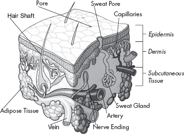
The skin consists of three layers. The epidermis, the outer layer, plays a big role in the Lancer Method. Below the epidermis is the middle layer of skin, called the dermis; the layer beneath that is called subcutaneous tissue or the hypodermis. Let’s examine how these layers interact.
The Hypodermis or Subcutaneous Layer
Starting from the deepest layer and moving up to the surface, we will first examine the hypodermis. This level is a network of collagen, connective tissue, and fat cells. The subcutaneous layer houses larger blood vessels and nerves that bring nourishment and sensation to the skin. Containing about 50 percent of the body’s fat, this layer varies in thickness from person to person. The subcutaneous tissue conserves body heat and acts as a shock absorber to protect your organs from injury. The hypodermis attaches the skin to the underlying bones and muscles.
The Dermis
There are many special cells and structures located in the middle layer, the dermis, which contains blood vessels, lymph vessels, hair follicles, sweat glands, sebaceous oil glands, scent glands, and the nerves responsible for the sensations of pain, itchiness, and temperature. Collagen, composed of bundles of protein, is the main component of the dermis and the most abundant protein in the body. The dermis is held together by collagen and elastin, another protein. These proteins keep your skin supple and resilient. Collagen fibers give support to the skin, and elastin provides flexibility and strength. Elastin gives your skin the bounce to return to its original position when pinched or poked. Collagen and elastin break down with age. When this happens, your skin loses volume and begins to sag and wrinkle.
The top level of the dermis is called the papillary region. The finger-like projections from this region extend to the epidermis and strengthen the connection between the two layers of skin. In the papillary region collagen is perpendicular to the skin’s surface and elastin is horizontal, forming a latticework. The reticular region below is thicker and dense with collagen and elastin. Both types of fibers are arranged parallel to the surface of the skin in this layer.
Most anti-aging and skin care programs try to fight aging by targeting the dermis, the location of collagen and elastin production. My skin care program instead focuses on the epidermis, particularly its outermost layer, and the stratum corneum. I have discovered that if you want to effect significant change, the dermis is not where the action is.
The dermis interacts with the epidermis via a signaling system that not only stimulates the immune system but also promotes the repair and remodeling of skin when injured. The Lancer Method uses this signaling system to restore your skin.
The Basement Membrane or the Dermal/Epidermal Junction
This porous membrane allows and controls the exchange of cells and fluid between the dermis and the epidermis. Holding the two layers together, the membrane is a highly irregular connection that undulates, increasing the surface area over which nutrients, oxygen, and waste products can be exchanged. The basement membrane has an important function in skin rejuvenation, because it regulates cell migration, an important process of regeneration that occurs in the epidermis.
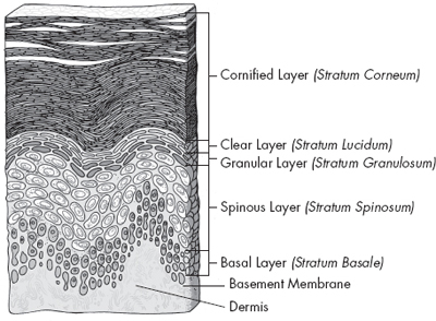
The Epidermis
We will examine the epidermis closely, because its actions are highly important to the Lancer Method. As you can see in the illustration on here, the outer layer of skin is composed of either four or five layers, depending on the location of the skin. From the bottom up, they are: the basal or germinal layer (stratum basale); the spinous layer (stratum spinosum); the granular layer (stratum granulosum); the clear layer (stratum lucidum), a translucent layer found only on the lips, palms, and soles of the feet; and the cornified or horny layer (stratum corneum). We are most concerned with the top layer, the cornified layer, for reasons I will explain.
Basal Layer
The basal layer contains Merkel cells, which are believed to be involved in the sensations we feel through our skin. Melanocytes are also found in the bottom layer of the epidermis. Those cells produce melanin, a brown pigment that gives skin its color and protects against ultraviolet rays.
The basal cells continually divide to form new keratinocytes that replace old cells, which are shed from the skin’s surface. Keratinocytes produce keratin, a long thread-like protein that makes the skin strong and has a protective role. Keratinocytes, which are shaped like columns, attach to the basement membrane zone. These cells divide and make the cells that move up to the outer layers of the epidermis.
Spinous Layer
The spinous layer is responsible for vitamin D synthesis. Langerhans cells found in this layer are the immune system’s front line of defense. As the new cells move through this layer, they become flatter.
Granular Layer
As they move through the spinous and granular layers, keratinocytes become increasingly flatter and their nuclei begin to degenerate. In this layer, they become cells composed of granules that are filled with proteins that promote hydration to keep the skin moist. The cells secrete a “cement” made of fats, fatty acids, and cholesterol, which fills the spaces between the cells and increases cohesion, making the epidermis an effective waterproof barrier.
Cornified Layer
By the time the granular cells reach the cornified layer, the completely flattened cells have become corneocytes without nuclei. Although these cells used to be considered dead, we have learned that they are still active and contain special organelles to help maintain stability. The compact layer, which is the lower, contains cells that are linked to one another. In the outer, sloughing layer, those links break down, and the cells gradually flake off.
This process of generation, maturation, and death of a cell takes about a month. As you age, cell renewal slows down and dead cells build up on the surface of the skin. This accumulation of dead cells makes skin appear dull.
Stratum Corneum Superstar: A New Assessment
In the past, the cornified layer was considered simply a dead layer of skin, a graveyard of the inactive skeletons of keratinocytes that had completed their upward journey from the basal layer. It turns out that this layer is more than a lattice of dead keratin. The significance of this outermost layer had been overlooked for decades. Researchers now know that the cornified layer has biological properties responsible for maintaining healthy skin. Rather than being inert, the cornified layer functions as a vital barrier that regulates water loss; prevents damage from external oxidants; limits infection through peptides that kill microorganisms; has hormone receptors; launches an immune response to invading microbes, antigens, and allergens; and protects cells below from damaging ultraviolet light. The Lancer Method focuses on the cornified layer’s role in stabilizing the skin and maintaining its structure. This is the secret at the heart of the Lancer Method, which uses the stratum corneum’s messaging system to revive your skin and give it a youthful appearance.
Cell Turnover in the Epidermis
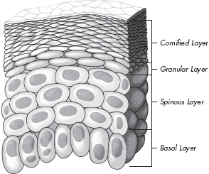
The epidermis normally never stops regenerating. Cells shaped like columns in the basal layer divide and push already formed cells into higher levels, where they flatten out. If you look at the shape of the cells, you will see that the closer they are to the skin’s surface, the flatter they become.
The skin, like the rest of the body, actively maintains homeostasis—in other words, a stable condition. With the dynamic and continual process of cell formation in the epidermis, the skin maintains a constant number of cells. When dead cells flake off the cornified layer, they are replaced by the newly formed cells moving up from the granular layer. Homeostasis is regulated in part by the dermis. Since the epidermis does not have blood vessels, the outer layer of skin cannot initiate the production of new cells. Feedback loops and sensors in the cornified layer detect when the balance is off or when injury occurs. Hormones, growth factors, and cytokines do the signaling to the dermis, which then drives cell production. The cornified layer communicates with the dermis through these feedback loops to stimulate the skin’s self-repair mechanism. The Lancer Method uses this natural process to speed up the production of new and healthy skin cells. The next chapter provides you with an in-depth explanation of how harnessing the skin’s own healing process actually works.
HOW THE SKIN STAYS HYDRATED
The epidermis is responsible for keeping skin hydrated. Water is vital for the proper functioning and maintenance of the epidermis, and affects the outward appearance of healthy skin. If there is too much water, skin cells break down because of increased permeability. As you can imagine, too little hydration results in dryness. The epidermis contains the underlying mechanism that controls hydration, maintaining a balance between water retention and water loss. This complex process is still not fully understood by modern scientists, but we are able to use what knowledge we do have to our benefit.
Throughout the body, there are channels called aquaporins that facilitate the passage of water and small molecules. There are thirteen different types of aquaporins in your body. We are concerned with aquaporin 3 (AQP3), which are abundantly found in the membranes of the keratinocytes in the epidermis, primarily in the stratum basale. Moving toward the skin’s surface, the number of these channels decreases with each layer of the epidermis. Water in the stratum corneum makes up only 10 to 15 percent of the content, compared with 75 percent in the underlying layers of the epidermis. This distribution allows living layers of the epidermis to remain well hydrated while water loss is minimized.
Skin Hydration through Aquaporin 3 Channel
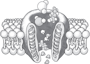
This closeup of an aquaporin 3 shows water and glycerol entering through a cell membrane.
AQP3 transport water and glycerol into the cells of the epidermis. Glycerol acts as a humectant, a substance that promotes the retention of water. As the glycerol diffuses into the stratum corneum and underlying epidermis, it pulls water with it, hydrating the skin. This action provides elasticity and helps barrier repair in the epidermis. Glycerol also affects metabolism in the stratum corneum and the composition of lipids. With age or chronic sun exposure, the number of aquaporins decreases, which accounts for the dryness visible in aging or sun-damaged skin.
The stratum corneum contains natural moisturizing factors, like urea, which contribute to maintaining the water level in the epidermis. Urea is a humectant that attracts water to the skin, particularly from the dermis. Most dermatologists focus on topical products to keep the skin hydrated. Glycerol and urea are ingredients in creams, lotions, shampoos, ointments, pastes, foams, and body washes. The Lancer Method not only prepares the skin to absorb moisturizing agents, but also increases the metabolism in the skin that helps to transport the active ingredients in topical treatments. As the Lancer Method speeds up production of new keratinocytes, the entire machine is upgraded. The aquaporins remain very active, and glycerol and urea continue to be produced, keeping the skin well hydrated.
You cannot be passive when it comes to taking care of your skin. Now that you have been introduced to the science behind the Lancer Method, it’s time to act. The next chapter will explain what you have to do to tap into your skin’s natural healing power to get the Lancer Glow. You will learn my simple technique—when and how to awaken your skin. Get ready to bring your skin back to life.