© C. GRIFFITHS
28 |
Text by Sanet Janse van Vuuren, Jonathan Taylor and Jenny Day |
|---|---|
Protista form a large group of diverse organisms that includes photosynthetic algae and heterotrophic (animal-like) protozoans. The relationships between these groups are not well understood and the term ‘protist’ is thus best used only as a common name. Protists are eukaryotic, meaning that their DNA is enclosed in a nucleus within the cell, and mitochondria are used for cellular respiration. Some obtain energy by photosynthesis while others feed on organic material. All require oxygen for respiration.
This diverse group of autotrophs (make their own food using light or chemical energy) is responsible for almost 50% of global photosynthesis. Sizes range from microscopic (<1mm) to metres in length (marine kelps). Unlike plants, algae lack transporting tissue and are usually associated with aquatic environments. Many are non-motile, but some smaller species can swim, glide or creep. There may be 30,000–1,000,000 species, or more. They occur almost everywhere and play a vital role, serving as the foundation for aquatic food chains.
© C. GRIFFITHS
Usually bright green in colour and often forming blooms (1) that turn the water green. Cells may be single, or clumped together to form colonies, filaments, or more complex structures. Cells are surrounded by cell walls. Chloroplasts usually contain storage structures with starch around them. Some are non-motile; others swim using flagella; a few glide. Asexually and sexually reproducing types occur. They are a major group of algae, both with respect to numbers of genera (500) and species (8,000), and in terms of their ecological importance.
Unicellular and colonial green algae
While the seaweeds are mostly marine, many microscopic forms, both solitary and colonial, are found in fresh waters.
2 Chlamydomonas
Chlamydomonas species

© S. JANSE VAN VUUREN
© P. SKALOUD

© P. SKALOUD
© S. JANSE VAN VUUREN

© A. KRUGER
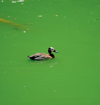
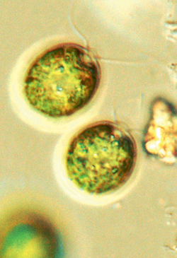
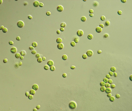
Unicellular; cells small, spherical, ovoid or ellipsoidal in lateral view and circular or slightly flattened in outline. Anterior end with two flagella of equal length, usually longer than cell. Single cup-shaped starch-containing chloroplast present, with red eyespot embedded within it. Species distinguished by position of chloroplasts and number and position of starch-storing organelles. Size: Cells mostly 5–15µm (micrometres) in length. Biology: Widely distributed in every kind of aquatic habitat. Form blooms, especially in nutrient-rich waters. RELATED GENERA: Chlorella species (2a) are similar, but lack flagella. Carteria species (2b) also similar, but possess four flagella. In species of Haematococcus (2c), the cytoplasm is connected to the cell wall with fine fibrils, and the space between cell wall and protoplast is filled with colourless watery mucilage. Cells normally green, but red pigmentation, which develops if conditions become unfavourable, may discolour bird baths (2d) and garden ponds.
3 Ankistrodesmus
Ankistrodesmus species
© S. JANSE VAN VUUREN
© S. JANSE VAN VUUREN
Cells solitary, loosely clustered or twisted around each other in bundles of 4–32 cells. Individual cells needle-like, tapering at both ends, and straight, curved or spiralling. Species distinguished mostly by cell shape and size. Size: 25–60µm in length × 1–6µm in width. Biology: Widespread and common in plankton of all types of water bodies. RELATED GENERA: Monoraphidium cells are strongly curved, while Ankistrodesmus cells are more or less straight, or slightly curved. Actinastrum hantzschii (3a) forms star-shaped colonies of 4, 8 or 16 cylindrical cells radiating from a central point of attachment. Cells shorter (10–25µm) than those of Ankistrodesmus.
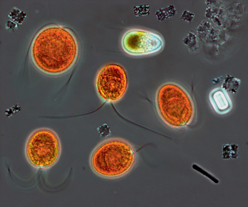
4 Salt lake alga
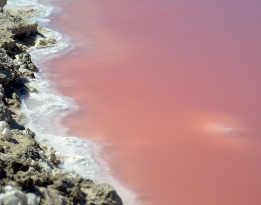
Dunaliella salina

© M. DYALL-SMITH
© M. GRIFFITHS
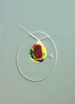
Cells solitary, spherical to pear-shaped, with two flagella. Characteristically bright orange or red, due to pigments in the cell. Size: 10–12µm in length. Biology: Cosmopolitan; confined to highly saline pans and salt works. Cells contain carotene pigment and glycerol, which protect against high salinity. Lakes or pans with tomato red water (4a) are sure to be hypersaline and to contain Dunaliella. (Purplish to pinkish waters are even saltier and contain purple photosynthesising bacteria).
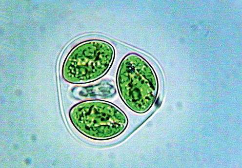
1 Oocystis
Oocystis species

© S. JANSE VAN VUUREN
Cells solitary or in oval to ellipsoidal colonies of 2–16 cells enclosed in mother cell wall from previous generation. Cells broad, ellipsoidal or oval, or with both ends slightly pointed; with one to many chloroplasts. Cell wall thick and smooth. Species differentiated by size and form, degree of thickening at poles, and number of chloroplasts and starch-storing organelles. Size: 12–50µm in length and 7–46µm in width, colonies up to 77µm in diameter. Biology: Planktonic; widespread and common in pools, ditches, lakes and slow-flowing rivers.
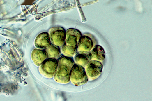
2 Pandorina
Pandorina species

© S. JANSE VAN VUUREN
Colony oval, ellipsoidal or spherical, surrounded by broad zone of clear mucilage, through which flagella protrude; 8–32 densely packed cells; no hollow at centre of colony. Cells triangular to pear-shaped, each resembling Chlamydomonas (p.314, 2). Size: Cells 8–20µm in length, colonies up to 100µm in diameter. Biology: Common in puddles, ponds, lakes and slow-flowing rivers.
3 Scenedesmus
Scenedesmus species


© S. JANSE VAN VUUREN
Non-motile colonies of 2, 4, 8 or 16 long cylindrical cells arranged in single or double rows. Cells with rounded or pointed ends. Terminal cells may have spines that decrease sinking rate and deter predation. Species distinguished by number, arrangement and size of cells and cell wall patterns. Size: Cells mostly 5–30µm in length and 2–10µm in width; spines up to 200µm. Biology: Common to abundant in phytoplankton of freshwater ponds, lakes and rivers.
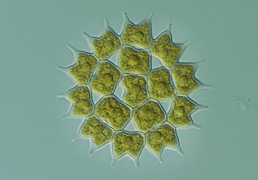
4 Pediastrum
Pediastrum species

© K. BRUUN
Disc-shaped non-motile colonies of 4–64 cells arranged in one layer. Peripheral cells may bear horn-like projections that decrease sinking rate and prevent predation. Cell walls smooth, finely reticulated or highly granulated, wrinkled or notched. Size: Cells 8–32µm in length, colonies up to 200µm in diameter. Biology: Widespread and common in standing and slow-flowing waters, where they are free-floating, often among plants or other algae. May form blooms in nutrient-rich environments.
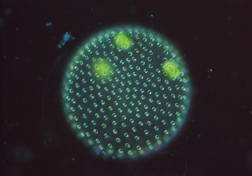
5 Volvox
Volvox species

© K. BRUUN
Free-swimming Volvox colonies can be seen with the naked eye. The hollow spherical colonies contain a peripheral layer of 500–50,000 Chlamydomonas-like cells. Daughter colonies develop inside the sphere. Size: Cells mostly 4–8µm in diameter, colonies 0.5–1.5mm in diameter. Biology: Widespread and common in slow-flowing nitrogen-rich water, especially during late summer. The largest free-swimming, colonial green algae; can impart a fishy odour to water.
Desmids
Green algae in which the cells are single but divided into two semicells (mirror images), with a prominent nucleus between them. Each semicell has at least one chloroplast. In acidic low-nutrient waters; sometimes in alkaline nutrient-rich waters.
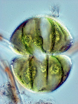
6 Cosmarium
Cosmarium species

© S. JANSE VAN VUUREN
© S. JANSE VAN VUUREN
Semicells rounded from the front and flattened, oval or elliptical from the side. Surface smooth or with granules, pits, or warts; no spines or apical indentations. Solitary, but sometimes in gelatinous macroscopic colonies. Size: Cells 10–200µm in length, 6–140µm in width. Biology: Very widespread and common, with >1,000 species. Mostly free-floating in lakes, reservoirs, ponds, sometimes rivers. RELATED GENERA: Micrasterias (6a) is most striking of all desmids. Edges of semicells variously incised. Large cells, up to 400µm long and 60–200µm in width.
7 Euastrum
Euastrum species

© S. JANSE VAN VUUREN
Semicells are oval, elliptical or pyramid-shaped and distinctly compressed when viewed from the side. Poles mostly with a deep median incision; margins usually incised. Most species identified by details of polar incision and protrusions on face of semicell. Size: Cells 10–200µm in length and 10–100µm in width. Biology: Present in the benthos of mildly acidic waters, mostly in weedy pools. Some related taxa found in alkaline waters.
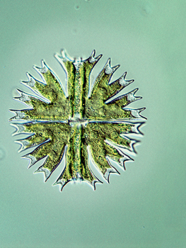
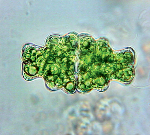
8 Staurastrum
Staurastrum species

© K. BRUUN
Each semicell has 2–10 or more hollow projections, ranging from short and stumpy to long and arm-like, often covered in warts or spines. Tip of each projection has short spines. The genus exhibits enormous variety in structure, and several attempts have been made to divide it into smaller genera. Size: Cells 15–120µm in length (excluding projections). Biology: Blooms of Staurastrum can impart a grassy odour to water.
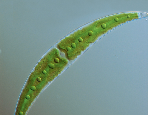
1 Closterium
Closterium species

© K. BRUUN
Cells elongated and crescent-, sickle- or bow-shaped, with varying degrees of curvature. Ends of cells usually tapered and may be acutely pointed, rounded or truncate. Cells always longer than wide. Median constriction, dividing cell into semicells, insignificant. Each end of cell has a conspicuous vacuole, within which one or more crystals of barium or calcium sulphate may be observed vibrating erratically; their function is unknown. Closterium can perform somersaulting movements by secreting mucilage from ends of the cell. Size: Cells 70–1,200µm in length and 4–50µm in width. Biology: Cosmpolitan; common in acidic oligotrophic lakes and ponds, rarely in more alkaline eutrophic environments.
2 Netrium
Netrium species

© S. JANSE VAN VUUREN
Cells are usually elongated and cylindrical with rounded ends, resembling a watermelon or cucumber. Each cell has 2–4 large chloroplasts that appear frilly due to their elaborate lobes and ridges. The cells can excrete large amounts of mucilage. Size: Cells 35–430µm in length. Biology: Cosmopolitan; abundant in acidic, oligotrophic, aquatic habitats or in Sphagnum (p.310, 2) bogs, with occasional records from more alkaline habitats.
Filamentous green algae (pond scums)
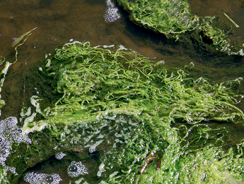
Many species of green algae form long filaments of cells attached end-to-end. They contribute to the biodiversity of ponds by providing refuge for numerous smaller algae, invertebrates and protozoans, but can become pests when abundant, usually the result of elevated nutrient concentrations in the water. Some, such as Spirogyra (see 3, below), remain submerged, but others may be forced by trapped bubbles of oxygen or methane to rise to the surface, where they can form thick, glutinous, slimy green mats, the surface cells of which whiten as they die in the sun. Submerged agglomerations may be silky, rough or slimy to the touch. A bale of barley straw has been shown to prevent growth of many filamentous species in ponds.
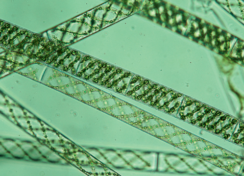
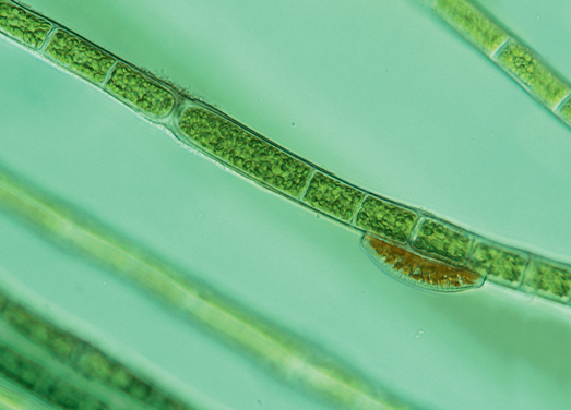
3 Water silk (Blanket weed)
Spirogyra species

© S. JANSE VAN VUUREN

© C. GRIFFITHS
© S. JANSE VAN VUUREN

© S. JANSE VAN VUUREN
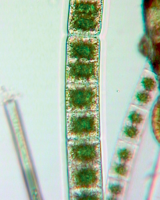
Long unbranched filaments that feel slimy to the touch. Cells cylindrical, with one or more spiral chloroplasts and numerous starch-storing structures. Edges of chloroplasts may be ruffled. Nucleus positioned in centre of cell, together with large central vacuole. Species differentiated by size and shape of cells, number of chloroplasts and reproductive details. Size: Cells up to 200µm in length and 10–150µm in width. Filament length varies, due to easy fragmentation. Biology: Widespread and common in standing water, but also present in streams of neutral to low pH. Filaments often form green clouds below water’s surface (3a). Blooms can impart a grassy odour to the water, block canals and clog filters. RELATED GENERA: Zygnema filaments (3b) are similar, but have two star-shaped chloroplasts per cell, while Mougeotia filaments (3c) have rectangular chloroplasts, able to rotate relative to light.
4 Cladophora
Cladophora species

© S. JANSE VAN VUUREN
© A. SWANEPOEL

© J. DAY
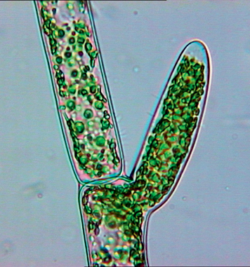
Unlike Spirogyra (see 3, above) filaments are branched and feel coarse (not slimy) to the touch. Filaments free-living, or attached by a disc-like holdfast or rhizoids. Cells cylindrical, with disc-shaped chloroplasts interconnected to form net-like pattern. Many nuclei per cell. Size: Cells 80–90µm in length, 25–80µm in diameter. Filaments may range in length, but growths as long as 20m have been observed. Biology: Filaments prefer nutrient-rich flowing water, where they adhere to hard surfaces, such as rocks, logs and concrete walls. Often form dense masses (4a) that can become a nuisance when decomposing, generating a septic odour that may be confused with raw sewage. Dry mats on desiccated temporary ponds appear as thick grey felt (4b). Cladophora glomerata is regarded as one of the worst nuisance algal species and forms large growths in irrigation canals all over South Africa.
1 Ulva (= Enteromorpha)
Ulva species

© C. GRIFFITHS
Macroscopic algae consisting of long hollow tubes that branch frequently. Free-floating or attached to submerged aquatic plants, sticks and stones in dams or slow-flowing streams. Cells compactly arranged in outer layer of tubular thallus. Each angular cell has a cup-shaped or plate-like chloroplast. Size: Cells 8–20µm in diameter, tubular body 2–20mm in diameter. Long trailing filaments may be up to 1.5m in length. Biology: Few freshwater representatives, most species being marine. Normally favour calcareous, brackish or saline waters. Thick mats, which sometimes clog streams, develop as a result of elevated nutrient levels. Enteromorpha has recently been incorporated into Ulva (sea lettuces), previous literature considering one-cell-layer-thick, tubular species, like this freshwater form, as belonging to Enteromorpha, and two-cell-layer-thick laminar forms to Ulva.
2 Microspora
Microspora species
© S. JANSE VAN VUUREN
Unbranched filaments, usually a single cell layer thick and not attached. Cells cylindrical, with thickened H-shaped cell walls. Chloroplast single and perforated, giving a net-like appearance. Size: Cells 10–20µm in diameter. Biology: Usually tangled among other filamentous algae in cool unpolluted streams. Found mostly in cooler seasons in acid waters.
Water nets
Extraordinary freshwater algae with extremely large cells, up to 15mm across, joined to form a network, each cell consisting of numerous nuclei, chloroplasts and starch grains. Reproduction asexual or sexual. Five species currently recognised.
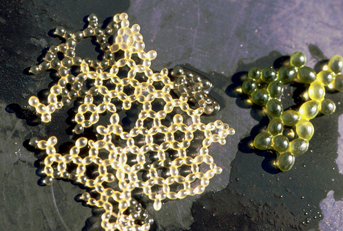
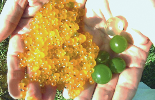
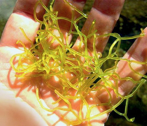
3 Cape water net
Hydrodictyon africanum
© M.A.P. JOSKA
© A. VISAGIE
A remarkable macroscopic alga consisting of a network of extremely large oval cells (3) swelling to form bright green grape-like spheres that turn orange-yellow with age (3a). Cells start off connected, but become separated as they grow. Size: Individual cells 10–15mm in length, swelling to spheres of the same diameter. Biology: In temporary still waters, surviving desiccation as resistant spores. Apparently endemic to the Cape Floristic Region. Very seldom encountered and, given threats to wetlands in the area, probably fits the IUCN Endangered category.
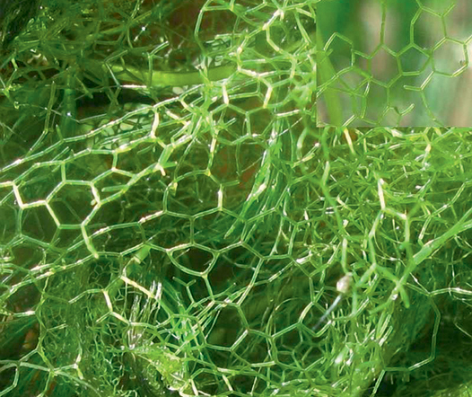
4 European water net
Hydrodictyon reticulatum
© N. JOB
© P.E. HANSEN (BOTH)
Large, oblong-oval cells joined end-to-end in pentagonal or hexagonal meshworks of connected rings, forming a ‘net’. Size: Individual cells 10–15mm, net (4a, inset) tens of centimetres across. Biology: New nets form within the mother cells. A highly invasive European species, its range is extending in many parts of the world. Apparently not found in temporary waters.
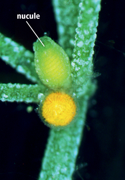
Large, bright green, complex algae resembling and often confused with plants. Some are rough and coarse to the touch, because they become encrusted with deposits of lime, hence the name ‘stonewort’; some also called musk grasses because of their foul, musky, garlic-like odour. They grow submerged and erect, anchored to bottom sediments with rhizoids (root-like structures). The central axis is divided into nodes and internodes, with whorls of side branches at the nodes. Most have both male and female structures, which are borne at junctions of main stem and side branches. Male structures (‘globules’) turn orange before sperm are released. Green female reproductive structures (‘nucules’) are situated above the globules and each carries a single egg cell.
5 Chara
Chara species

© R. VAN DER HOFF
© S. JANSE VAN VUUREN
Side branches not themselves branched. Below each node are unicellular outgrowths (stipulodes), varying in length and size, situated in one or two rows. At apical end of nucule (5a) is a crown of five cells. Size: 5–100cm or more in length. Biology: Most species confined to high pH, calcareous waters, and their remains contribute to calcium-rich sediments.
6 Nitella
Nitella species


© C. GRIFFITHS
Resembles Chara in appearance, size and biology, but may occur in acidic peaty waters. Central axis without a distinct outer layer and no stipulodes present below nodes. Side branches often also branched. Apical end of nucule consists of 10 crown cells, arranged in two tiers of five cells each.
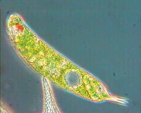
Unicellular organisms, usually with numerous bright green chloroplasts and a conspicuous red eyespot anteriorly. Motile cells with two flagella inserted anteriorly, one being short and inconspicuous. Several euglenoids are known for their characteristic undulating, shape-changing motion. Food reserves stored in round or rod-shaped granules in the cells. Useful bio-indicators of high nutrient levels. Exhibit only asexual reproduction. About 40 genera and 800 species in the phylum.
1 Euglena
Euglena species

© S. JANSE VAN VUUREN
© ROGELIO MORENO G
Single elongate cells, rounded anteriorly and pointed posteriorly. Move slowly, using a single emergent flagellum implanted into gullet. Some species can change shape. Size: Cell 20–540µm long and 5–50µm wide. Biology: Widespread and common, mostly in water rich in organic materials. Some species heterotrophic (engulfing organic matter or absorbing nutrients from surrounding medium). RELATED GENERA: Phacus (1a) cells are flattened with a straight or slightly bent tail of variable length; cell cannot change shape.
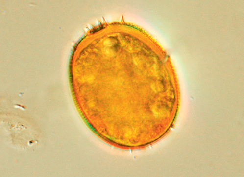
2 Trachelomonas
Trachelomonas species
© S. JANSE VAN VUUREN
Similar to Euglena, but in a protective case (lorica), varying in colour from yellow to dark brown or red. The lorica may have different shapes in various species and genera, sometimes with a distinct collar arising near apical pore, and its wall may be smooth, or ornamented with spines, warts or pits. Size: Cell 5–100μm in diameter. Biology: Common in acidic to neutral waters (pH 4.5–7), often in peaty pools and other habitats rich in iron and manganese.
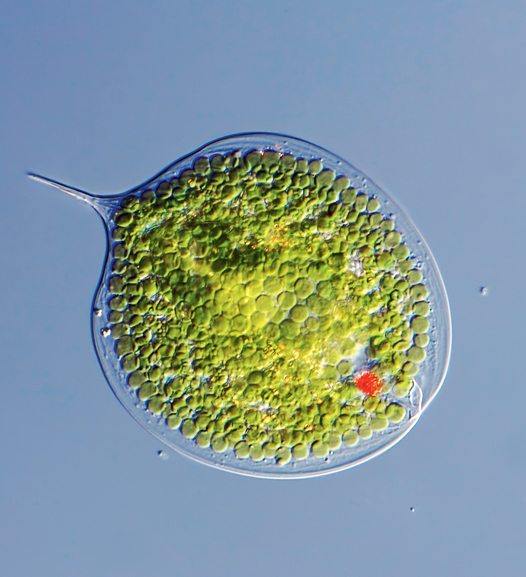
Unicellular; photosynthetic (with golden-brown chloroplasts) or non-photosynthetic. Cells motile, with two flagella located inside grooves on cell surface. Cells naked, without plates or covered with conspicuous plates: presence, thickness and arrangement of plates used in classification. Reproduction usually asexual. More than 2,000 known species, 90% of which are marine.
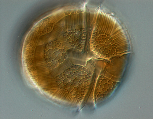
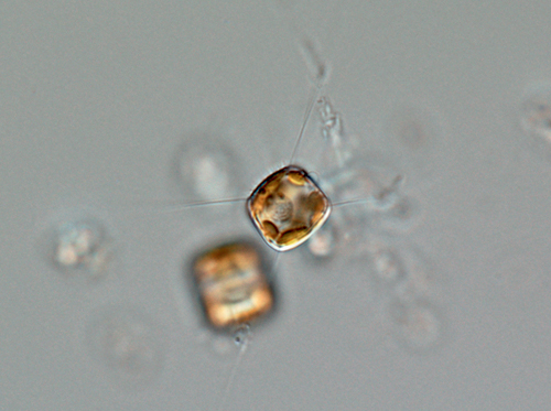
3 Ceratium
Ceratium species


© S. JANSE VAN VUUREN
© K. BRUUN
Most common of all freshwater dinoflagellates; easily identified by their shape: cells large, strongly flattened and curved if viewed from the end, and with one anterior and 1–3 posterior horns. Narrow grooves house two flagella, allowing Ceratium to swim slowly. Size: Cells up to 450µm long and 30–100µm wide. Biology: Common in nutrient-rich freshwater habitats. During blooms water often coloured brown (similar to ‘red tides’ in the ocean, but not toxic). RELATED GENERA: Peridinium species (3a) are similar, but lack horns.
Most red algae are marine and benthic, but a few species have been reported from freshwater habitats, mainly in running waters. Although the prefix ‘rhodo’ refers to the colour red, freshwater representatives are commonly grey, violet-green or tan. Most are multicellular and macroscopic, consisting of simple or branched filaments. They do not have flagellated cells, are structurally complex, and have complicated life cycles.
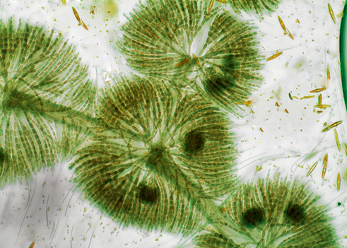
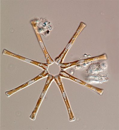
4 Frog-spawn alga
Batrachospermum species

© C. CARTER

© M. KELLY
One of few genera of freshwater red algae, ranging in colour from olive green, violet or grey to reddish brown. Extremely slimy to the touch. Main axis consists of a single column of cylindrical cells bearing dense whorls of short branches (4) that often contain even denser masses of branched filaments, giving appearance of a bird’s nest. The branches resemble beads on a string and have a slippery texture, hence the name ‘frog-spawn alga’. Size: Length usually 1–10cm, may reach 40cm; strands up to 2mm wide. Biology: Widespread; sometimes attached to stones in cold lakes, but more often at bottom of cold-water streams (4a). Usually associated with good environmental conditions, such as low nutrients, clear, cool water and diverse invertebrate communities, but occasionally proliferating in enriched conditions. Many species recently transferred to a new genus, Sheathia.

© J. TAYLOR

© J. TAYLOR
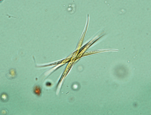
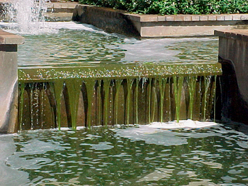
A group of algae characterised mainly by possession of a unique silica cell wall divided into two halves and connected by a series of silica bands forming a girdle. Each half cell wall is termed a ‘valve’ and the complete cell a ‘frustule’. Diatoms are ubiquitous, occupying most damp and aquatic habitats globally. They are among the most common species of protists in benthic and planktonic communities. Cells may be solitary and free-moving, and/or attached to the substrate by means of mucilaginous stalks. When attached to rocks and other solid substrata they may form a thick golden-brown layer (1, 1a). The cells may also form chains, or colonies of various other shapes, able to float in the water column as part of the phytoplankton. Diatoms are divided into two groups, based on ornamentation and symmetry of the cells – centric diatoms are round with radial symmetry, while pennate diatoms have an elongated shape and bilateral symmetry. Pennate diatoms often possess a slit-like structure in the cell wall, known as the ‘raphe’, which allows them to perform gliding motions. Individual cells can range in size from just a few micrometres (µm) to 500µm (0.5mm) and have a large variety of shapes. The view of cells from the top is known as the ‘valve view’, while that from the side is known as ‘girdle view’. Reproduction can be asexual or sexual. Diatoms are important sources of food for aquatic grazers, such as amoebas (see pp.326–328) and other protozoans, and are important primary producers in many ecosystems. When the cells are cleaned of their organic content, tiny perforations are revealed in the cell wall, often forming highly complex and beautiful patterns (e.g. Cymbella, p.326, 2). As a result, diatoms have always been popular subjects for microscopic study. Each diatom species has very specific water chemistry requirements, often tolerating rather narrow ranges of aspects such as salinity, pH and nutrient concentrations. As a result, diatoms are excellent bio-indicators of various aspects of water chemistry and are used in South Africa in routine assessments of the ‘state of the rivers’. Because of their hardness, diatom frustules can remain in sediments for many hundreds or thousands of years and are used to study changes in environmental conditions in lakes and wetlands over time. Approximately 11,000 freshwater species have been described, with perhaps 25,000–100,000 additional species yet to be described. About 1,800 species are known from South Africa.
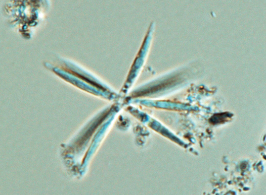
2 Asterionella
Asterionella formosa

© J. TAYLOR
Star-shaped colonies, with each cell attached at base to its neighbor by a mucilaginous pad. Individual cells swollen at each apex. Each cell contains a number of disc-shaped plastids (chloroplasts). Size: 30–160µm long and 1.3–6µm wide. Biology: Commonly found in the plankton of reservoirs slightly enriched with nitrogen and phosphorus and also en masse at some times of year in large slow-flowing rivers, such as the Vaal.
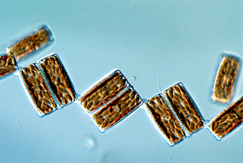
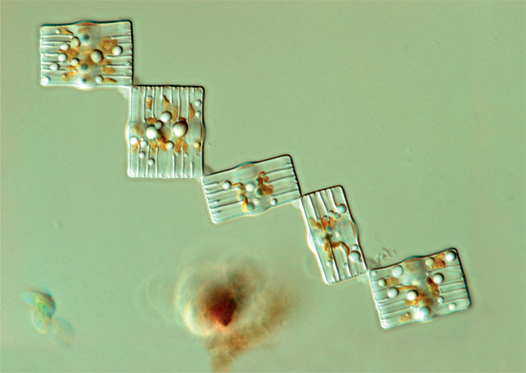
3 Diatoma
Diatoma vulgaris

© J. TAYLOR

© J. TAYLOR

© J. TAYLOR
Cells rectangular in girdle view and elliptical in valve view, most commonly encountered in girdle view, as cells are linked by corners to form zig-zag chains, similar to Tabellaria (see 5, below). Valve face crossed from side to side by thickenings or ribs, which can be seen in living cells. Plastids numerous and oval. No septa on girdle bands. Size: 8–60µm long and 7–18µm wide. Biology: Often occur en masse (3a) during winter months in nutrient-rich streams around South Africa. May completely cover hard substrata and even submerged tree branches (3b).
4 Cyclotella
Cyclotella meneghiniana

© J. TAYLOR
© J. TAYLOR
Circular in valve view (4), rectangular in girdle view (4a), usually occurring as solitary cells or pairs. Plastids small, numerous, disc-shaped. Cell maintains position in water column by extruding chitinous threads, many times longer than the cell, through specialised pores. Differentiated from other centric genera by lines on the valve face separated by ribs arranged radially around the periphery of an unornamented central area. Size: 5–43µm in diameter. Biology: Occurs chiefly in plankton of large, nutrient-rich, slow-flowing rivers; may also be found in the benthos. Common, cosmopolitan species.
5 Tabellaria
Tabellaria flocculosa
© J. TAYLOR
Cells tablet-shaped, most often seen from the side in girdle view and linked at their corners, forming a zig-zag colony, which may contain many tens of cells. Each connecting band within the cell has a septum, which appears as a slight thickening. Plastids numerous, disc-shaped. Size: 6–130µm in length and 3.8–8.5µm in width. Biology: Common in standing oligotrophic waters and slightly acidic mountain streams.
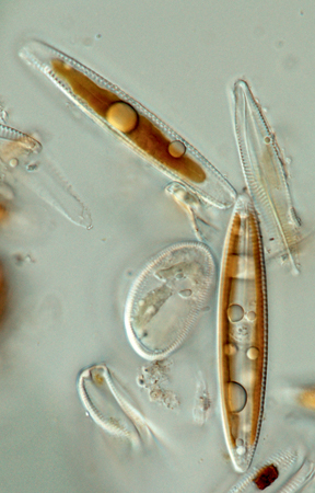
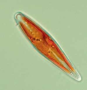
1 Navicula
Navicula tripunctata

© J. TAYLOR
Cells solitary, or in mucilaginous tubes. Valves elongate, bilaterally symmetrical around apical axis. Two plastids, one pressed to each side of valve margins; nuclear material at centre of cell in a cytoplasmic bridge. Size: 5–100µm in length and 3–25µm in width. Biology: Most commonly in the benthos, though cells may be washed into the plankton. Motile, performing gliding movements by means of a long slit, running along length of cell. RELATED SPECIES: A large genus, representatives occurring in a variety of water quality conditions ranging from fresh to brackish and from acidic (e.g. N. angusta) to alkaline (e.g. N. rostellata).
2 Cymbella
Cymbella aspera

© J. TAYLOR

© J. TAYLOR
Valves dorsoventrally symmetrical, with arched dorsal margin and more-or-less straight ventral margin. Plastid single, large, H-shaped; globules of stored lipids often visible. Size: 15–200μm long and 8–35μm wide. Biology: Solitary and motile, or attached to a substratum by means of a long mucilage stalk. Blooms may occur in winter months, forming layers of cells up to several centimetres thick (2a). The mucilaginous stalks may grow very long and look like cotton wool. Rarely found in heavily polluted waters.
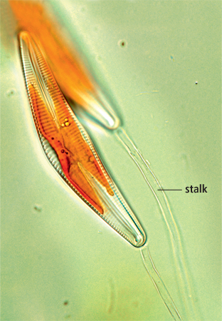
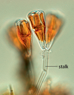
3 Gomphonema
Gomphonema species

© J. TAYLOR
© J. TAYLOR
Cells with narrow foot pole (point of attachment to mucilaginous stalk) and broader head pole; may have undulate margins and be swollen in centre, or at poles, or both. Stalk (3a) extruded through specialised pores. Plastid H-shaped; similar to that of Cymbella (see 2, above). Size: 10–90µm in length and 4–15µm in width. Biology: Cells solitary and motile, or attached to a substrate. Occur in a range of ecological conditions, including heavily polluted waters.
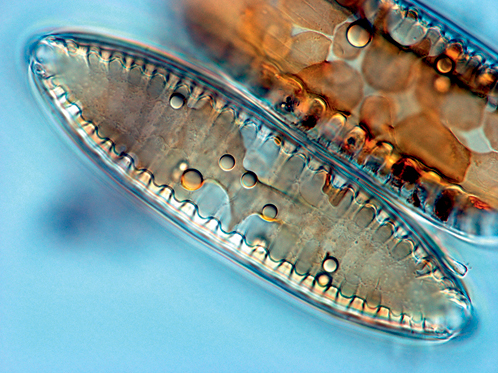
4 Tryblionella
Tryblionella gracilis

© J. TAYLOR
Large, bilaterally symmetrical cells. Raphe slit on margin of cell, supported by thickenings. Thickened ribs extend across undulating valve face. Two plastids located near each apex (in Navicula, see 1 above, they are side by side). Size: 20–180µm in length and 4.5–35µm in width. Biology: Cells solitary and motile. Occurs in a range of ecological conditions, but usually associated with relatively high salinities.
5 Surirella
Surirella species

© J. TAYLOR

© J. TAYLOR
Large cells, often asymmetrical. Raphe follows margins of valve. Two multilobed plastids, one pressed to each valve face. Size: 12–120µm in length and 6–45µm in width. Biology: Cells solitary and motile. Occurs in a range of ecological conditions and may be large enough to host other attached algae (5a).
The common name ‘protozoan’ is used for motile, unicellular or colonial eukaryotic organisms, such as Amoeba and Paramecium. They are animal-like in that they consume food and almost certainly include the forebears of the Kingdom Animalia. Confusingly, though, some can become photosynthetic under certain circumstances, while other closely related species are always photosynthetic, and might therefore be classified as algae. Classification schemes for these groups are complex and change over time, so here we use only common names for higher categories. Most protozoans are too small to be seen without a compound microscope. The taxonomic identity of most southern African species remains uncertain. Protozoans are classified mainly by the type of locomotion they undergo. Some are propelled by cilia, others by flagella and some glide using temporary extensions of the cell body called ‘pseudopodia’. Parasitic forms (e.g. Plasmodium, which causes malaria, and Trypanosoma, which causes sleeping sickness) are non-motile. All can reproduce by asexual fission, but some also undergo sexual reproduction.
Amoebas
Of no fixed shape, gliding by means of pseudopodia. Some have a hard external ‘shell’ or ‘test’. All feed on bacteria, other protists, or particles of organic matter, which they engulf with their pseudopodia. Very few undergo sexual reproduction. The group has not been adequately studied in southern Africa.
6 Naked amoebas
Amoeba species

© C. REED
© F. SIEMENSMA
© F. SIEMENSMA
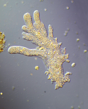
Unicellular, transparent. Amoeba (6) has several pseudopodia at any one time while Pelomyxa (6a) is slug-like with only one or two. Size: Mostly 200–300µm in diameter, but some such as the aptly named Chaos chaos (6b) up to 500µm, just visible to the naked eye. Biology: Feed primarily on unicellular algae. Probably several species in southern Africa.
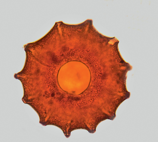
1 Testate amoebas
Arcella species
© F. SIEMENSMA
© F. SIEMENSMA
Enclosed in mushroom-shaped chitinous shell or test, pale brown when new, but darkening to dark brown (1). Pseudopodia (1a) extend from single central aperture. Size: Up to 300µm. Biology: Can float to the surface using vacuoles of carbon dioxide in cytoplasm; usually in small freshwater wetlands, wet foliage or mosses. Most species supposedly cosmopolitan and probably several species in southern Africa.
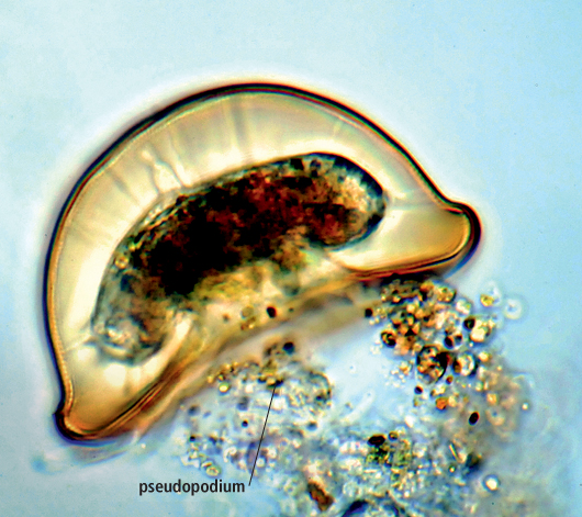
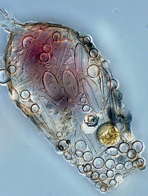
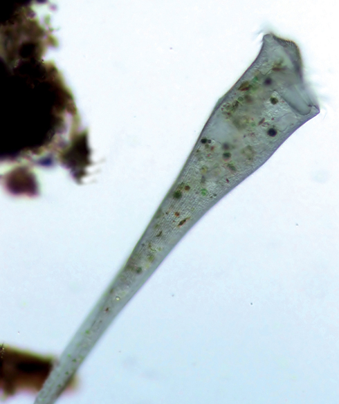
2 Testate amoebas
Difflugia species
© THIERRY ARNET/CC BY SA 3.0
Similar to Arcella (see 1, above), but test made of small pieces of mineral material (even diatom frustules) cemented together. Depending on species, test may be spherical or elongate, and may have projections or a collar, or both. Size: About 250µm. Biology: Some species contain endo-symbiotic algae (e.g. Chlorella, p.314, 2). Usually found in wetlands. Probably several species in southern Africa.
Heliozoans
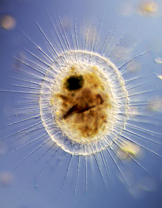
Radially symmetrical, planktonic amoebozoans with needle-like pseudopodia that extend radially from a more-or-less spherical cell body. Cell naked, or with a few silicious plates. Feed on organic particles, sometimes nearly as large as themselves.
3 Sun animalcule
Actinosphaerium species

© F. SIEMENSMA
Multinucleate, with a bubbly outer layer and numerous delicate spindle-shaped pseudopodia. Size: About 200µm. Biology: Some species stalked. One of few groups of amoebozoans in which sexual reproduction is known to occur: individual cells divide to form identical gametes, which fuse to form a new individual. Found in still-water wetlands. Probably several species in southern Africa.
Flagellates
An amorphous group bearing one or more slender, whip-like locomotory organelles called flagella. Some are autotrophic with distinct chloroplasts (and included in the Euglenophyta, p.322), others colourless and heterotrophic, while a few can switch between green and colourless forms as conditions dictate. Photosynthetic phytoflagellates are dealt with in the section on algae, and animal-like ones here. Most zooflagellates are commensal (e.g. in the guts of termites) or parasitic (e.g. Trypanosoma, which causes sleeping sickness), but a few are free-living.
4 Choanoflagellates
Salpinoeca species


© W. BETTIGHOFER
Small, colourless, attached flagellates recognised by presence in each cell of a collar around base of the single flagellum. Size: About 10µm long. Biology: Feed on minute particles in the water, beating of the flagellum drawing a food current towards the collar, where particles are trapped. Reproduction asexual, by binary fission. Of biological interest, as likely forebears of sponges and the Kingdom Animalia. Image from Europe; none yet studied in southern Africa.
5 Pathogenic flagellate
Naegleria fowleri


© CDC / DR GOVINDA S. VISVESVARA
Small cells free-living as flagellates, but able to infect human brains by entering nasal passage, losing their flagella and becoming amoeboid. Image (false colour) shows flagellated and amoeboid individuals. Size: 8–15µm. Biology: Cause fatal primary amoebic meningoencephalitis. Occur worldwide in warm standing waters, but uncommon.
Ciliates
By far the most morphologically and physiologically advanced protozoans. Although the body consists of a single cell, the internal structure is complex. Locomotion is by means of co-ordinated beating of a series of minute ‘hairs’ called ‘cilia’. In some forms these are arranged in rows down the length of the cell body but in others, cilia are confined to areas around the ‘mouth’ or are clumped to form stiff bristles. Ciliary locomotion is characteristically a smooth gliding, often accompanied by a corkscrewing or spiralling motion. Reproduction is routinely by binary fission, but a complex form of sexual reproduction also occurs, two individuals fusing and exchanging chromosomes. Many species are free-living in fresh waters or the sea, but some are symbionts, for example in the guts of mammals such as cattle. Larger ciliates are often bigger than rotifers (p.266) gastrotrichs (p.266) and the miracidia of flukes (p.270) and are easily confused with them.
1 Trumpet animalcule
Stentor species
© USER MICROPIX/CC BY SA 3.0
© C. REED
Relatively large cone- or trumpet-shaped ciliates, flared anterior of body forming wide ‘mouth’ surrounded by cilia, while narrow end acts as an anchor to substratum. When swimming, body oval in shape. May be colourless or pink, mauve or turquoise; bright green forms contain symbiotic Chlorella (p.314, 2). Size: Among the largest protozoans, reaching 2mm in length. Biology: Found in freshwater wetlands, often en masse (1a) where they feed mostly on living protozoans. Sexual reproduction occurs and some species produce desiccation-resistant cysts. Several species exist – S. coeruleus is blue or mauve.
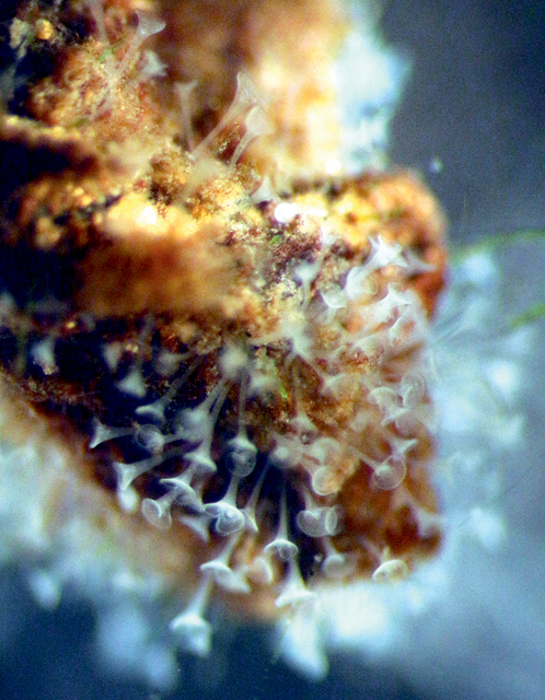
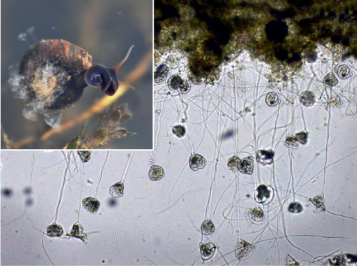
2 Vorticella
Vorticella campanula

© AIWOK/CC BY SA 3.0
© C. REED
Stalked ciliates, their shape reminiscent of wine glasses. Ciliated ‘rim’ or ‘mouth’ of cell produces food currents. Stalk contractile and, when stimulated, shortens rapidly, causing it to coil like a spring. Size: Cell body 150µm, stalk to 1mm. Biology: Sessile and attached to small objects, even snails (inset), on the bottoms of freshwater wetlands, but able to detach and swim. Feed mainly on bacteria. Reproduction mostly by binary fission, but sexual reproduction by conjugation also occurs.
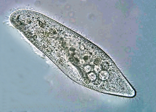
3 Slipper animalcule
Paramecium species

© DEUTEROSTOME/CC BY SA 3.0
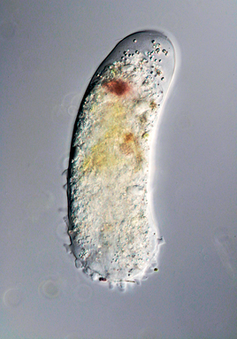
Mobile; footprint-shaped body covered by cilia that beat to create a characteristic smooth spiralling movement. Colourless, or coloured by food particles within the body, or sometimes green due to unicellular algae (see Chlorella, p314, 2) within the body, allowing both heterotrophic and photosynthetic modes of nutrition. Size: 50–330µm in length. Biology: Mostly in benthic debris in shallow still waters. Feed on small organic fragments, including living bacteria and yeasts. Reproduction both asexual, by binary fission, and sexual. Several ‘species’ may be part of a species complex.
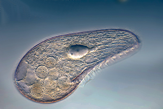
4 Blepharisma
Blepharisma species

© FRANK FOX/CC BY SA 3.0
Shape differs according to species, from teardrop-shaped to elongate. Body covered with cilia. Often reddish or pinkish because of a photosensitive pigment that allows them to seek darkness. Size: Variable, usually about 2–300µm. Biology: Predatory; if fed on smaller specimens of Blepharisma they may become cannibalistic giants, much larger than normal. Many species, but southern African forms not described.
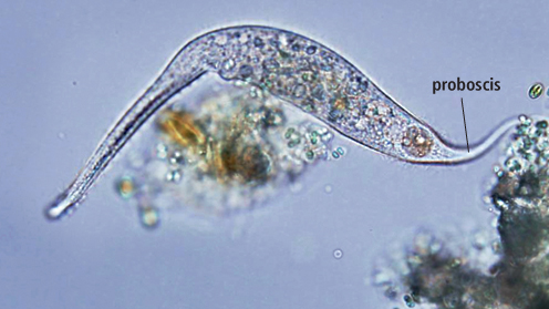
5 Dileptus
Dileptus margaritifer

© AZBIOSTUDENT/CC BY SA 3.0
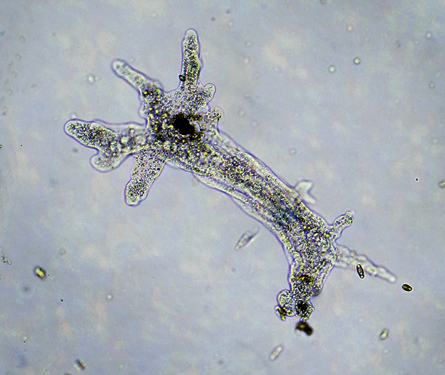
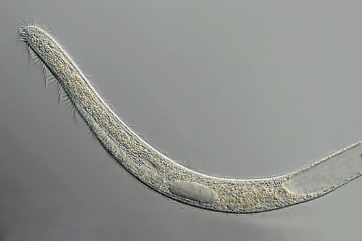
Long, flexible, cylindrical ciliates with numerous nuclei. Anteriorly, a mobile proboscis is constantly curling around objects and searching for food. Posterior narrow and tail-like. Size: Up to 450µm or more. Biology: Voracious predators, stunning prey with toxic darts.
6 Spirostomum
Spirostomum species
© C. KREBS
Among the largest ciliates, visible to the naked eye. Numerous nuclei like a bead necklace. Worm-like, sinuous, gliding movement; able to contract extremely fast if disturbed. Size: Up to 4mm. Biology: Common in fresh to brackish water even where oxygen levels are low. Several species seem to occur in region.