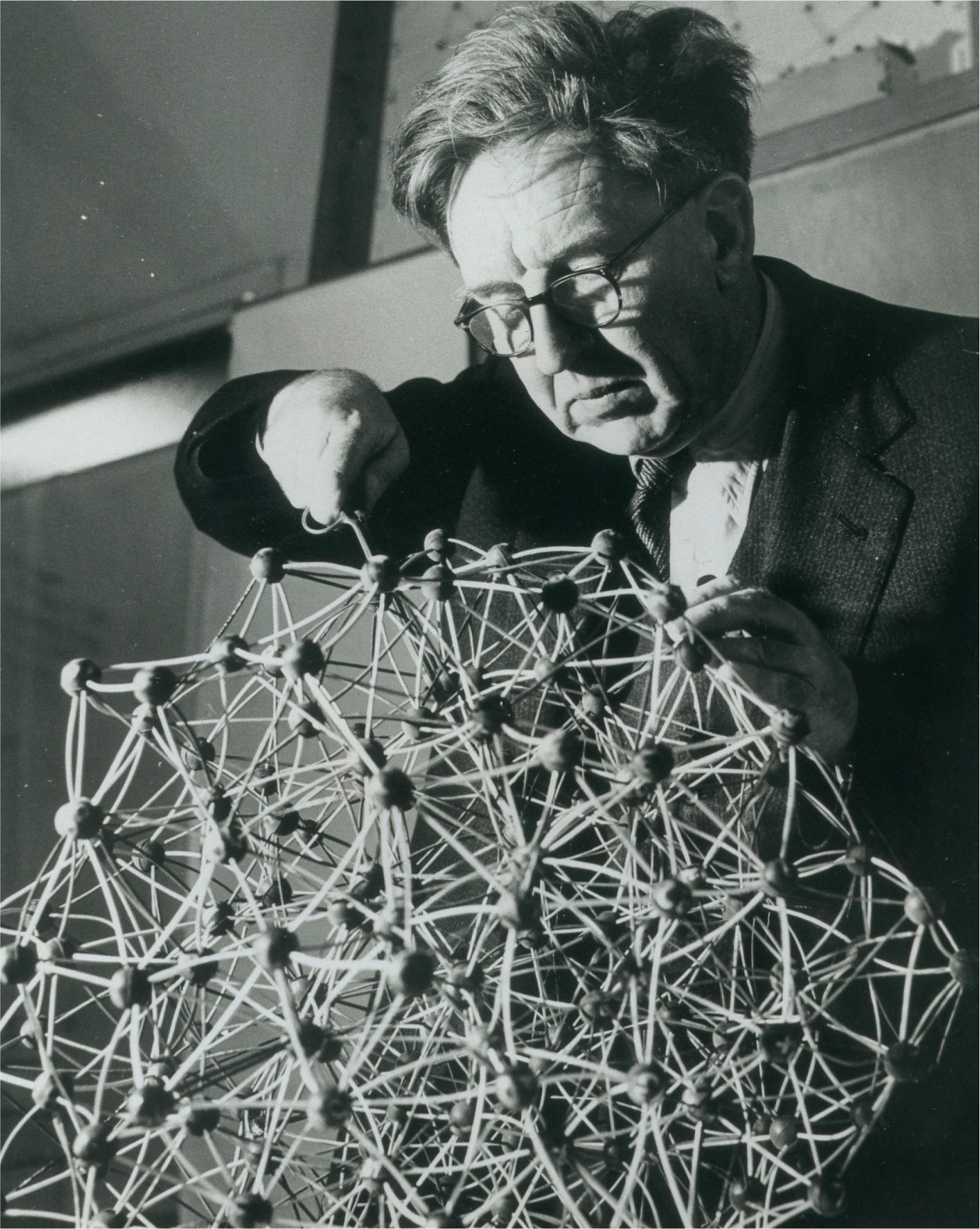
J. D. Bernal with a model of a protein molecule.
The way improvements in technology lead to advances in science is highlighted by the way X-ray crystallography (see here) was applied to the study of biological molecules. The first biomolecules to be investigated in this way were the proteins, complex molecules which were thought, in the 1930s, to be the key carriers of biological information, as well as providing structural material for the body. Proteins are long chains made up of sub-units called amino acids. All proteins in all living things on Earth are made up of different combinations of just a score of these units, arranged in different ways. Some idea of their complexity is provided by the fact that while weights of amino acids are typically about 100 units on a scale where an atom of hydrogen has one mass unit, molecular weights of proteins range from a few thousand to several million units. It was crystallography that would eventually reveal that the key feature of these long chain molecules is that they fold up into complex shapes, and these shapes determine their biological properties.
The first steps towards this understanding were taken by J. D. Bernal and his colleagues in Cambridge in 1934. Bernal had worked with William Bragg in the 1920s, and cut his scientific teeth by using X-ray crystallography to determine the structures of graphite and bronze. At the Cavendish laboratory, he turned his attention to organic molecules, but ran into a problem with proteins because when these were dried out ready for study their structure collapsed, like a structured house of cards collapsing into a disordered heap.
The standard way of preparing crystals is to grow them in a concentrated solution in water, known as the ‘mother liquor’. To crystallize a protein, the purified protein is allowed to settle out of such a concentrated solution. The individual protein molecules align themselves in a repeating series of ‘unit cells’ with a regular pattern, forming a crystalline ‘lattice’. John Philpott, a researcher based in Uppsala, in Sweden, was doing just this with the protein pepsin in the mid-1930s (pepsin is a digestive enzyme that breaks down other proteins in our food). When he went on a skiing holiday he left some crystals in their mother liquor in the refrigerator in his lab, and when he got back was surprised to find how much they had grown. A visitor from Cambridge, Glen Millikan, took some of these crystals, still in a tube containing the mother liquor, back to the Cavendish with him in his coat pocket, and gave them to Bernal.

Working with Dorothy Crowfoot (who later married and became known as Dorothy Crowfoot Hodgkin), Bernal first studied a crystal exposed to air while being irradiated, but found no diffraction. He had, however, also been monitoring the crystal through a microscope using polarized light. When the crystal was fresh and damp, this showed a feature known as birefringence, which indicates an ordered crystal structure, but the birefringence disappeared as the crystal dried out. So they carried out more experiments with both the crystal and its mother liquor sealed inside a thin-walled glass capillary tube. This provided the first X-ray diffraction photograph of single pepsin crystals, still in their mother liquor, in 1934. Bernal’s sealed capillary technique became the standard way to collect diffraction data on large molecules of this kind for the next fifty years.
Bernal realized that photographs like this might be interpreted to reveal the structure of the protein molecules themselves. As Bernal and Crowfoot said when describing their experiment in the journal, Nature: ‘Now that a crystalline protein has been made to give X-ray photographs, it is clear that we have the means of checking them and, by examining the structure of all crystalline proteins, arriving at far more detailed conclusions about protein structure than previous physical or chemical methods have been able to give.’39
Dorothy Crowfoot Hodgkin went on to play a major role in the development of the application of X-ray diffraction crystallography to the study of biologically important molecules over the next few decades (see here). But this was an immensely difficult task, for reasons that slowly became clear in the years that followed. What is called the primary structure of a protein is the order of the amino acids along a chain. These chains can then be twisted around to make, for example, a helix, which is the secondary structure. And then the helix is twisted into a kind of knot in three dimensions, the tertiary structure. By 1971, only seven proteins had had their structure fully worked out. Since then, improving technology, including powerful computers, has led to the detailed structural analysis of more than 30,000 proteins.