
Top texture: © Laguna Design / Science Source;
Chapter 22: Translation

Chapter Opener: Pasieka/Getty Images.
22.1 Introduction
A messenger RNA (mRNA) transcript carries a series of codons that interact with the anticodons of aminoacyl-tRNAs so that a corresponding series of amino acids is incorporated into a polypeptide chain. The ribosome provides the environment for controlling the interaction between mRNA and aminoacyl-tRNA. The ribosome behaves like a small migrating factory that travels along the mRNA template, engaging in rapid cycles of peptide bond synthesis to build a polypeptide. Aminoacyl-tRNAs shoot into the ribosome at an incredibly fast rate to deposit amino acids, and elongation factor proteins cyclically associate with and dissociate from the ribosome. Together with its accessory factors, the ribosomal structure provides the full range of activities required for all the steps of translation.
Figure 22.1 shows the relative dimensions of the components of the translation apparatus. The ribosome consists of two subunits (“large” and “small”) that have specific roles in translation. Messenger RNA is associated with the small subunit; approximately 35 bases of the mRNA are bound at any time during translation. The mRNA threads its way along the surface close to the junction of the two subunits. Two tRNA molecules are active in translation at any moment, so polypeptide elongation involves reactions taking place at just 2 of the approximately 10 codons associated with the ribosome. The two tRNAs are inserted into internal binding sites that stretch across the two ribosomal subunits. A third tRNA remains on the ribosome after it has been used in translation before being recycled.

FIGURE 22.1 The ribosome is large enough to bind several tRNAs and an mRNA.
The basic structure of the ribosome has been conserved during evolution, but there are appreciable variations in the overall size and proportions of RNAs and proteins in the ribosomes of prokaryotes and the eukaryotic cytosol, mitochondria, and chloroplasts. Figure 22.2 compares the components of bacterial and mammalian ribosomes. Both are ribonucleoprotein particles that contain more RNA than protein. The ribosomal proteins are known as r-proteins.

FIGURE 22.2 Ribosomes are large ribonucleoprotein particles that contain more RNA than protein and are composed of a large and a small subunit.
Each of the ribosomal subunits contains a major rRNA and a number of small proteins. The large subunit may also contain smaller RNA(s). In Escherichia coli, the small (30S) subunit consists of the 16S rRNA and 21 r-proteins. The large (50S) subunit contains the 23S rRNA, the small 5S RNA, and 31 r-proteins. With the exception of one protein that is present in four copies per ribosome, there is one copy of each protein. The major RNAs constitute the larger part of the mass of the bacterial ribosome. Their presence is pervasive so that most or all of the r-proteins actually contact rRNA. Thus, the major rRNAs form what is sometimes considered the “backbone” of each subunit—a continuous thread whose presence dominates the structure and determines the positions of the ribosomal proteins.
The ribosomes in the cytosol of eukaryotes are larger than those of prokaryotes. The total content of both RNA and protein is greater, the major RNA molecules are longer (called 18S and 28S rRNAs), and there are more proteins. RNA is still the predominant component by mass.
The ribosomes of eukaryotic mitochondria and chloroplasts are distinct from the ribosomes of the cytosol, and they take varied forms. In some cases, they are almost the size of prokaryotic ribosomes and have about 70% RNA; in other cases, they are only 60S and have less than 30% RNA.
The ribosome possesses several active centers, each of which is constructed from a group of proteins associated with a region of ribosomal RNA. The active centers require the direct participation of rRNA in a structural or even catalytic role (where the RNA functions as a ribozyme) with proteins supporting these functions in secondary roles. Some catalytic functions require individual proteins, but none of the activities can be reproduced by isolated proteins or groups of proteins; they function only in the context of the ribosome.
Two experimental approaches can be taken in analyzing the functions of structural components of the ribosome. In one approach, the effects of mutations in genes for particular ribosomal proteins or at specific positions in rRNA genes shed light on the participation of these molecules in particular reactions. In a second approach, structural analysis, including direct modification of components of the ribosome and comparisons to identify conserved features in rRNA, identifies the physical locations of components involved in particular functions.
22.2 Translation Occurs by Initiation, Elongation, and Termination
An amino acid is brought to the ribosome by an aminoacyl-tRNA. Its addition to the growing polypeptide chain occurs by an interaction with the tRNA that brought the previous amino acid. Each of these tRNAs lies in its own distinct site on the ribosome. Figure 22.3 shows that the two sites have different features:
Except for the initiator tRNA, an incoming aminoacyl-tRNA binds to the A site. Prior to the entry of aminoacyl-tRNA, the site exposes the mRNA codon representing the next amino acid to be added to the chain.
The codon representing the most recent amino acid to have been added to the nascent polypeptide chain lies in the P site. This site is occupied by peptidyl-tRNA, a tRNA carrying the nascent polypeptide chain.

FIGURE 22.3 The ribosome has two sites for binding charged tRNA.
Figure 22.4 shows that the aminoacyl end of the tRNA is located on the large subunit, whereas the anticodon at the other end of the tRNA interacts with the mRNA bound by the small subunit. Thus, the P and A sites each extend across both ribosomal subunits.

FIGURE 22.4 The P and A sites position the two bound tRNAs across both ribosomal subunits.
For a ribosome to form a peptide bond, it must be in the state shown in step 1 in Figure 22.3, when peptidyl-tRNA is in the P site and aminoacyl-tRNA is in the A site. Peptide bond formation occurs when the polypeptide carried by the peptidyl-tRNA is transferred to the amino acid carried by the aminoacyl-tRNA. This step requires correct positioning of the aminoacyl-ends of the two tRNAs within the large subunit. This reaction is catalyzed by the large subunit of the ribosome.
Transfer of the polypeptide generates the ribosome shown in step 2 of Figure 22.3, in which the deacylated tRNA, lacking any amino acids, lies in the P site, and a new peptidyl-tRNA is in the A site. The peptide on this peptidyl-tRNA is one amino acid residue longer than the one that was carried on the peptidyl-tRNA that had been in the P site in step 1.
The ribosome now moves one triplet along the messenger RNA. This stage is called translocation. The movement transfers the deacylated tRNA out of the P site and moves the peptidyl-tRNA into the P site (see step 3 in Figure 22.3). The next codon to be translated now lies in the A site, ready for a new aminoacyl-tRNA to enter, when the cycle will be repeated. Figure 22.5 summarizes the interaction between tRNAs and the ribosome.

FIGURE 22.5 Aminoacyl-tRNA enters the A site, receives the polypeptide chain from peptidyl-tRNA, and is transferred into the P site for the next cycle of elongation.
The deacylated tRNA leaves the ribosome via another tRNA-binding site, the E site. This site is transiently occupied by the tRNA en route between leaving the P site and being released from the ribosome into the cytosol. Thus, the route of tRNA through the ribosome is into the A site, through the P site, and out through the E site (see also Figure 22.28 in the section later in this chapter titled Translocation Moves the Ribosome). Figure 22.6 compares the movement of tRNA and mRNA, which may be considered a sort of ratchet in which the reaction is driven by the codon–anticodon interaction.

FIGURE 22.6 tRNA and mRNA move through the ribosome in the same direction.
Translation is divided into the three stages shown in Figure 22.7:
Initiation involves the reactions that precede formation of the peptide bond between the first two amino acids of the polypeptide. It requires the ribosome to bind to the mRNA, which forms an initiation complex that contains the first aminoacyl-tRNA. This is a relatively slow step in translation and usually determines the rate at which an mRNA is translated.
Elongation includes all the reactions from the formation of the first peptide bond to the addition of the last amino acid. Amino acids are added to the chain one at a time; the addition of an amino acid is the most rapid step in translation.
Termination encompasses the steps that are needed to release the completed polypeptide chain; at the same time, the ribosome dissociates from the mRNA.
Different sets of accessory protein factors assist the ribosome at each stage. Energy is provided at various stages by the hydrolysis of guanine triphosphate (GTP).

FIGURE 22.7 Translation has three stages.
During initiation, the small ribosomal subunit binds to mRNA and then is joined by the large subunit. During elongation, the mRNA moves through the ribosome and is translated in nucleotide triplets. (Although the ribosome is usually referred to as moving along mRNA, it is more accurate to say that the mRNA is pulled through the ribosome.) At termination, the polypeptide is released, the mRNA is released, and the individual ribosomal subunits dissociate and can be used again.
22.3 Special Mechanisms Control the Accuracy of Translation
The general accuracy of translation is confirmed by the consistency that is found when determining the amino acid sequence of a polypeptide. Few detailed measurements of the error rate in vivo are available, but it is generally thought to be in the range of one error for every 104 to 105 amino acids incorporated. Considering that most polypeptides are produced in large quantities, this means that the error rate is too low to have much effect on the phenotype of the cell.
It is not immediately obvious how such a low error rate is achieved. In fact, an error can be made at several steps in gene expression:
The enzymes that synthesize RNA may insert a base that is not complementary to the base on the template strand.
Synthetases may attach the wrong tRNA to an amino acid or the wrong amino acid to a tRNA.
A ribosome may allow binding of a tRNA that does not correspond to the codon in the A site.
Each case represents a similar problem for the mechanism: how to distinguish one particular member from the entire set, all of which share the same general features.
Probably any substrate can initially contact the active center by a random-hit process, but then the wrong substrates are rejected and only the correct one is accepted. The correct substrate is always rare (e.g., 1 of 4 bases, 1 of 20 amino acids, 1 of about 30 to 50 tRNAs), so the criteria for discrimination must be strict. The point is that the enzyme or ribozyme must have some mechanism for discriminating among substrates that are structurally very similar.
Figure 22.8 summarizes the error rates at the steps that can affect the accuracy of translation. Errors in transcribing mRNA are rare, probably less than 10−6. This is an important stage for accuracy because a single mRNA molecule can be translated into many polypeptide copies. The mechanisms that ensure transcriptional accuracy are discussed in the chapter titled Prokaryotic Transcription.

FIGURE 22.8 Errors occur at rates ranging from 10−6 to 5 × 10⁻⁴ at different stages of translation.
The ribosome can make two types of errors in translation. It may cause a frameshift by skipping a base when it reads the mRNA (or, in the reverse direction, by reading a base twice—once as the last base of one codon, and then again as the first base of the next codon or twice within the same codon). These errors are rare, occurring at a rate of about 10−5. Or, it may allow an incorrect aminoacyl-tRNA to (mis)pair with a codon, so that the wrong amino acid is incorporated. This is probably the most common error in translation, occurring at a rate of about 5 × 10⁻⁴. This rate is determined by ribosome structure and dissociation kinetics (see the chapter titled Using the Genetic Code).
An aminoacyl-tRNA synthetase can make two types of errors: It can place the wrong amino acid on its tRNA, or it can charge its amino acid with the wrong tRNA (see the chapter titled Using the Genetic Code). The incorporation of the wrong amino acid is more common, probably because the tRNA offers a larger surface with which the enzyme can make many more contacts to ensure specificity. Aminoacyl-tRNA synthetases have specific mechanisms to correct errors before a mischarged tRNA is released (see the chapter titled Using the Genetic Code).
22.4 Initiation in Bacteria Needs 30S Subunits and Accessory Factors
Prokaryotic ribosomes engaged in elongating a polypeptide chain exist as 70S particles. At termination, they are released from the mRNA as free ribosomes or ribosomal subunits. In growing bacteria, the majority of ribosomes are synthesizing polypeptides; the free pool is likely to contain about 20% of the ribosomes.
Ribosomes in the free pool can dissociate into separate subunits; this means that 70S ribosomes are in dynamic equilibrium with 30S and 50S subunits. Initiation of translation is not a function of intact ribosomes, but is undertaken by the separate subunits. These subunits reassociate during the initiation reaction. Figure 22.9 summarizes the ribosomal subunit cycle during translation in bacteria.

FIGURE 22.9 Initiation requires free ribosome subunits. When ribosomes are released at termination, the 30S subunits bind initiation factors and dissociate to generate free subunits. When subunits reassociate to produce a functional ribosome at initiation, they release these factors.
Initiation occurs at a special sequence on mRNA called the ribosome-binding site (including the Shine–Dalgarno sequence, which is discussed in the next section). This is a short sequence of bases that is positioned upstream from the coding region and is complementary to a portion of the 16S rRNA (see the section later in this chapter titled 16S rRNA Plays an Active Role in Translation). The small and large subunits associate at the ribosome-binding site to form an intact ribosome. The reaction occurs in two steps:
Recognition of mRNA occurs when a small subunit binds to form an initiation complex at the ribosome-binding site.
A large subunit then joins the complex to generate a complete ribosome.
Although the 30S subunit is involved in initiation, it is not sufficient by itself to bind mRNA and tRNA; this requires additional proteins called initiation factors (IFs). These factors are found only on 30S subunits, and they are released when the 30S subunits associate with 50S subunits to generate 70S ribosomes. This action distinguishes initiation factors from the structural proteins of the ribosome. The initiation factors are solely concerned with formation of the initiation complex; they are absent from 70S ribosomes and they play no part in the stages of elongation. Figure 22.10 summarizes the stages of initiation.

FIGURE 22.10 Initiation factors stabilize free 30S subunits and bind initiator tRNA to the 30S–mRNA complex.
Prokaryotes use three initiation factors, numbered IF-1, IF-2, and IF-3. They are needed for both mRNA and tRNA to enter the initiation complex:
IF-3 has multiple functions: It is needed to stabilize (free) 30S subunits and to inhibit the premature binding of the 50S subunit; it enables 30S subunits to bind to initiation sites in mRNA; and, as part of the 30S-mRNA complex, it checks the accuracy of recognition of the first aminoacyl-tRNA.
IF-2 binds a special initiator tRNA and controls its entry into the ribosome.
IF-1 binds to 30S subunits as a part of the complete initiation complex. It binds in the vicinity of the A site and prevents aminoacyl-tRNA from entering. Its location also may impede the 30S subunit from binding to the 50S subunit.
Numerous structural studies indicate that IF-3 has two distinct, largely globular domains, with the C-terminal domain at the 50S contact site on the 30S subunit and the N-terminal domain in the vicinity of the 30S E site. This broad positioning of IF-3 on the 30S subunit is consistent with its multiple functions.
The first function of IF-3 is control of the equilibrium between ribosomal states, as shown in Figure 22.11. IF-3 binds to free 30S subunits that are released from the pool of 70S ribosomes. The presence of IF-3 prevents the 30S subunit from reassociating with a 50S subunit. IF-3 can interact directly with 16S rRNA, and significant overlap exists between the bases in 16S rRNA protected by IF-3 and those protected by binding of the 50S subunit, suggesting that it physically prevents junction of the subunits. IF-3 therefore behaves as an anti-association factor that causes a 30S subunit to remain in the pool of free subunits. The reaction between IF-3 and the 30S subunit is stoichiometric: One molecule of IF-3 binds per subunit. Because of the relatively small amount of IF-3, its availability determines the number of free 30S subunits.

FIGURE 22.11 Initiation requires 30S subunits that carry IF-3.
The second function of IF-3 controls the ability of 30S subunits to bind to mRNA. Small subunits must have IF-3 in order to form initiation complexes with mRNA. IF-3 must be released from the 30S-mRNA complex in order for the 50S subunit to join. On its release, IF-3 immediately recycles by finding another 30S subunit.
Finally, IF-3 checks the accuracy of recognition of the first aminoacyl-tRNA and helps to direct it to the P site of the 30S subunit. The former has been attributed to the C-terminal domain of IF-3 (see the section later in this chapter titled Use of fMet-tRNAf Is Controlled by IF-2 and the Ribosome). By comparison, the N-terminal domain of IF-3 is positioned to help direct the aminoacyl-tRNA into the P site of the 30S subunit by blocking the E site at the same time that IF-1 is blocking the A site.
IF-2 has a ribosome-dependent GTPase activity: It sponsors the hydrolysis of GTP in the presence of ribosomes, releasing the energy stored in the high-energy bond. The GTP is hydrolyzed when the 50S subunit joins to generate a complete ribosome. The GTP cleavage could be involved in changing the conformation of the ribosome, so that the joined subunits are converted into an active 70S ribosome.
22.5 Initiation Involves Base Pairing Between mRNA and rRNA
The signal for initiating a polypeptide chain is a special initiation codon that marks the start of the reading frame. Usually the initiation codon is the triplet AUG, but in bacteria GUG or UUG may also be used.
An mRNA may contain many AUG triplets, so how is the correct initiation codon recognized as the starting point for translation? The sites on mRNA where translation is initiated can be identified by binding the ribosome to mRNA under conditions that block elongation so that the ribosome remains at the initiation site. When ribonuclease is added to the blocked initiation complex, all the regions of mRNA outside the ribosome are degraded, but those actually bound to it are protected, as illustrated in Figure 22.12. The protected fragments can then be recovered and characterized.
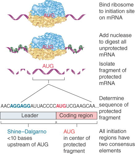
FIGURE 22.12 Ribosome-binding sites on mRNA can be identified by studying initiation complexes. They include the upstream Shine–Dalgarno sequence and the initiation codon.
The initiation sequences protected by prokaryotic ribosomes are approximately 30 bases long. The ribosome-binding sites of different bacterial mRNAs display two common features:
The AUG (or less often, GUG or UUG) initiation codon is always included within the protected sequence.
Approximately 10 bases upstream of the initiation codon is a sequence that corresponds to part or all of the hexamer:
5′ … A G G A G G … 3′
This polypurine stretch is known as the Shine–Dalgarno sequence. It is complementary to a highly conserved sequence close to the 3′ end of the 16S rRNA. (The extent of complementarity differs among individual mRNAs and ranges from a four-base core sequence GAGG to a nine-base sequence extending beyond each end of the hexamer.) Written in reverse direction, the rRNA sequence is the hexamer:
3′ … U C C U C C … 5′
Does the Shine–Dalgarno sequence pair with its rRNA complement during mRNA–ribosome binding? Mutations of either sequence demonstrate its importance in initiation. Point mutations in the Shine–Dalgarno sequence can prevent an mRNA from being translated. In addition, the introduction of mutations into the complementary sequence in the rRNA is deleterious to the cell and changes the pattern of translation. The decisive confirmation of the base-pairing reaction is that a mutation in the Shine–Dalgarno sequence of an mRNA can be suppressed by a mutation in the rRNA that restores base pairing.
The sequence at the 3′ end of the rRNA is conserved among prokaryotes and eukaryotes, except that in all eukaryotes there is a deletion of the five-base sequence CCUCC that is the principal complement to the Shine–Dalgarno sequence. Base pairing does not appear to occur between eukaryotic mRNAs and the 18S rRNA. This is a significant difference between prokaryotes and eukaryotes in the mechanism of initiation.
In bacteria, a 30S subunit binds directly to a ribosome-binding site. As a result, the initiation complex forms at a sequence surrounding the AUG initiation codon. When the mRNA is polycistronic (see the section later in this chapter titled The Cycle of Bacterial Messenger RNA), each coding region starts with a ribosome-binding site.
The nature of bacterial gene expression means that translation of a polycistronic bacterial mRNA proceeds sequentially through each of its cistrons (coding regions). At the time when ribosomes attach to the first coding region, the subsequent coding regions have not yet been transcribed. By the time the second ribosomal binding site is available, translation through the first cistron is well under way.
What happens between the coding regions varies among individual polycistronic mRNAs. In most cases, the ribosomes probably bind independently at the beginning of each cistron. The most common series of events is illustrated in Figure 22.13. When synthesis of the first polypeptide terminates, the ribosomes leave the mRNA and dissociate into subunits. Then a new ribosome must assemble at the next coding region and begin translation of the next cistron.

FIGURE 22.13 Initiation occurs independently at each cistron in a polycistronic mRNA. When the intercistronic region is longer than the span of sequence interacting with the ribosome, dissociation at the termination site is followed by independent reinitiation at the next cistron.
In some polycistronic bacterial mRNAs, translation between adjacent cistrons is directly linked, because ribosomes gain access to the initiation codon of the second cistron as they complete translation of the first cistron. This requires the distance between the two coding regions to be small. It may depend on the high local density of ribosomes, or the juxtaposition of termination and initiation sites could allow some of the usual intercistronic events to be bypassed. A ribosome physically spans about 30 bases of mRNA, so it can simultaneously contact a termination codon and the next initiation site if they are separated by only a few bases.
22.6 A Special Initiator tRNA Starts the Polypeptide Chain
Synthesis of all polypeptides starts with the same amino acid—methionine. tRNAs recognizing the AUG codon carry methionine, and two types of tRNA can carry this amino acid. One is used for initiation, the other for recognizing AUG codons during elongation.
In bacteria, mitochondria, and chloroplasts, the initiator tRNA carries a methionine residue that has been formylated on its amino group, forming a molecule of N-formyl-methionyl-tRNA. The tRNA is known as tRNAf-Met. The name of the aminoacyl-tRNA is usually abbreviated to fMet-tRNAf.
The initiator tRNA gains its modified amino acid in a two-stage reaction. First, it is charged with the amino acid to generate Met-tRNAf, and then the formylation reaction shown in Figure 22.14 blocks the free amino (–NH2) group. Although the blocked amino acid group would prevent the initiator from participating in chain elongation, it does not interfere with the ability to initiate a polypeptide.

FIGURE 22.14 The initiator N-formyl-methionyl-tRNA (fMet-tRNAf) is generated by formylation of methionyl-tRNA using formyl-tetrahydrofolate as a cofactor.
This tRNA is used only for initiation. It recognizes the codons AUG or GUG (or occasionally UUG). The codons are not recognized equally well; the extent of initiation declines by about half when AUG is replaced by GUG, and declines by about half again when UUG is used.
The tRNA type responsible for recognizing only AUG codons following the initiation codon is tRNAmMet. Its methionine cannot be formylated.
What features distinguish the fMet-tRNAf initiator and the Met-tRNAm elongator? Some characteristic features of the tRNA sequence are important, as summarized in Figure 22.15. Some of these features are needed to prevent the initiator from being used in elongation, whereas others are necessary for it to function in initiation:
Formylation is not strictly necessary because nonformylated Met-tRNAf can function as an initiator. However, formylation improves the efficiency with which the Met-tRNAf is used because it is one of the features recognized by IF-2, which binds the initiator tRNA.
The bases that face one another at the last position of the stem to which the amino acid is connected are paired in all tRNAs except tRNAf Met. Mutations that create a base pair in this position of tRNAfMet allow it to function in elongation. Therefore, the absence of this pair is important in preventing tRNAfMet from being used in elongation. It is also needed for the formylation reaction.
A series of three G-C pairs in the stem that precedes the loop containing the anticodon is unique to tRNAfMet. These base pairs are required to allow the fMet-tRNAf to be inserted directly into the P site.

FIGURE 22.15 fMet-tRNAf has unique features that distinguish it as the initiator tRNA.
In bacteria and mitochondria, the formyl residue on the initiator methionine is removed from the protein by a specific deformylase enzyme to generate a normal NH2 terminus. If methionine is to be the N-terminal amino acid of the protein, this is the only necessary step. In about half of the polypeptides, the methionine at the terminus is removed by an aminopeptidase, which creates a new terminus from R2 (originally the second amino acid incorporated into the chain). When both steps are necessary, they occur sequentially. The removal reaction(s) occur(s) rather rapidly when the nascent polypeptide chain has reached a length of about 15 amino acids.
22.7 Use of fMet-tRNAf Is Controlled by IF-2 and the Ribosome
In bacterial translation, the meaning of the AUG and GUG codons depends on their context. When the AUG codon is used for initiation, a formyl-methionine begins the polypeptide; when it is used within the coding region, methionine is added to the polypeptide. The meaning of the GUG codon is even more dependent on its location. When present as the first codon, formyl-methionine is added, but when present within a gene it is bound by Val-tRNA, one of the regular members of the tRNA set, to provide valine as specified by the genetic code.
How is the context of AUG and GUG codons interpreted? Figure 22.16 illustrates the decisive role of the ribosome when acting in conjunction with accessory factors.

FIGURE 22.16 Only fMet-tRNAf can be used for initiation by 30S subunits; other aminoacyl-tRNAs (aa-tRNAs) must be used for elongation by 70S ribosomes.
In an initiation complex, the small subunit alone is bound to mRNA. The initiation codon lies within the part of the P site carried by the small subunit. The only aminoacyl-tRNA that can become part of the initiation complex is the initiator, which has the unique property of being able to enter directly into the partial P site to bind to its complementary codon.
When the large subunit joins the complex, the partial tRNA-binding sites are converted into the intact P and A sites. The initiator fMet-tRNAf occupies the P site, and the A site is available for entry of the aminoacyl-tRNA complementary to the second codon of the mRNA. The first peptide bond forms between the initiator and the next aminoacyl-tRNA.
Initiation occurs when an AUG (or GUG) codon lies within a ribosome-binding site because only the initiator tRNA can enter the partial P site formed when the 30S subunit binds de novo to the mRNA. During elongation only the regular aminoacyl-tRNAs can enter the complete A site.
Accessory factors are critical for the binding of aminoacyl-tRNAs. All aminoacyl-tRNAs associate with the ribosome by binding to an accessory factor. The factor used in initiation is IF-2 (see the section earlier in this chapter titled Initiation in Bacteria Needs 30S Subunits and Accessory Factors). The accessory factor used at elongation, EF-Tu, is discussed in the section later in this chapter titled Elongation Factor Tu Loads Aminoacyl-tRNA into the A Site.
The initiation factor IF-2 places the initiator tRNA into the P site. By forming a complex specifically with fMet-tRNAf, IF-2 ensures that only the initiator tRNA, and none of the regular aminoacyl-tRNAs, participates in the initiation reaction. Conversely, EF-Tu, which places aminoacyl-tRNAs in the A site, cannot bind fMet-tRNAf, which is therefore excluded from use during elongation.
The accuracy of initiation is also assisted by IF-3, which stabilizes binding of the initiator tRNA by recognizing correct base pairing with the second and third bases of the AUG initiation codon.
Figure 22.17 details the series of events by which IF-2 places the fMet-tRNAf initiator in the P site. IF-2, bound to GTP, associates with the P site of the 30S subunit. At this point, the 30S subunit carries all the initiation factors. fMet-tRNAf then binds to the IF-2 on the 30S subunit, and IF-2 transfers the tRNA into the partial P site.

FIGURE 22.17 IF-2 is needed to bind fMet-tRNAf to the 30S–mRNA complex. After 50S binding, all IFs are released and GTP is cleaved.
22.8 Small Subunits Scan for Initiation Sites on Eukaryotic mRNA
Initiation of translation in eukaryotic cytoplasm resembles the process that occurs in bacteria, but the order of events is different and the number of accessory factors is greater. Some of the differences in initiation are related to a difference in the way that bacterial 30S and eukaryotic 40S subunits find their binding sites for initiating translation on mRNA. In eukaryotes, small subunits first recognize the 5′ cap at the end of the mRNA and then move to the initiation site, where they are joined by large subunits. (In prokaryotes, small subunits bind directly to the initiation site.)
Virtually all eukaryotic mRNAs are monocistronic, but each mRNA usually is substantially longer than the sequence that encodes its polypeptide. The average mRNA in eukaryotic cytoplasm is 1,000 to 2,000 bases long, has a methylated cap at the 5′ terminus, and carries 100 to 200 adenine bases at the 3′ terminus.
The untranslated 5′ leader is relatively short, usually less than 100 bases. The length of the coding region is determined by the size of the polypeptide product. The untranslated 3′ trailer is often rather long, at times reaching lengths of up to about 1,000 bases.
The first feature to be recognized during translation of a eukaryotic mRNA is the methylated cap at the 5′ end. mRNAs whose caps have been removed are not translated efficiently in vitro. Binding of 40S subunits to mRNAs requires several initiation factors, including proteins that recognize the structure of the cap.
Modification at the 5′ end occurs in almost all cellular or viral mRNAs and is essential for their translation in eukaryotic cytoplasm (although it is not needed in mitochondria or chloroplasts). The sole exception to this rule is provided by a few viral mRNAs (such as those of poliovirus) that are not capped; only these exceptional viral mRNAs can be translated in vitro without caps. They use an alternative pathway that bypasses the need for the cap.
We have dealt with the process of initiation as though the initiation site is always freely available. However, its availability may be impeded by the mRNA’s secondary structure. The recognition of mRNA requires several additional factors; an important part of their function is to remove any secondary structure in the mRNA.
In some mRNAs, the AUG initiation codon lies within 40 bases of the 5′ terminus of the mRNA, so that both the cap and AUG lie within the span of ribosome binding. However, in many mRNAs the cap and AUG are farther apart; in extreme cases, they can be as much as 1,000 bases away from each other. Yet the presence of the cap is still necessary for a stable complex to be formed at the initiation codon. How can the ribosome rely on two sites so far apart for mRNA recognition?
Figure 22.18 illustrates the “scanning” model, which has the 40S subunit initially recognizing the 5′ cap and then “migrating” along the mRNA. Scanning from the 5′ end is a linear process. When 40S subunits scan the leader region, they can melt secondary structure hairpins with stabilities less than −30 kcal, but hairpins of greater stability impede or prevent migration.

FIGURE 22.18 Eukaryotic ribosomes migrate from the 5′ end of mRNA to the ribosome binding site, which includes an AUG initiation codon.
Migration stops when the 40S subunit encounters the AUG initiation codon. Usually, though not always, the first AUG triplet sequence to be encountered will be the initiation codon. However, the AUG triplet by itself is not sufficient to halt migration; it is recognized efficiently as an initiation codon only when it is in the right context. The most important determinants of context are the bases in positions −4 and +1. An initiation codon may be recognized in the sequence NNNPuNNAUGG by the small ribosomal subunit using the Met-tRNA anticodon. The purine (A or G) three bases before the AUG codon and the G immediately following it can influence the efficiency of translation by 10 times. When the leader sequence is long, further 40S subunits can recognize the 5′ end before the first has left the initiation site, creating a queue of subunits proceeding along the leader to the initiation site.
It is usually true that the initiation codon is the first AUG to be encountered in the most efficiently translated mRNAs. However, what happens when there is an AUG triplet in the 5′ untranslated region (UTR)? Two escape mechanisms are possible for a ribosome that starts scanning at the 5′ end. The most common is that scanning is leaky; that is, a ribosome may continue past a noninitiation AUG because it is not in the right context. In the rare case that it does recognize the AUG, it may initiate translation but terminate before the proper initiation codon, after which it resumes scanning.
The majority of eukaryotic initiation events involve scanning from the 5′ cap, but there is an alternative means of initiation, used especially by certain viral RNAs, in which a 40S subunit associates directly with an internal site called an internal ribosome entry site (IRES). In this case, any AUG codons that may be in the 5′ UTR are bypassed entirely. There are few sequence homologies between known IRES elements. Three types of IRESs can be identified based on their interaction with the 40S subunit:
The most common type of IRES includes the AUG initiation codon at its upstream boundary. The 40S subunit binds directly to it, using a subset of the same factors that are required for initiation at 5′ ends.
Another type of IRES is located as much as 100 nucleotides upstream of the AUG, requiring a 40S subunit to migrate, again probably by a scanning mechanism.
An exceptional type of IRES in hepatitis C virus can bind a 40S subunit directly, without requiring any initiation factors. The order of events is different from all other eukaryotic initiation. Following 40S-mRNA binding, a complex containing initiator factors and the initiator tRNA binds.
Use of the IRES is especially important in picornavirus infection, where it was first discovered, because the virus inhibits host translation by destroying cap structures and inhibiting the initiation factors that bind them. One such target is subunit eIF4G (see the next section, Eukaryotes Use a Complex of Many Initiation Factors), which binds the 5′ end of mRNA. Thus, infection prevents translation of host mRNAs but allows viral mRNAs to be translated because they use the IRES.
Ribosome binding is stabilized at the initiation site. When the 40S subunit is joined by a 60S subunit, the intact ribosome is located at the site identified by the protection assay. A 40S subunit protects a region of up to 60 bases; when the 60S subunits join the complex the protected region contracts to about the same length of 30 to 40 bases seen in prokaryotes.
22.9 Eukaryotes Use a Complex of Many Initiation Factors
Initiation in eukaryotes has the same general features as in prokaryotes in using a specific initiation codon and initiator tRNA. Initiation in eukaryotic cytoplasm uses AUG as the initiator codon. The initiator tRNA is a distinct type, but its methionine does not become formylated, as in prokaryotes. It is called tRNAiMet. Thus, the difference between the initiating and elongating Met-tRNAs lies solely in the tRNA portion of the complex, with Met-tRNAi used for initiation and Met-tRNAm used for elongation.
At least two features are unique to the initiator tRNAiMet in yeast: It has an unusual tertiary structure, and it is modified by phosphorylation of the 2′-ribose position on base 64 (if this modification is prevented, the initiator can be used in elongation). Thus, a distinction between initiator and elongator Met-tRNAs is maintained in eukaryotes, but its structural basis is different from that in prokaryotes.
Eukaryotic cells have more initiation factors than prokaryotic cells do: The current list includes about a dozen factors that are directly or indirectly required for initiation. The factors are named similarly to those in prokaryotes (sometimes by analogy with the bacterial factors) and are given the prefix “e” to indicate their eukaryotic origin. They act at all stages of the process, including:
Forming an initiation complex with the 5′ end of mRNA
Forming a complex with Met-tRNAi
Binding the mRNA-factor complex to the Met-tRNAi-factor complex
Enabling the ribosome to scan mRNA from the 5′ end to the first AUG
Detecting binding of initiator tRNA to AUG at the start site
Mediating joining of the 60S subunit
Figure 22.19 summarizes the stages of initiation and shows which initiation factors are involved at each stage. eIF2, together with Met-tRNAi, eIF3, eIF1, and eIF1A, binds to the 40S ribosome subunit to form the 43S preinitiation complex. eIF4A, eIF4B, eIF4E, and eIF4G bind to the 5′ end of the mRNA to form the cap-binding complex. This complex associates with 3′ end of the mRNA via eIF4G, which interacts with poly(A) binding protein (PABP). The 43S complex binds the initiation factors at the 5′ end of the mRNA and scans for the initiation codon. It can be isolated as the 48S initiation complex.

FIGURE 22.19 Some eukaryotic initiation factors bind to the 40S ribosome subunit to form the 43S preinitiation complex; others bind to mRNA. When the 43S complex binds to mRNA, it scans for the initiation codon and can be isolated as the 48S complex.
The subunit eIF2 is the key factor in binding Met-tRNAi. Unlike prokaryotic IF2, which is a monomeric GTP-binding protein, eIF2 is a heterotrimeric GTP-binding protein consisting of α, β, and γ subunits, none of which is homologous to bacterial IF2 (see Table 22.1 in the section later in this chapter titled Termination Codons Are Recognized by Protein Factors). eIF2 is active when bound to GTP and inactive when bound to guanine diphosphate (GDP). Figure 22.20 shows that the eIF2-GTP binds to Met-tRNAi. The product is sometimes called the ternary complex (after its three components, eIF2, GTP, and Met-tRNAi). Assembly of the ternary complex is regulated by the guanine nucleotide exchange factor (GEF) eIF2B, which exchanges GDP for GTP following hydrolysis of GTP by eIF2.

FIGURE 22.20 In eukaryotic initiation, eIF-2 forms a ternary complex with Met-tRNAi and GTP. The ternary complex binds to free 40S subunits, which attach to the 5′ end of mRNA.
Figure 22.21 shows that the ternary complex places Met-tRNAi onto the 40S subunit. Along with factors eIF1, eIF1A, and eIF3, this generates the 43S preinitiation complex. The reaction is independent of the presence of mRNA. In fact, the Met-tRNAi initiator must be present in order for the 40S subunit to bind to mRNA. eIF3, which is required to maintain 40S subunits in their dissociated state, is a very large factor, with 8 to 10 subunits. eIF1 and eIF1A, which is homologous to bacterial IF1, appear to enhance eIF3’s dissociation activity.
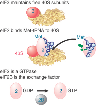
FIGURE 22.21 Initiation factors bind the initiator Met-tRNA to the 40S subunit to form a 43S complex. Later in the reaction, GTP is hydrolyzed and eIF2 is released in the form of eIF2-GDP. eIF2B regenerates the active form.
Figure 22.22 shows the group of factors that bind to the 5′ end of mRNA. The factor eIF4F is a protein complex that contains three of the initiation factors. It appears that they preassemble as a complex before binding to mRNA. The complex includes the cap-binding subunit eIF4E, the helicase eIF4A, and the “scaffolding” subunit eIF4G. After eIF4E binds the cap, eIF4A unwinds any secondary structure that exists in the first 15 bases of the mRNA. Energy for the unwinding is provided by hydrolysis of ATP. Unwinding of the structure further along the mRNA is accomplished by eIF4A together with another factor, eIF4B. The main role of eIF4G is to link other components of the initiation complex.

FIGURE 22.22 The heterotrimer eIF4F binds to the 5′ end of mRNA as well as to other factors.
The subunit eIF4E is a focus for regulation. Its activity is increased by phosphorylation, which is triggered by stimuli that increase translation and reversed by stimuli that repress translation. The subunit eIF4F has a kinase activity that phosphorylates eIF4E. The availability of eIF4E is also controlled by proteins that bind to it (called 4E-BP1, -2, and -3), to prevent it from functioning in initiation.
The presence of a poly(A) tail on the 3′ end of the mRNA stimulates the formation of the initiation complex at the 5′ end. PABP binds to the eIF4G scaffolding protein, bringing about a circular organization of the mRNA with both the 5′ and 3′ ends held in this complex. The formation of this closed loop stimulates translation; PABP is required for this effect, meaning that PABP effectively serves as an initiation factor. The PABP–eIF4G interaction on the mRNA promotes the recruitment of the 43S complex to the mRNA, as well as the joining of the 60S subunit.
Figure 22.23 shows that the interactions involved in binding the mRNA to the 43S complex are not completely defined, but appear to involve eIF4G and eIF3, as well as the mRNA and 40S subunit. The subunit eIF4G binds to eIF3. This provides the means by which the 40S ribosomal subunit binds to eIF4F and thus is recruited to the complex. In effect, eIF4F functions to get eIF4G in place so that it can attract the small ribosomal subunit.
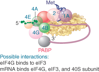
FIGURE 22.23 Interactions involving initiation factors are important when mRNA binds to the 43S complex.
When the small subunit has bound to the mRNA, it (usually) migrates to the first AUG codon using the Met-tRNA anticodon to find it. Scanning is assisted by the factors eIF1 and eIF1A. This process requires expenditure of energy in the form of ATP, and thus factors associated with ATP hydrolysis (eIF4A, IF4B, and eIF4F) also play a role in this step. Figure 22.24 shows that the small subunit stops when it reaches the initiation site, at which point the initiator tRNA base pairs with the AUG initiation codon, forming a stable 48S complex.

FIGURE 22.24 eIF1 and eIF1A help the 43S initiation complex to “scan” the mRNA until it reaches an AUG codon. eIF2 hydrolyzes its GTP to enable its release together with IF3. eIF5B mediates joining of the 60S and 40S subunits.
Joining of the 60S subunit with the initiation complex cannot occur until eIF2 and eIF3 have been released from the initiation complex. This is mediated by eIF5 and causes eIF2 to hydrolyze its GTP. The reaction occurs on the 40S subunit and requires the base pairing of the initiator tRNA with the AUG initiation codon. All of the remaining factors are likely released when the complete 80S ribosome is formed.
Finally, the initiation factor eIF5B enables the 60S subunit to join the complex, forming an intact ribosome that is ready to start elongation. eIF5B has a similar sequence to the prokaryotic initiation factor IF2, which has a similar role in hydrolyzing GTP (in addition to its role in binding the initiator tRNA).
Once the factors have been released, they can associate with the initiator tRNA and ribosomal subunits in another initiation cycle. The subunit eIF2 has hydrolyzed its GTP; as a result, the active form must be regenerated. This is accomplished by the guanosine exchange factor (GEF), eIF2B, which displaces the GDP so that it can be replaced by GTP.
The subunit eIF2 is a target for regulation. Several regulatory kinases act on the α subunit of eIF2. Phosphorylation prevents eIF2B from regenerating the active form, which limits the action of eIF2B to one cycle of initiation and thereby inhibits translation.
22.10 Elongation Factor Tu Loads Aminoacyl-tRNA into the A Site
Once the complete ribosome is formed at the initiation codon, the stage is set for an elongation cycle in which an aminoacyl-tRNA enters the A site of a ribosome whose P site is occupied by a peptidyl-tRNA. Any aminoacyl-tRNA except the initiator can enter the A site; the one that does enter is determined by the mRNA codon in the A site. Its entry is mediated by an elongation factor (EF-Tu in bacteria). The process is similar in eukaryotes. EF-Tu is a highly conserved protein among bacteria and mitochondria and is homologous to its eukaryotic counterpart.
Just like its counterpart in the initiation stage (IF-2), EF-Tu is associated with the ribosome only during the process of aminoacyl-tRNA entry. Once the aminoacyl-tRNA is in place EF-Tu leaves the ribosome to work again with another aminoacyl-tRNA. Thus, it displays the cyclic association with, and dissociation from, the ribosome that is the hallmark of the accessory factors.
Figure 22.25 depicts the role of EF-Tu in bringing aminoacyl-tRNA to the A site. EF-Tu is a monomeric GTP-binding protein that is active when bound to GTP and inactive when bound to guanine diphosphate (GDP). The binary complex of EF-Tu–GTP binds to aminoacyl-tRNA to form a ternary complex of aminoacyl-tRNA–EF-Tu–GTP. The ternary complex binds only to the A site of ribosomes whose P site is already occupied by peptidyl-tRNA. This is the critical reaction in ensuring that the aminoacyl-tRNA and peptidyl-tRNA are correctly positioned for peptide bond formation.

FIGURE 22.25 EF-Tu–GTP places aminoacyl-tRNA on the A site of ribosome and then is released as EF-Tu–GDP. EF-Ts is required to mediate the replacement of GDP by GTP. The reaction consumes GTP and releases GDP. The only aminoacyl-tRNA that cannot be recognized by EF-Tu–GTP is fMet-tRNAf, whose failure to bind prevents it from responding to internal AUG or GUG codons.
Aminoacyl-tRNA is loaded into the A site in two stages. First, the anticodon end binds to the A site of the 30S subunit. Then, codon–anticodon base pairing triggers a change in the conformation of the ribosome. This stabilizes tRNA binding and causes EF-Tu to hydrolyze its GTP. The CCA end of the tRNA now moves into the A site on the 50S subunit. The binary complex EF-Tu–GDP is released. This form of EF-Tu is inactive and does not bind aminoacyl-tRNA effectively.
The guanine nucleotide exchange factor, EF-Ts, mediates the regeneration of the inactive form EF-Tu–GDP into the active form EF-Tu–GTP. First, EF-Ts displaces the GDP from EF-Tu, forming the combined factor EF-Tu–EF-Ts. Then the EF-Ts is, in turn, displaced by GTP, reforming EF-Tu–GTP. The active binary complex binds to an aminoacyl-tRNA, and the released EF-Ts can recycle.
Each cell has about 70,000 molecules of EF-Tu (which is about 5% of the total amount of bacterial protein), which approaches the number of aminoacyl-tRNA molecules. This implies that most aminoacyl-tRNAs are likely to be in ternary complexes. Each cell has only about 10,000 molecules of EF-T, about the same as the number of ribosomes. The kinetics of the interaction between EF-Tu and EF-Ts suggest that the EF-Tu–EF-Ts complex exists only transiently, so that the EF-Tu is very rapidly converted to the GTP-bound form, and then to a ternary complex.
The role of GTP in the ternary complex has been studied by substituting an analog that cannot be hydrolyzed. The compound GMP-PCP has a methylene bridge in place of the oxygen that links the β and γ phosphates in GTP. In the presence of GMP-PCP, a ternary complex that binds aminoacyl-tRNA to the ribosome can be formed. However, the peptide bond cannot be formed, so the presence of GTP is needed for aminoacyl-tRNA to be bound at the A site. The hydrolysis is not required until later.
Kirromycin is an antibiotic that inhibits the function of EF-Tu. When EF-Tu is bound by kirromycin, it remains able to bind aminoacyl-tRNA to the A site. However, the EF-Tu–GDP complex cannot be released from the ribosome. Its continued presence prevents formation of the peptide bond between the peptidyl-tRNA and the aminoacyl-tRNA. As a result, the ribosome becomes “stalled” on the mRNA, bringing translation to a halt.
This effect of kirromycin demonstrates that inhibiting one step in translation blocks the next step. The reason is that the continued presence of EF-Tu prevents the aminoacyl end of aminoacyl-tRNA from entering the A site on the 50S subunit. Thus, the release of EF-Tu–GDP is needed for the ribosome to undertake peptide bond formation. The same principle is seen at other stages of translation: One reaction must be properly completed before the next can occur.
The interaction with EF-Tu also plays a role in quality control. Aminoacyl-tRNAs are brought into the A site without regard for whether their anticodons will fit the codon. The hydrolysis of EF-Tu–GTP is relatively slow; it takes longer than the time required for an incorrect aminoacyl-tRNA to dissociate from the A site, so most incorrect aminoacyl-tRNAs are removed at this stage. The release of EF-Tu–GDP after hydrolysis is also slow, so any remaining incorrect aminoacyl-tRNAs may dissociate at this stage. The basic principle is that the reactions involving EF-Tu occur slowly enough to allow incorrect aminoacyl-tRNAs to dissociate before they become “trapped” in translation.
In eukaryotes, the factor eEF1a is responsible for bringing aminoacyl-tRNA to the ribosome, also in a reaction that involves cleavage of a high-energy bond in GTP. Like its prokaryotic homolog (EF-Tu), it is abundant in the cell. After hydrolysis of GTP, the active form is regenerated by the factor eEF1βγ, a counterpart to EF-Ts.
22.11 The Polypeptide Chain Is Transferred to Aminoacyl-tRNA
The ribosome remains in place while the polypeptide chain is elongated by transferring the polypeptide attached to the tRNA in the P site to the aminoacyl-tRNA in the A site. The reaction is shown in Figure 22.26. The component responsible for synthesis of the peptide bond is called peptidyl transferase. It is a function of the large (50S or 60S) ribosomal subunit. The reaction is triggered when EF-Tu releases the aminoacyl end of its tRNA, which then swings into a location close to the end of the peptidyl-tRNA. This site has a peptidyl transferase activity that essentially ensures a rapid transfer of the peptide chain to the aminoacyl-tRNA. Both rRNA and 50S subunit proteins are necessary for this activity, but the actual act of catalysis is a property of the ribosomal RNA of the 50S subunit (see the section later in this chapter titled 23S rRNA Has Peptidyl Transferase Activity).

FIGURE 22.26 Peptide bond formation takes place by a reaction between the polypeptide of peptidyl-tRNA in the P site and the amino acid of aminoacyl-tRNA in the A site.
The nature of the transfer reaction is revealed by the ability of the antibiotic puromycin to inhibit translation. Puromycin resembles an amino acid attached to the terminal adenosine of tRNA. Figure 22.27 shows that puromycin has a nitrogen instead of the oxygen that joins an amino acid to a tRNA. The antibiotic is treated by the ribosome as though it were an incoming aminoacyl-tRNA, after which the polypeptide attached to peptidyl-tRNA is transferred to the –NH2 group of the puromycin.
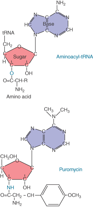
FIGURE 22.27 Puromycin mimics aminoacyl-tRNA because it resembles an aromatic amino acid linked to a sugar-base moiety.
The puromycin moiety is not anchored to the A site of the ribosome; as a result, the polypeptidyl-puromycin adduct is released from the ribosome in the form of polypeptidyl-puromycin. This premature termination of translation is responsible for the lethal action of the antibiotic.
22.12 Translocation Moves the Ribosome
The cycle of addition of amino acids to the growing polypeptide chain is completed by translocation, when the ribosome advances three nucleotides along the mRNA. Figure 22.28 shows that translocation expels the uncharged tRNA from the P site, allowing the new peptidyl-tRNA to enter. The ribosome then has an empty A site ready for entry of the aminoacyl-tRNA corresponding to the next codon. As the figure shows, in bacteria the discharged tRNA is transferred from the P site to the E site (from which it is then expelled directly into the cytosol). The A and P sites straddle both the large and small subunits; the E site (in bacteria) is located largely on the 50S subunit, but has some contacts in the 30S subunit.

FIGURE 22.28 A bacterial ribosome has three tRNA-binding sites. Aminoacyl-tRNA enters the A site of a ribosome that has peptidyl-tRNA in the P site. Peptide bond synthesis deacylates the P site tRNA and generates peptidyl-tRNA in the A site. Translocation moves the deacylated tRNA into the E site and moves peptidyl-tRNA into the P site.
Evidence suggests that translocation follows the hybrid state model, which has translocation occurring in two stages. Figure 22.29 shows that first there is a shift of the 50S subunit relative to the 30S subunit, followed by a second shift that occurs when the 30S subunit moves along mRNA to restore the original conformation. The basis for this model was the observation that the pattern of contacts that tRNA makes with the ribosome (measured by chemical footprinting) changes in two stages. When puromycin is added to a ribosome that has an aminoacylated tRNA in the P site, the contacts of tRNA on the 50S subunit change from the P site to the E site, but the contacts on the 30S subunit do not change. This suggests that the 50S subunit has moved to a posttransfer state, but that the 30S subunit has not moved.

FIGURE 22.29 The hybrid state model for translocation involves two stages. First, at peptide bond formation the aminoacyl end of the tRNA in the A site becomes relocated in the P site. Second, the anticodon end of the tRNA becomes relocated in the P site.
The interpretation of these results is that first the aminoacyl ends of the tRNAs (located in the 50S subunit) move into the new sites (while the anticodon ends remain bound to their anticodons in the 30S subunit). At this stage, the tRNAs are effectively bound in hybrid sites, consisting of the 50S E/30S P and the 50S P/30S A sites. Then movement is extended to the 30S subunits, so that the anticodon–codon pairing region finds itself in the right site. The most likely means of creating the hybrid state is by a movement of one ribosomal subunit relative to the other so that translocation in effect involves two stages, with the normal structure of the ribosome being restored by the second stage.
The ribosome faces an interesting dilemma at translocation. It needs to break many of its contacts with tRNA in order to allow movement. However, at the same time it must maintain pairing between tRNA and the anticodon, breaking the pairing of the deacylated tRNA only at the right moment. One likely possibility is that the ribosome switches between alternative, discrete conformations, essentially acting as a Brownian motor. The switch could consist of changes in rRNA base pairing. The accuracy of translation is influenced by certain mutations that influence alternative base-pairing arrangements. The most likely interpretation is that the effect is mediated by the strengths of the alternative ribosome conformations in binding to tRNA, with elongation factors acting to stabilize certain conformations.
22.13 Elongation Factors Bind Alternately to the Ribosome
Translocation requires GTP and another elongation factor, EF-G. (The eukaryotic homolog of EF-G is eEF2.) This factor is a major constituent of the cell; it is present at a level of about 1 copy per ribosome (20,000 molecules per cell).
Ribosomes cannot bind EF-Tu and EF-G simultaneously, so translation follows the cycle illustrated in Figure 22.30, in which the factors are alternately bound to and released from the ribosome. Thus, EF-Tu–GDP must be released before EF-G can bind, and then EF-G must be released before aminoacyl-tRNA–EF-Tu–GTP can bind.

FIGURE 22.30 Binding of factors EF-Tu and EF-G alternates as ribosomes accept new aminoacyl-tRNAs, form peptide bonds, and translocate.
Does the ability of each elongation factor to exclude the other rely on an allosteric effect on the overall conformation of the ribosome or on direct competition for overlapping binding sites? Figure 22.31 shows an extraordinary similarity between the structures of the ternary complex of aminoacyl-tRNA–EF-Tu–GDP and EF-G. The structure of EF-G mimics the overall structure of EF-Tu bound to the amino acceptor stem of aminoacyl-tRNA. This suggests that they compete for the same binding site (presumably in the vicinity of the A site). The need for each factor to be released before the other can bind ensures that the events of translation proceed in an orderly manner.

FIGURE 22.31 The structure of the ternary complex of aminoacyl-tRNA–EF-Tu–GTP (left) resembles the structure of EF-G (right). Structurally conserved domains of EF-Tu and EF-G are in red and green; the tRNA and the domain resembling it in EF-G are in purple.
Photo courtesy of Poul Nissen, University of Aarhus, Denmark.
Both elongation factors are monomeric GTP-binding proteins that are active when bound to GTP but inactive when bound to GDP. The triphosphate form is required for binding to the ribosome, which ensures that each factor obtains access to the ribosome only in the company of the GTP that it needs to fulfill its function.
EF-G binds to the ribosome to facilitate translocation and then is released following ribosome movement. EF-G can still bind to the ribosome when GMP-PCP is substituted for GTP, so the presence of a guanine nucleotide is needed for binding, but its hydrolysis is not absolutely essential for translocation (though translocation is much slower in the absence of GTP hydrolysis). The hydrolysis of GTP is needed to release EF-G.
The need for EF-G release was discovered by the effects of the steroid antibiotic fusidic acid, which “jams” the ribosome in its posttranslocation state. In the presence of fusidic acid, one round of translocation occurs; EF-G binds to the ribosome, GTP is hydrolyzed, and the ribosome moves over by three nucleotides. However, fusidic acid stabilizes the ribosome–EF-G–GDP complex so that EF-G and GDP remain on the ribosome instead of being released. As a result, the ribosome cannot bind aminoacyl-tRNA, and no further amino acids can be added to the chain.
Translocation is an intrinsic property of the ribosome that requires a major change in structure (see the section later in this chapter titled Ribosomes Have Several Active Centers). This intrinsic translocation is activated by EF-G in conjunction with GTP hydrolysis, which occurs before translocation and accelerates the ribosomal movement. The most likely mechanism is that GTP hydrolysis causes a change in the structure of EF-G, which, in turn, forces a change in the ribosome structure. An extensive reorientation of EF-G occurs at translocation. Before translocation, it is bound across the two ribosomal subunits. Most of its contacts with the 30S subunit are made by a region called domain 4, which is inserted into the A site. This domain could be responsible for displacing the tRNA. After translocation, domain 4 is instead oriented toward the 50S subunit.
The eukaryotic counterpart to EF-G is the protein eEF2, which functions in a similar manner to a translocase dependent on GTP hydrolysis. Its action also is inhibited by fusidic acid. A stable complex of eEF2 with GTP can be isolated and the complex can bind to ribosomes with consequent hydrolysis of its GTP.
A unique property of eEF2 is its susceptibility to diphtheria toxin. The toxin uses nicotinamide adenine dinucleotide (NAD) as a cofactor to transfer an adenosine diphosphate ribosyl (ADPR) moiety onto the eEF2. The ADPR–eEF2 conjugate is inactive in translation. The substrate for the attachment is an unusual amino acid that is produced by modifying a histidine; it is common to the eEF2 of many species.
The ADP-ribosylation is responsible for the lethal effects of diphtheria toxin. The reaction is extremely effective: A single molecule of toxin can modify enough eEF2 molecules to kill a cell.
22.14 Three Codons Terminate Translation
Only 61 of the 64 possible nucleotide triplets specify amino acids. The other three triplets are termination codons (also known as nonsense codons or stop codons), which end translation. They have casual names from the history of their discovery. The UAG triplet is called the amber codon, UAA is the ochre codon, and UGA is the opal codon.
The nature of these triplets was originally shown by a genetic test that distinguished two types of point mutations:
A point mutation that changes a codon to represent a different amino acid is called a missense mutation. One amino acid replaces the other in the polypeptide; the effect on protein function depends on the site of mutation and the nature of the amino acid replacement.
A point mutation that changes a codon to one of the three termination codons is called a nonsense mutation. It causes premature termination of translation at the mutant codon. Only the first part of the polypeptide is made in the mutant cell. This is likely to abolish protein function (depending, of course, on how far along the polypeptide the mutant site is located).
In every gene that has been sequenced, one of the termination codons lies immediately downstream from the codon representing the C-terminal amino acid of the wild-type sequence. Nonsense mutations show that any one of the three codons is sufficient to terminate translation within a gene. The UAG, UAA, and UGA triplet sequences are therefore necessary and sufficient to end translation, whether they occur naturally at the end of an open reading frame (ORF) or are created by nonsense mutations within coding sequences. (Sometimes the term nonsense codon is used to describe the termination triplets. Nonsense is really a term that describes the effect of a mutation in a gene rather than the meaning of the codon for translation. Stop codon is a better term.)
In bacterial genes, UAA is the most commonly used termination codon. UGA is used more frequently than UAG, although there appear to be more errors reading UGA. (An error in reading a termination codon—when an aminoacyl-tRNA improperly recognizes it—results in the continuation of translation until another termination codon is encountered or the ribosome reaches the 3′ end of the mRNA, which may result in other problems. For this circumstance, bacteria have a special RNA.)
22.15 Termination Codons Are Recognized by Protein Factors
Two stages are involved in ending translation. The termination reaction itself involves release of the polypeptide chain from the last tRNA. The posttermination reaction involves release of the tRNA and mRNA and dissociation of the ribosome into its subunits.
None of the termination codons normally have tRNAs that can pair with them. They function in an entirely different manner from other codons and are recognized directly by protein factors. (The reaction does not depend on codon–anticodon recognition, so there seems to be no particular reason why it should require a triplet sequence. Presumably this is an evolutionary consequence of the genetic code.)
Termination codons are recognized by class 1 release factors (RFs). In E. coli, two class 1 release factors are specific for different codons. RF1 recognizes UAA and UAG, and RF2 recognizes UGA and UAA. The factors act at the ribosomal A site and require polypeptidyl-tRNA in the P site. The reading frames are present at much lower levels than initiation or elongation factors, with about 600 molecules of each per cell, equivalent to one reading frame per 10 ribosomes. At one time there was probably only a single release factor that recognized all termination codons, which later evolved into two factors with specificities for particular codons. Eukaryotes have a single class 1 release factor, eRF. The efficiency with which the bacterial factors recognize their target codons is influenced by the bases on the 3′ side.
The class 1 release factors are assisted by class 2 release factors, which are not codon specific. The class 2 factors are GTP-binding proteins. In E. coli, the role of the class 2 factor, RF3, is to release the class 1 factor from the ribosome. RF3 is a GTP-binding protein that is related to the elongation factors.
Although the general mechanism of termination is similar in prokaryotes and eukaryotes, the interactions between the class 1 and class 2 factors have some differences.
The class 1 factors RF1 and RF2 recognize the termination codons and activate the ribosome to hydrolyze the peptidyl tRNA. Cleavage of polypeptide from tRNA takes place by a reaction analogous to the usual peptidyl transfer, except that the acceptor is H2O instead of aminoacyl-tRNA.
At this point RF1 or RF2 is released from the ribosome by the class 2 factor RF3, which is related to EF-G. RF3-GDP binds to the ribosome before the termination reaction occurs, and the GDP is replaced by GTP. This enables RF3 to contact the ribosomal GTPase center, where it causes RF1 or RF2 to be released when the polypeptide chain is terminated.
RF3 resembles the GTP-binding domains of EF-Tu and EF-G, and RF1 and RF2 resemble the C-terminal domain of EF-G, which mimics tRNA. This suggests that the release factors utilize the same site that is used by the elongation factors. Figure 22.32 illustrates the basic idea that these factors all have the same general shape and bind to the ribosome successively at the same site (basically the A site or a region extensively overlapping with it).

FIGURE 22.32 Molecular mimicry enables the EF-Tu–tRNA complex, the translocation factor EF-G, and the release factors RF1/2-RF3 to bind to the same ribosomal site. RRF is the ribosome recycling factor.
The eukaryotic class 1 release factor, eRF1, is a single protein that recognizes all three termination codons. Its sequence is unrelated to the bacterial factors. It can terminate translation in vitro without the class 2 factor, eRF2, although eRF2 is essential in yeast in vivo. The structure of eRF1 follows a familiar theme; Figure 22.33 shows that it consists of three domains that mimic the structure of tRNA.
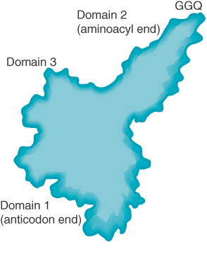
FIGURE 22.33 The eukaryotic termination factor eRF1 has a structure that mimics tRNA. The motif GGQ at the tip of domain 2 is essential for hydrolyzing the polypeptide chain from tRNA.
An essential motif of three amino acids, GGQ, is exposed at the top of domain 2. Its position in the A site corresponds to the usual location of an amino acid on an aminoacyl-tRNA. This positions it to use the glutamine (Q) to position H2O to substitute for the amino acid of aminoacyl-tRNA in the peptidyl transfer reaction. Figure 22.34 compares the termination reaction with the usual peptide transfer reaction. Termination transfers a hydroxyl group from H2O, thus effectively hydrolyzing the peptide–tRNA bond.

FIGURE 22.34 Peptide transfer and termination are similar reactions in which a base in the peptidyl transfer center triggers a transesterification reaction by attacking an N–H or O–H bond, releasing the N or O to attack the link to tRNA.
Mutations in the RF genes reduce the efficiency of termination, as seen by an increased ability to continue translation past the termination codon. Overexpression of RF1 or RF2 increases the efficiency of termination at the codons on which it acts. This suggests that codon recognition by RF1 or RF2 competes with aminoacyl-tRNAs that erroneously pair with the termination codons. The release factors recognize their target sequences very efficiently.
The termination reaction releases the completed polypeptide but leaves a deacylated tRNA and the mRNA still associated with the ribosome. Figure 22.35 shows that the dissociation of the remaining components (tRNA, mRNA, 30S, and 50S subunits) requires the ribosome recycling factor (RRF). RRF acts together with EF-G in a reaction that uses hydrolysis of GTP. As for the other factors involved in release, RRF has a structure that mimics tRNA, except that it lacks an equivalent for the 3′ amino acid–binding region. IF-3 is also required. RRF acts on the 50S subunit and IF-3 acts to remove deacylated tRNA from the 30S subunit. Once the subunits have separated, IF-3 remains necessary, of course, to prevent their reassociation.

FIGURE 22.35 The RF (release factor) terminates translation by releasing the polypeptide chain. The RRF (ribosome recycling factor) releases the last tRNA, and EF-G releases RRF, causing the ribosome to dissociate.
Table 22.1 compares the functional and sequence homologies of the prokaryotic and eukaryotic translation factors.
TABLE 22.1 Functional homologies of prokaryotic and eukaryotic translation factors.
| Initiation Factors | |||
| Prokaryotic | Eukaryotic | General Function | Notes |
| IF-1 | eIF1A | Blocks A site | eIF1A assists eIF2 in promoting Met-tRNAiMet to bind to 40S; also promotes subunit dissociation. |
| IF-2*† | eIF2, eIF3, eIF5B* | Entry of initiator tRNA | eIF2 is a GTPase. eIF3 stimulates formation of the ternary complex, its binding to 40S, and binding and scanning of mRNA. eIF5B is involved in initiator tRNA entry and is a GTPase. |
| IF-3 | eIF1, eIF4 complex, eIF3 | Small subunit binding to mRNA | eIF4 complex functions in cap binding. |
| Elongation Factors | |||
| Prokaryotic | Eukaryotic | General Function | |
| EF-Tu†‡, EF-G† | eEF1α‡ | GTP-binding | |
| EF-Ts | eEF1β, eEF1γ | GDP-exchanging | |
| EF-G§ | eEF2§ | Ribosome translocation | |
| Release Factors | |||
| Prokaryotic | Eukaryotic | General Function | |
| RF1 | eRF1 | UAA/UAG recognition | |
| RF2 | eRF1 | UAA/UGA recognition | |
| RF3† | eRF3 | Stimulation of other RF(s) | |
* IF-2 and eIF5B have sequence homology. † IF-2, EF-Tu, EF-G, and RF3 have sequence homology. |
|||
22.16 Ribosomal RNA Is Found Throughout Both Ribosomal Subunits
Two-thirds of the mass of the bacterial ribosome is made up of rRNA. The most revealing approach to analyzing secondary structure of large RNAs is to compare the sequences of homologous rRNAs in related organisms. Those regions that are important in the secondary structure retain the ability to interact by base pairing. Thus, if a base pair is required, it can form at the same relative position in each rRNA. This approach has enabled detailed models of 16S and 23S rRNA to be constructed.
Each of the major rRNAs has a secondary structure with several discrete domains. Four general domains are formed by 16S rRNA, in which just under half of the sequence is base paired. Six general domains are formed by 23S rRNA. The individual double-helical regions tend to be short (fewer than 8 bp). Frequently the duplex regions are not perfect and contain bulges of unpaired bases. Comparable models have been drawn for mitochondrial rRNAs (which are shorter and have fewer domains) and for eukaryotic cytosolic rRNAs (which are longer and have more domains). The greater length of eukaryotic rRNAs is due largely to the acquisition of sequences representing additional domains. The crystal structure of the ribosome shows that in each subunit the domains of the major rRNA fold independently and have discrete locations.
Differences in the ability of 16S rRNA to react with chemical agents are found when 30S subunits are compared with 70S ribosomes; there also are differences between separate ribosomal subunits and those engaged in translation. Changes in the reactivity of the rRNA occur when mRNA is bound, when the subunits associate, or when tRNA is bound. Some changes reflect a direct interaction of the rRNA with mRNA or tRNA, whereas others are caused indirectly by other changes in ribosome structure. The main point is that ribosome conformation is flexible during translation, particularly that of the small subunit, because it must physically check the accuracy of codon–anticodon pairing.
A feature of the primary structure of rRNA is the presence of methylated residues. There are about 10 methyl groups in 16S rRNA (located mostly toward the 3′ end of the molecule) and about 20 in 23S rRNA. In mammalian cells, the 18S and 28S rRNAs carry 43 and 74 methyl groups, respectively, so about 2% of the nucleotides are methylated (about three times the proportion of methylated nucleotides in bacterial rRNAs).
The large ribosomal subunit also contains a molecule of a 120-base 5S RNA (in all ribosomes except those of mitochondria). The sequence of 5S RNA is less well conserved than those of the major rRNAs. All 5S RNA molecules display a highly base-paired structure.
In eukaryotic cytosolic ribosomes, another small RNA is present in the large subunit, the 5.8S RNA. Its sequence corresponds to the 5′ end of the prokaryotic 23S rRNA.
Some ribosomal proteins bind strongly to isolated rRNAs. Others do not bind to free rRNAs, but can bind after other proteins have bound. This suggests that the conformation of the rRNA is important in determining whether binding sites exist for some proteins. As each protein binds, it induces conformational changes in the rRNA that make it possible for other proteins to bind. In E. coli, virtually all the 30S ribosomal proteins interact (albeit to varying degrees) with 16S rRNA. The binding sites on the proteins show a wide variety of structural features, suggesting that protein–RNA recognition mechanisms may be diverse.
The 70S ribosome has an asymmetric structure. Figure 22.36 shows a schematic of the structure of the 30S subunit, which is divided into four regions: the head, neck, body, and platform. Figure 22.37 shows a similar representation of the 50S subunit, where two prominent features are the central protuberance (where 5S rRNA is located) and the stalk (made of multiple copies of protein L7). Figure 22.38 shows that the platform of the small subunit fits into the notch of the large subunit. A cavity (resembling a doughnut, but not visible in the figure) between the subunits contains some of the important sites.
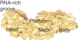
FIGURE 22.36 The 30S subunit has a head separated by a neck from the body, with a protruding platform.

FIGURE 22.37 The 50S subunit has a central protuberance where 5S rRNA is located, separated by a notch from a stalk made of copies of the protein L7.

FIGURE 22.38 The platform of the 30S subunit fits into the notch of the 50S subunit to form the 70S ribosome.
The structure of the 30S subunit follows the organization of 16S rRNA, with each structural feature corresponding to a domain of the rRNA. The body is based on the 5′ domain, the platform on the central domain, and the head on the 3′ region. Figure 22.39 shows that the 30S subunit has an asymmetric distribution of RNA and protein. One important feature is that the platform of the 30S subunit that provides the interface with the 50S subunit is composed almost entirely of RNA. At most, two proteins (a small part of S7 and possibly part of S12) lie near the interface. This means that the association and dissociation of ribosomal subunits must depend on interactions with the 16S rRNA. Subunit association is affected by a mutation in a loop of 16S rRNA (at position 791) that is located at the subunit interface, and other nucleotides in 16S rRNA have been shown to be involved by modification/interference experiments. This observation supports the idea that the evolutionary origin of the ribosome may have been as a particle consisting solely of RNA rather than of both RNA protein.
FIGURE 22.39 The 30S ribosomal subunit is a ribonucleoprotein particle. Ribosomal proteins are white and rRNA is light blue.
Courtesy of Dr. Kalju Kahn.
The 50S subunit has a more even distribution of components than the 30S does, with long rods of double-stranded RNA crisscrossing the structure. The RNA forms a mass of tightly packed helices. The exterior surface largely consists of protein, except for the peptidyl transferase center (see the section later in this chapter titled 23S rRNA Has Peptidyl Transferase Activity). Almost all segments of the 23S rRNA interact with protein, but many of the proteins are relatively unstructured.
The junction of subunits in the 70S ribosome involves contacts between 16S rRNA (many in the platform region) and 23S rRNA. A few interactions also occur between rRNAs of each subunit with proteins in the other and a few protein–protein contacts. Figure 22.40 identifies the contact points on the rRNA structures. Figure 22.41 opens out the structure (imagine the 50S subunit rotated counterclockwise and the 30S subunit rotated clockwise around the axis shown in the figure) to show the locations of the contact points on the face of each subunit.

FIGURE 22.40 Contact points between the rRNAs are located in two domains of 16S rRNA and one domain of 23S rRNA.
Laguna Design/Getty Images.

FIGURE 22.41 Contacts between the ribosomal subunits are mostly made by RNA (shown in purple). Contacts involving proteins are shown in yellow. The two subunits are rotated away from one another to show the faces where contacts are made; from a plane of contact perpendicular to the screen, the 50S subunit is rotated 90° counterclockwise, and the 30S is rotated 90° clockwise (this shows it in the reverse of the usual orientation).
Photos courtesy of Harry Noller, University of California, Santa Cruz.
22.17 Ribosomes Have Several Active Centers
The basic ribosomal feature is that it is a cooperative structure that depends on changes in the relationships among its active sites during translation. The active sites are not small, discrete regions like the active centers of enzymes. Rather, they are large regions whose construction and activities may depend just as much on the rRNA as on the ribosomal proteins. The crystal structures of the individual subunits and bacterial ribosomes give us a good impression of the overall organization and emphasize the role of the rRNA. The 2.8 Å–resolution structure clearly identifies the locations of the tRNAs and the functional sites. Many ribosomal functions can now be accounted for in terms of its structure.
Ribosomal functions are centered around the interactions with tRNAs. Figure 22.42 shows the 70S ribosome with the positions of tRNAs in the three binding sites. The tRNAs in the A and P sites are nearly parallel to one another. All three tRNAs are aligned with their anticodon loops bound to the mRNA in the groove on the 30S subunit. The rest of each tRNA is bound to the 50S subunit. The environment surrounding each tRNA is mostly provided by rRNA. In each site, the rRNA contacts the tRNA at parts of the structure that are universally conserved.

FIGURE 22.42 The 70S ribosome consists of the 50S subunit (white) and the 30S subunit (purple), with three tRNAs located superficially: yellow in the A site, blue in the P site, and green in the E site.
Photo courtesy of Harry Noller, University of California, Santa Cruz.
Before a high-resolution structure of the ribosome was available, it was a puzzle to understand how two bulky tRNAs could fit next to one another in reading adjacent codons. The crystal structure shows a 45° kink in the mRNA between the P and A sites, which allows the tRNAs to fit, as shown in the expansion of Figure 22.43. The tRNAs in the P and A sites are angled at 26° relative to each other at their anticodons. The closest approach between the backbones of the tRNAs occurs at the 3′ ends, where they converge to within 5 Å (perpendicular to the plane of the page). This allows the peptide chain to be transferred from the peptidyl-tRNA in the P site to the aminoacyl-tRNA in the A site.

FIGURE 22.43 Three tRNAs have different orientations on the ribosome. mRNA turns between the P and A sites to allow aminoacyl-tRNAs to bind adjacent codons.
Photo courtesy of Harry Noller, University of California, Santa Cruz.
Aminoacyl-tRNA is inserted into the A site by EF-Tu, and its pairing with the codon is necessary for EF-Tu to hydrolyze GTP and be released from the ribosome (see the section earlier in this chapter titled Elongation Factor Tu Loads Aminoacyl-tRNA into the A Site). EF-Tu initially places the aminoacyl-tRNA into the small subunit, where the anticodon pairs with the codon. Movement of the tRNA is required to bring it fully into the A site, when its 3′ end enters the peptidyl transferase center on the large subunit. Different models have been proposed for how this process may occur. One suggests that the entire tRNA swivels so that the elbow in the L-shaped structure made by the D and TψC arms moves into the ribosome, enabling the TψC arm to pair with rRNA. Another suggests that the internal structure of the tRNA changes, using the anticodon loop as a hinge, with the rest of the tRNA rotating from a position in which it is stacked on the 3′ side of the anticodon loop to one in which it is stacked on the 5′ side. Following the transition, EF-Tu hydrolyzes GTP, allowing peptide bond formation to proceed.
Translocation involves large movements in the positions of the tRNAs within the ribosome. The anticodon end of tRNA moves about 28 Å from the A site to the P site, and then moves an additional 20 Å from the P site to the E site. As a result of the angle of each tRNA relative to the anticodon, the bulk of the tRNA moves much larger distances: 40 Å from the A site to the P site and 55 Å from the P site to the E site. This suggests that translocation requires a major reorganization of structure.
For many years, it was thought that translocation could occur only in the presence of the factor EF-G. However, the antibiotic sparsomycin (which inhibits peptidyl transferase activity) triggers translocation. This suggests that the energy to drive translocation is actually stored in the ribosome after peptide bond formation has occurred. Usually EF-G acts on the ribosome to release this energy and enable it to drive translocation, but sparsomycin can play the same role. Sparsomycin inhibits peptidyl transferase by binding to the peptidyl-tRNA, blocking its interaction with aminoacyl-tRNA. It probably creates a conformation that resembles the usual posttranslocation conformation, which, in turn, promotes movement of the peptidyl-tRNA. The conclusion is that translocation is an intrinsic property of the ribosome.
The hybrid state model suggests that translocation may take place in two stages, with one ribosomal subunit moving relative to the other to create an intermediate stage in which there are hybrid tRNA-binding sites (50S E/30S P and 50S P/30S A). Comparisons of the ribosome structure between pre- and posttranslocation states, and comparisons in 16S rRNA conformation between free 30S subunits and 70S ribosomes, suggest that mobility of structure is especially marked in the head and platform regions of the 30S subunit. An interesting insight into the hybrid state model is provided by the fact that many bases in rRNA involved in subunit association are close to bases involved in interacting with tRNA. This suggests that tRNA-binding sites are close to the interface between subunits and carries the implication that changes in subunit interaction could be connected with movement of tRNA.
Much of the structure of the bacterial ribosome is occupied by its active centers. The schematic view of the ribosomal sites in Figure 22.44 shows they comprise about two-thirds of the ribosomal structure. A tRNA enters the A site, is transferred by translocation into the P site, and then leaves the ribosome by the E site. The A and P sites extend across both ribosome subunits; tRNA is paired with mRNA in the 30S subunit, but peptide transfer takes place in the 50S subunit. The A and P sites are adjacent, enabling translocation to move the tRNA from one site into the other. The E site is located near the P site (representing a position en route to the surface of the 50S subunit). The peptidyl transferase center is located on the 50S subunit, close to the aminoacyl ends of the tRNAs in the A and P sites (see the next section, 16S rRNA Plays an Active Role in Translation).

FIGURE 22.44 The ribosome has several active centers. It may be associated with a membrane. mRNA takes a turn as it passes through the A and P sites, which are angled with regard to each other. The E site lies beyond the P site. The peptidyl transferase site (not shown) stretches across the tops of the A and P sites. Part of the site bound by EF-Tu/G lies at the base of the A and P sites.
All of the GTP-binding proteins that function in translation (EF-Tu, EF-G, IF-2, RF1, RF2, and RF3) bind to the same factor-binding site (sometimes called the GTPase center), which probably triggers their hydrolysis of GTP. This site is located at the base of the stalk of the large subunit, which consists of the proteins L7 and L12. (L7 is a modification of L12 and has an acetyl group on the N-terminus.) In addition to this region, the complex of protein L11 with a 58-base stretch of 23S rRNA provides the binding site for some antibiotics that affect GTPase activity. Neither of these ribosomal structures actually possesses GTPase activity, but they are both necessary for it. The role of the ribosome is to trigger GTP hydrolysis by factors bound in the factor-binding site.
Initial binding of 30S subunits to mRNA requires protein S1, which has a strong affinity for single-stranded nucleic acid. It is responsible for maintaining the single-stranded state in mRNA that is bound to the 30S subunit. This action is necessary to prevent the mRNA from taking up a base-paired conformation that would be unsuitable for translation. S1 has an extremely elongated structure and associates with S18 and S21. The three proteins constitute a domain that is involved in the initial binding of mRNA and in binding initiator tRNA. This locates the mRNA-binding site in the vicinity of the cleft of the small subunit. The 3′ end of rRNA, which pairs with the mRNA initiation site, is located in this region.
The initiation factors bind in the same region of the ribosome. IF-3 can be crosslinked to the 3′ end of the rRNA, as well as to several ribosomal proteins, including those probably involved in binding mRNA. The role of IF-3 could be to stabilize mRNA–30S subunit binding; then it would be displaced when the 50S subunit joins.
The incorporation of 5S RNA into 50S subunits that are assembled in vitro depends on the ability of three proteins—L5, L8, and L25—to form a stoichiometric complex with it. The complex can bind to 23S rRNA, although none of the isolated components can do so. It lies in the vicinity of the P and A sites.
A nascent polypeptide extends through the ribosome, away from the active sites, into the region in which ribosomes may be attached to membranes. A polypeptide chain emerges from the ribosome through an exit channel, which leads from the peptidyl transferase site to the surface of the 50S subunit. The tunnel is composed mostly of rRNA. It is quite narrow—only 1 to 2 nm wide—and is about 10 nm long. The nascent polypeptide emerges from the ribosome about 15 Å away from the peptidyl transferase site. The tunnel can hold about 50 amino acids and probably constrains the polypeptide chain so that it cannot completely fold until it leaves the exit domain, though some limited secondary structures may form.
22.18 16S rRNA Plays an Active Role in Translation
The ribosome was originally viewed as a collection of proteins with various catalytic activities held together by protein–protein interactions and RNA–protein interactions. However, the discovery of RNA molecules with catalytic activities (see the RNA Splicing and Processing chapter) immediately suggests that rRNA might play a more active role in ribosome function. Evidence now suggests that rRNA interacts with mRNA or tRNA at each stage of translation and that the proteins are necessary to maintain the rRNA in a structure in which it can perform the catalytic functions. Several interactions involve specific regions of rRNA:
The 3′ terminus of the 16S rRNA interacts directly with mRNA at initiation.
Specific regions of 16S rRNA interact directly with the anticodon regions of tRNAs in both the A site and the P site. Similarly, 23S rRNA interacts with the CCA terminus of peptidyl-tRNA in both the P site and A site.
Subunit interaction involves interactions between 16S and 23S rRNAs (see the section earlier in this chapter titled Ribosomal RNA Is Found Throughout Both Ribosomal Subunits).
A lot of information about the individual steps of bacterial translation has been obtained by using antibiotics that inhibit the process at particular stages. The target for the antibiotic can be identified by the component in which resistant mutations occur. Some antibiotics act on individual ribosomal proteins, but several act on rRNA, which suggests that the rRNA is involved with many or even all of the functions of the ribosome.
Two types of approaches have been used to investigate the functions of rRNA. Structural studies show that particular regions of rRNA are located in important sites of the ribosome and that chemical modifications of these bases impede particular ribosomal functions. In addition, mutations identify nucleotides in rRNA that are required for particular ribosomal functions. Figure 22.45 summarizes the sites in 16S rRNA that have been identified by these means.
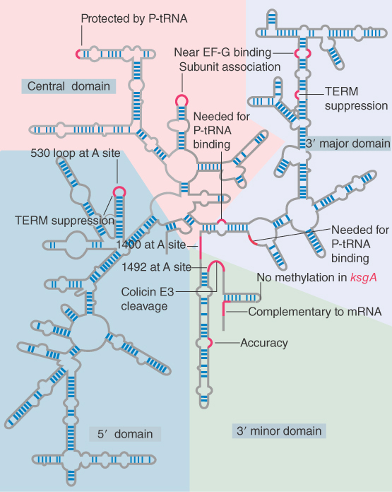
FIGURE 22.45 Some sites in 16S rRNA are protected from chemical probes when 50S subunits join 30S subunits or when aminoacyl-tRNA binds to the A site. Others are the sites of mutations that affect translation. TEM suppression sites may affect termination at some or several termination codons. The large colored blocks indicate the four domains of the rRNA.
An indication of the importance of the 3′ end of 16S rRNA is given by its susceptibility to the lethal agent colicin E3. Produced by some bacteria, colicin cleaves about 50 nucleotides from the 3′ end of the 16S rRNA of E. coli. The cleavage entirely abolishes initiation of translation. The region that is cleaved has several important functions: binding the factor IF-3, recognition of mRNA, and binding of tRNA.
The 3′ end of the 16S rRNA is directly involved in the initiation reaction by pairing with the Shine–Dalgarno sequence in the ribosome-binding site of mRNA. Another direct role for the 3′ end of 16S rRNA in translation is shown by the properties of kasugamycin-resistant mutants, which lack certain modifications in 16S rRNA. Kasugamycin blocks initiation of translation. Resistant mutants (called ksgA) lack a methylase enzyme that introduces four methyl groups into two adjacent adenines at a site near the 3′ terminus of the 16S rRNA. The methylation generates the highly conserved sequence G–m26A–m26A, which is found in both prokaryotic and eukaryotic small rRNAs. The methylated sequence is involved in the joining of the 30S and 50S subunits, which, in turn, is connected also with the retention of initiator tRNA in the complete ribosome. Kasugamycin causes fMet-tRNAf to be released from the sensitive (methylated) ribosomes, but the resistant ribosomes are able to retain the initiator.
Changes in the structure of 16S rRNA occur when ribosomes are engaged in translation, as seen by protection of particular bases against chemical attack. The individual sites fall into a few groups that are concentrated in the 3′ minor and central domains. Although the locations are dispersed in the linear sequence of 16S rRNA, it seems likely that base positions involved in the same function are actually close together in the tertiary structure.
Some of the changes in 16S rRNA are triggered by joining with 50S subunits, binding of mRNA, or binding of tRNA. They indicate that these events are associated with changes in ribosome conformation that affect the exposure of rRNA. They do not necessarily indicate direct participation of rRNA in these functions. One change that occurs during translation is shown in Figure 22.46; it involves a local movement to change the nature of a short duplex sequence.

FIGURE 22.46 A change in conformation of 16S rRNA may occur during translation.
The 16S rRNA is involved in both A site and P site function, and significant changes in its structure occur when these sites are occupied. Certain distinct regions are protected by tRNA bound in the A site. One is the 530 loop (which also is the site of a mutation that prevents termination at the UAA, UAG, and UGA codons). The other is the 1400 to 1500 region (so called because bases 1399 to 1492 and the adenines at 1492 and 1493 are two single-stranded stretches that are connected by a long hairpin). All of the effects that tRNA binding has on 16S rRNA can be produced by the isolated oligonucleotide of the anticodon stem-loop, thus tRNA–30S subunit binding must involve this region.
The adenines at 1492 and 1493 provide a mechanism for detecting properly paired codon–anticodon complexes. The principle of the interaction is that the structure of the 16S rRNA responds to the structure of the first two base pairs in the minor groove of the duplex formed by the codon–anticodon interaction. Modification of the N1 position of either base 1492 or 1493 in rRNA prevents tRNA from binding in the A site. However, mutations at 1492 or 1493 can be suppressed by the introduction of fluorine at the 2′ position of the corresponding bases in mRNA (which restores the interaction). Figure 22.47 shows that codon–anticodon pairing allows the N1 of each adenine to interact with the 2′–OH in the mRNA backbone. The interaction stabilizes the association of tRNA with the A site. When an incorrect tRNA enters the A site, the structure of the codon–anticodon complex is distorted, and this interaction cannot occur.

FIGURE 22.47 Codon–anticodon pairing supports interaction with adenines 1492 and 1493 of 16S rRNA, but mispaired tRNA–mRNA cannot interact.
A variety of bases in different positions of 16S rRNA are protected by tRNA in the P site; most likely the bases lie near one another in the tertiary structure. In fact, there are more contacts with tRNA when it is in the P site than when it is in the A site. This may be responsible for the increased stability of peptidyl-tRNA compared with aminoacyl-tRNA. This makes sense; once the tRNA has reached the P site, the ribosome has determined that it is correctly bound, whereas in the A site the assessment of binding is still being made. The 1400 region can be directly crosslinked to peptidyl-tRNA, which suggests that this region is a structural component of the P site.
The general conclusion of these results is that rRNA has many interactions with both tRNA and mRNA and that these interactions recur in each cycle of peptide bond formation.
22.19 23S rRNA Has Peptidyl Transferase Activity
The sites involved in the functions of 23S rRNA are less well identified than those of 16S rRNA, but the same general pattern is observed: Bases at certain positions affect specific functions. Bases at some positions in 23S rRNA are affected by the conformation of the A site or the P site. In particular, oligonucleotides derived from the 3′ CCA terminus of tRNA protect a set of bases in 23S rRNA that essentially are the same as those protected by peptidyl-tRNA. This suggests that the major interaction of 23S rRNA with peptidyl-tRNA in the P site involves the 3′ end of the tRNA.
The tRNA makes contact with the 23S rRNA in both the P and A sites. At the P site, G2552 of 23S rRNA base pairs with C74 of the peptidyl tRNA. A mutation in the G in the rRNA prevents interaction with tRNA, but interaction is restored by a compensating mutation in the C of the amino acceptor end of the tRNA. At the A site, G2553 of the 23S rRNA base pairs with C75 of the aminoacyl-tRNA. Thus, rRNA plays a close role in both the tRNA-binding sites. As structural studies continue to emerge, the movements of tRNA between the A and P sites in terms of making and breaking contacts with rRNA will be elucidated.
Another site that binds tRNA is the E site, which is localized almost exclusively on the 50S subunit. Bases affected by its conformation can be identified in 23S rRNA.
What is the nature of the site on the 50S subunit that provides peptidyl transferase function? A long search for ribosomal proteins that might possess the catalytic activity was unsuccessful and led to the discovery that the ribosomal RNA of the large subunit can catalyze the formation of a peptide bond between peptidyl-tRNA and aminoacyl-tRNA. The involvement of rRNA was first indicated because a region of the 23S rRNA is the site of mutations that confer resistance to antibiotics that inhibit peptidyl transferase. Extraction of almost all the protein content of 50S subunits leaves the 23S rRNA largely associated with fragments of proteins, amounting to less than 5% of the mass of the ribosomal proteins. This preparation retains peptidyl transferase activity. Treatments that damage the RNA abolish the catalytic activity.
Following from these results, 23S rRNA prepared by transcription in vitro can catalyze the formation of a peptide bond between Ac-Phe-tRNA and Phe-tRNA. The yield of Ac-Phe-Phe is very low, suggesting that the 23S rRNA requires proteins in order to function at a high efficiency. However, given that the rRNA has the basic catalytic activity, the role of the proteins must be indirect, serving to fold the rRNA properly or to present the substrates to it. The reaction also works, although less effectively, if the domains of 23S rRNA are synthesized separately and then combined. In fact, some activity is shown by domain V alone, which has the catalytic center. Activity is abolished by mutations in position 2252 of domain V that lies in the P site.
The crystal structure of an archaeal 50S subunit shows that the peptidyl transferase site basically consists of 23S rRNA. No protein exists within 18 Å of the active site where the transfer reaction occurs between peptidyl-tRNA and aminoacyl-tRNA!
Peptide bond synthesis requires an attack by the amino group of one amino acid on the carboxyl group of another amino acid. Catalysis requires a basic residue to accept the hydrogen atom that is released from the amino group, as shown in Figure 22.48. If rRNA is the catalyst, it must provide this residue, but it is not known how this happens. The purine and pyrimidine bases are not basic at physiological pH. A highly conserved base (at position 2451 in E. coli) had been implicated but appears now neither to have the right properties nor to be crucial for peptidyl transferase activity.

FIGURE 22.48 Peptide bond formation requires acid–base catalysis in which an H atom is transferred to a basic residue.
The catalytic activity of isolated rRNA is quite low, and proteins that are bound to the 23S rRNA outside of the peptidyl transfer region are almost certainly required to enable the rRNA to form the proper structure in vivo. The idea that rRNA is the catalytic component is consistent with the results discussed in the RNA Splicing and Processing chapter, which identify catalytic properties in RNA that are involved with several RNA-processing reactions. It fits with the notion that the modern ribosome evolved from a prototype originally composed solely of RNA.
22.20 Ribosomal Structures Change When the Subunits Come Together
A body of indirect evidence suggests that the structures of the individual subunits change significantly when they join together to form a complete ribosome. Differences in the susceptibilities of the rRNAs to outside agents are one of the strongest indicators (see the section earlier in this chapter titled 16S rRNA Plays an Active Role in Translation). More directly, comparisons of the high-resolution crystal structures of the individual subunits with the lower-resolution structure of the intact ribosome suggest the existence of significant differences. These ideas have been confirmed by a crystal structure of the E. coli ribosome at 3.5 Å, which furthermore identifies two different conformations of the ribosome, possibly representing different stages in translation.
The crystal contains two ribosomes per unit, each with a different conformation. The differences are due to changes in the positioning of domains within each subunit, the most important being that in one conformation the head of the small subunit has swiveled 6° around the neck region toward the E site. Also, a 6° rotation in the opposite direction is seen in the (low-resolution) structures of Thermus thermophilus ribosomes that are bound to mRNA and have tRNAs in both A and P sites, suggesting that the head may swivel overall by 12° depending on the stage of translation. The rotation of the head follows the path of tRNAs through the ribosome, raising the possibility that its swiveling controls movement of mRNA and tRNA.
The changes in conformation that occur when subunits join together are much more marked in the 30S subunit than in the 50S subunit. The changes are probably involved with controlling the position and movement of mRNA. The most significant change in the 50S subunit concerns the peptidyl transferase center. The 50S subunits are about 1,000 times less effective in catalyzing peptide bond synthesis than complete ribosomes; the reason may be a change in structure that positions the substrate more effectively in the active site in the complete ribosome.
One of the main features emerging from the structure of the complete ribosome is the very high density of solvent contacts at their interface; this may help in the making and breaking of contacts that are essential for subunit association and dissociation and may also be involved in structural changes that occur during translocation.
22.21 Translation Can Be Regulated
Control over which and how much protein is made occurs first at the level of transcription control (as discussed in The Operon chapter); then through RNA-processing control (rare in bacteria, but common in eukaryotes); and, finally, translation-level control, which is examined here. (Refer to The Operon chapter for detail on the lac operon and its regulation.)
The lac repressor is encoded by the lacI gene; this is an unregulated gene that is continuously transcribed, but from a poor promoter. Also, the coding region of the lac repressor is in a very “poor” mRNA, meaning that the 5′ UTR of the mRNA has a poor sequence context that does not allow rapid ribosome binding or movement onto the ORF. Just as promoters can be “good” or “poor,” so can mRNAs. Together, this means that ribosomes do not translate the small amount of mRNA at the same level as the lacZYA polycistronic mRNA. Thus, very little lac repressor is found in a cell—only about 10 tetramers.
A second way that translation can be modulated is by codon usage. Multiple codons exist for most of the amino acids. These codons are not utilized equally by tRNAs; some have abundant tRNAs, others do not. An ORF consisting of codons with abundant tRNAs can be rapidly translated, whereas another ORF that contains codons with less-abundant tRNAs will be translated more slowly.
Additionally, more active mechanisms exist for translation-level control. One mechanism for controlling gene expression at the level of translation parallels the use of a repressor to prevent transcription. Translational repression occurs when a protein binds to a target region on mRNA to prevent ribosomes from recognizing the initiation region. Formally, protein–mRNA binding is equivalent to a repressor protein binding to DNA to prevent polymerase from utilizing a promoter. Polycistronic RNA allows coordinate regulation of translation, analogous to transcription repression of an operon. Figure 22.49 illustrates the most common form of this interaction, in which the regulator protein binds directly to a sequence that includes the AUG initiation codon, thereby preventing the ribosome from binding.
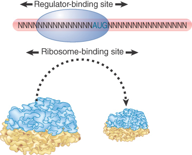
FIGURE 22.49 A regulator protein may block translation by binding to a site on mRNA that overlaps the ribosome-binding site at the initiation codon.
Some examples of translational repressors and their targets are summarized in Table 22.2. A classic example of how the product of translation can directly control the translation of its mRNA is the coat protein of the RNA phage R17; it binds to a hairpin that encompasses the ribosome-binding site in the phage mRNA. Similarly, the phage T4 RegA protein binds to a consensus sequence that includes the AUG initiation codon in several T4 early mRNAs, and T4 DNA polymerase binds to a sequence in its own mRNA that includes the Shine–Dalgarno element needed for ribosome binding.
TABLE 22.2 Proteins that bind to sequences within the initiation regions of mRNAs may function as translational repressors.
| Repressor | Target Gene | Site of Action |
|---|---|---|
| R17 coat protein | R17 replicase | Hairpin that includes ribosome-binding site |
| T4 RegA | Early T4 mRNAs | Various sequences, including initiation codon |
| T4 DNA polymerase | T4 DNA polymerase | Shine–Dalgarno sequence |
| T4 p32 | Gene 32 | Singe-stranded 5′ leader |
Another form of translational control occurs when translation of one gene requires changes in secondary structure that depend on translation of an immediately preceding gene. This happens during translation of the RNA phages, whose genes always are expressed in a set order. Figure 22.50 shows that the phage RNA takes up a secondary structure in which only one initiation sequence is accessible; the second cannot be recognized by ribosomes because it is base paired with other regions of the RNA. However, translation of the first gene disrupts the secondary structure, allowing ribosomes to bind to the initiation site of the next gene. In this mRNA, secondary structure controls translatability.

FIGURE 22.50 Secondary structure can control initiation. Only one initiation site is available in the RNA phage, but translation of the first gene changes the conformation of the RNA so that other initiation site(s) become available.
22.22 The Cycle of Bacterial Messenger RNA
Messenger RNA has the same function in all cells, but there are important differences in the details of the synthesis and in the structures of prokaryotic and eukaryotic mRNAs.
A major difference in the production of mRNA depends on the cellular locations where transcription and translation occur:
In bacteria, mRNA is transcribed and translated in the single cellular compartment; the two processes are so closely linked that they occur simultaneously. Ribosomes attach to bacterial mRNA even before its transcription has been completed so the polysome is likely to still be attached to DNA. Bacterial mRNA is usually unstable and is therefore translated into polypeptides for only a few minutes. This process is called coupled transcription/translation.
In a eukaryotic cell, synthesis and maturation of mRNA occur exclusively in the nucleus. Only after these events are completed is the mRNA exported to the cytoplasm, where it is translated by ribosomes. A typical eukaryotic mRNA is often intrinsically stable and continues to be translated for several hours, though there is a great deal of variation in the stability of specific mRNAs, in some cases due to stability or instability sequences in the 5′ or 3′ UTRs.
Figure 22.51 shows that transcription and translation are intimately related in bacteria. Transcription begins when the enzyme RNA polymerase binds to DNA and then moves along, making a copy of one strand. Soon after transcription begins, ribosomes attach to the 5′ end of the mRNA and start translation, even before the rest of the mRNA has been synthesized. Multiple ribosomes move along the mRNA while it is being synthesized. The 3′ end of the mRNA is generated when transcription terminates. Ribosomes continue to translate the mRNA while it persists, but it is degraded in the overall 5′ to 3′ direction quite rapidly. The mRNA is synthesized, translated by the ribosomes, and degraded, all in rapid succession. An individual molecule of mRNA persists for only a matter of minutes at most.

FIGURE 22.51 mRNA is transcribed, translated, and degraded simultaneously in bacteria.
Bacterial transcription and translation take place at similar rates. At 37°C, transcription of mRNA occurs at a rate of about 40 to 50 nucleotides per second. This is very close to the rate of polypeptide synthesis, which is roughly 15 amino acids per second. It therefore takes about 1 minute to transcribe and translate an mRNA of 2,500 nucleotides, corresponding to a 90-kD polypeptide. When expression of a new gene is initiated, its mRNA will typically appear in the cell within about 1.5 minutes. The corresponding polypeptide will appear within another 30 seconds.
Bacterial translation is very efficient, and most mRNAs are translated by a large number of tightly packed ribosomes. In one example, trp mRNA, about 15 initiations of transcription occur every minute and each of the 15 mRNAs is probably translated by about 30 ribosomes in the interval between its transcription and degradation.
The instability of most bacterial mRNAs is striking. Degradation of mRNA closely follows its translation and likely begins within 1 minute of the start of transcription. The 5′ end of the mRNA starts to decay before the 3′ end has been synthesized or translated. Degradation seems to follow the last ribosome of the convoy along the mRNA. However, degradation proceeds more slowly, probably at about half the speed of transcription or translation.
The stability of mRNA has a major influence on the amount of polypeptide that is produced. It is usually expressed in terms of the half-life. The mRNA representing any particular gene has a characteristic half-life, but the average is about 2 minutes in bacteria.
Of course, this series of events is only possible because transcription, translation, and degradation all occur in the same direction. The dynamics of gene expression have been “caught in the act” in the electron micrograph of Figure 22.52. In these (unknown) transcription units, several mRNAs are undergoing synthesis simultaneously, and each carries many ribosomes engaged in translation. (This corresponds to the stage shown in the second panel in Figure 22.51.) An RNA whose synthesis has not yet been completed is called a nascent RNA.

FIGURE 22.52 Transcription units can be visualized in bacteria.
© Prof. Oscar L. Miller/Photo Researchers, Inc.
Bacterial mRNAs vary greatly in the number of proteins that they encode. Some mRNAs carry only a single ORF; they are monocistronic. Others (the majority) carry sequences encoding several polypeptides; they are polycistronic. In these cases, a single mRNA is transcribed from a group of adjacent cistrons. (Such a cluster of cistrons constitutes an operon that is controlled as a single genetic unit; see The Operon chapter.)
All mRNAs contain three regions. The coding region, or open reading frame (ORF), consists of a series of codons representing the amino acid sequence of the polypeptide, starting (usually) with AUG and ending with one of the three termination codons. However, the mRNA is always longer than the coding region as extra regions are present at both ends. An additional sequence at the 5′ end, upstream of the coding region, is described as the leader or 5′ UTR. An additional sequence downstream from the termination signal, forming the 3′ end, is called the trailer or 3′ UTR. Although they do not encode a polypeptide, these sequences may contain important regulatory instructions, especially in eukaryotic mRNAs.
A polycistronic mRNA also contains intercistronic regions, as illustrated in Figure 22.53. They vary greatly in size. They may be as long as 30 nucleotides in bacterial mRNAs (and even longer in phage RNAs), or they may be very short, with as few as one or two nucleotides separating the termination codon for one polypeptide from the initiation codon for the next. In an extreme case, two genes actually overlap, so that the last base of one coding region is also the first base of the next coding region.

FIGURE 22.53 Bacterial mRNA includes untranslated as well as translated regions. Each coding region has its own initiation and termination signals. A typical mRNA may have several coding regions (ORFs).
The number of ribosomes engaged in translating a particular cistron depends on the efficiency of its initiation site in the 5′ UTR. The initiation site for the first cistron becomes available as soon as the 5′ end of the mRNA is synthesized. How are subsequent cistrons translated? Are the several coding regions in a polycistronic mRNA translated independently, or is their expression connected? Is the mechanism of initiation the same for all cistrons, or is it different for the first cistron and the downstream cistrons?
Translation of a bacterial mRNA proceeds sequentially through its cistrons. At the time when ribosomes attach to the first coding region, the subsequent coding regions have not yet been transcribed. By the time the second ribosomal binding site is available, translation is well under way through the first cistron. Typically, ribosomes terminate translation at the end of each cistron, and then a new ribosome assembles independently at the start of the next coding region. This is influenced by the intercistronic region and the density of ribosomes on the mRNA.
Summary
A codon in an mRNA is recognized by an aminoacyl-tRNA, which has an anticodon complementary to the codon and carries the amino acid corresponding to the codon. A special initiator tRNA (fMet-tRNAf in prokaryotes or Met-tRNAi in eukaryotes) recognizes the AUG codon, which is used to start most coding sequences. (In prokaryotes, GUG is also used.) Only the termination (or stop or nonsense) codons—UAA, UAG, and UGA—are not recognized by aminoacyl-tRNAs.
Ribosomes are released from translation to enter a pool of free ribosomes that are in equilibrium with separate small and large subunits. Small subunits bind to mRNA and then are joined by large subunits to generate an intact ribosome that undertakes translation. Recognition of a prokaryotic initiation site involves binding of a sequence at the 3′ end of rRNA to the Shine–Dalgarno sequence, which lies upstream from the AUG (or GUG) codon in the mRNA. Recognition of a eukaryotic mRNA involves binding of the small ribosomal subunit to the 5′ cap; the subunit then migrates to the initiation site by scanning for AUG codons. When it recognizes an appropriate AUG initiation codon (usually, but not always, the first it encounters), it is joined by a large subunit.
A ribosome can carry at least two aminoacyl-tRNAs simultaneously; its P site is occupied by a polypeptidyl-tRNA, which carries the polypeptide chain synthesized so far, whereas the A site is used for entry by an aminoacyl-tRNA carrying the next amino acid to be added to the chain. Ribosomes also have an E site, through which deacylated tRNA passes before it is released after being used in translation. The polypeptide chain in the P site is transferred to the aminoacyl-tRNA in the A site, creating a deacylated tRNA in the P site and a peptidyl-tRNA in the A site.
Following peptide bond synthesis, the ribosome translocates one codon along the mRNA, moving deacylated tRNA into the E site and peptidyl-tRNA from the A site into the P site. Translocation is catalyzed by the elongation factor EF-G and, like several other stages of ribosome function, requires hydrolysis of GTP. During translocation, the ribosome passes through a hybrid stage in which the 50S subunit moves relative to the 30S subunit.
Translation is an energetically expensive process. ATP is used to provide energy at several stages, including the charging of tRNA with its amino acid and the unwinding of mRNA. It has been estimated that up to 90% of all the ATP molecules synthesized in a rapidly growing bacterium are consumed in assembling amino acids into protein!
Additional factors are required at each stage of translation. They are defined by their cyclic association with, and dissociation from, the ribosome. Initiation factors are involved in prokaryotic initiation. IF-3 is needed for 30S subunits to bind to mRNA and also is responsible for maintaining the 30S subunit in a free form. IF-2 is needed for fMet-tRNAf to bind to the 30S subunit and is responsible for excluding other aminoacyl-tRNAs from the initiation reaction. GTP is hydrolyzed after the initiator tRNA has been bound to the initiation complex. The initiation factors must be released in order to allow a large subunit to join the initiation complex.
Eukaryotic initiation involves a greater number of protein factors. Some of them are involved in the initial binding of the 40S subunit to the capped 5′ end of the mRNA, at which point the initiator tRNA is bound by another group of factors. After this initial binding, the small subunit scans the mRNA until it recognizes the correct AUG initiation codon. At this point, initiation factors are released and the 60S subunit joins the complex.
Prokaryotic elongation factors are involved in elongation. EF-Tu binds aminoacyl-tRNA to the 70S ribosome. GTP is hydrolyzed when EF-Tu is released, and EF-Ts is required to regenerate the active form of EF-Tu. EF-G is required for translocation. Binding of the EF-Tu and EF-G factors to ribosomes is mutually exclusive, which ensures that each step must be completed before the next can be started.
Termination occurs at any one of the three special codons: UAA, UAG, and UGA. Class 1 release factors that specifically recognize the termination codons activate the ribosome to hydrolyze the peptidyl-tRNA. A class 2 release factor is required to release the class 1 release factor from the ribosome. The GTP-binding factors IF-2, EF-Tu, EF-G, and RF3 all have similar structures, with the latter two mimicking the RNA–protein structure of the first two when they are bound to tRNA. They all bind to the same ribosomal site, the A site.
Ribosomes are ribonucleoprotein particles in which a majority of the mass is provided by rRNA. The shapes of all ribosomes are generally similar, and those of both bacteria (70S) and eukaryotes (80S) have been characterized in detail. In bacteria, the small (30S) subunit has a squashed shape, with a “body” containing about two-thirds of the mass divided from the “head” by a cleft. The large (50S) subunit is more spherical, with a prominent “stalk” on the right and a “central protuberance.” Approximate locations of all proteins in the small subunit are known.
Each subunit contains a single major rRNA: 16S and 23S in prokaryotes and 18S and 28S in eukaryotes. The large subunit also has minor rRNAs, most notably 5S rRNA. Both major rRNAs have extensive base pairing, mostly in the form of short, imperfectly paired duplex stems with single-stranded loops. Conserved features in the rRNA can be identified by comparing sequences and the secondary structures that can be drawn for rRNA of a variety of organisms. The 16S rRNA has four distinct domains; the 23S rRNA has six distinct domains. Eukaryotic rRNAs have additional domains.
The crystal structure shows that the 30S subunit has an asymmetric distribution of RNA and protein. RNA is concentrated at the interface with the 50S subunit. The 50S subunit has a surface of protein, with long rods of double-stranded RNA crisscrossing the structure. Joining of the 30S subunit to the 50S subunit involves contacts between 16S rRNA and 23S rRNA. The interface between the subunits is very rich in contacts for solvent. Structural changes occur in both subunits when they join to form a complete ribosome.
Each subunit has several active centers, which are concentrated in the translational domain of the ribosome where polypeptides are synthesized. Polypeptides leave the ribosome through the exit domain, which can associate with a membrane. The major active sites are the P and A sites, the E site, the EF-Tu and EF-G binding sites, peptidyl transferase, and the mRNA-binding site. Ribosome conformation may change at stages during translation; differences in the accessibility of particular regions of the major rRNAs have been detected.
The tRNAs in the A and P sites are parallel to one another. The anticodon loops are bound to mRNA in a groove on the 30S subunit. The rest of each tRNA is bound to the 50S subunit. A conformational shift of tRNA within the A site is required to bring its aminoacyl end into juxtaposition with the end of the peptidyl-tRNA in the P site. The peptidyl transferase site that links the P- and A-binding sites is a domain of the 23S rRNA, which has the peptidyl transferase catalytic activity, though proteins are probably needed to acquire the correct structure.
An active role for the rRNAs in translation is indicated by mutations that affect ribosomal function, interactions with mRNA or tRNA that can be detected by chemical crosslinking, and the requirement to maintain individual base-pairing interactions with the tRNA or mRNA. The 3′-terminal region of the rRNA base pairs with mRNA at initiation. Internal regions make individual contacts with the tRNAs in both the P and A sites. Ribosomal RNA is the target for some antibiotics or other agents that inhibit translation.
Gene expression may be modulated at the level of translation by the ability of an mRNA to attract a ribosome and by the abundance of specific tRNAs that recognize different codons. More active mechanisms that regulate at the level of translation are also found. Translation may be regulated by a protein that can bind to the mRNA to prevent the ribosome from binding.
References
22.4 Initiation in Bacteria Needs 30S Subunits and Accessory Factors
Reviews
Maitra, U. (1982). Initiation factors in protein biosynthesis. Annu. Rev. Biochem. 51, 869–900.
Noller, H. F. (2007). Structure of the bacterial ribosome and some implications for translational regulation. In Translational Control in Biology and Medicine. (Mathews, M. B., Sonenberg, N., and Hershey, J. W. B., Eds.), pp. 87–128. New York: Cold Spring Harbor Laboratory Press.
Research
Carter, A. P., Clemons, W. M., Brodersen, D. E., Morgan-Warren, R. J., Hartsch, T., Wimberly, B. T., and Ramakrishnan, V. (2001). Crystal structure of an initiation factor bound to the 30S ribosomal subunit. Science 291, 498–501.
Dallas, A., and Noller, H. F. (2001). Interaction of translation initiation factor 3 with the 30S ribosomal subunit. Mol. Cell 8, 855–864.
Moazed, D., Samaha, R. R., Gualerzi, C., and Noller, H. F. (1995). Specific protection of 16S rRNA by translational initiation factors. J. Mol. Biol. 248, 207–210.
22.6 A Special Initiator tRNA Starts the Polypeptide Chain
Research
Lee, C. P., Seong, B. L., and RajBhandary, U. L. (1991). Structural and sequence elements important for recognition of E. coli formylmethionine tRNA by methionyl-tRNA transformylase are clustered in the acceptor stem. J. Biol. Chem. 266, 18012–18017.
Marcker, K., and Sanger, F. (1964). N-Formylmethionyl-S-RNA. J. Mol. Biol. 8, 835–840.
Sundari, R. M., Stringer, E. A., Schulman, L. H., and Maitra, U. (1976). Interaction of bacterial initiation factor 2 with initiator tRNA. J. Biol. Chem. 251, 3338–3345.
22.8 Small Subunits Scan for Initiation Sites on Eukaryotic mRNA
Reviews
Hellen, C. U., and Sarnow, P. (2001). Internal ribosome entry sites in eukaryotic mRNA molecules. Genes Dev. 15, 1593–1612.
Kozak, M. (1978). How do eukaryotic ribosomes select initiation regions in mRNA? Cell 15, 1109–1123.
Kozak, M. (1983). Comparison of initiation of protein synthesis in prokaryotes, eukaryotes, and organelles. Microbiol. Rev. 47, 1–45.
Research
Kaminski, A., Howell, M. T., and Jackson, R. J. (1990). Initiation of encephalomyocarditis virus RNA translation: the authentic initiation site is not selected by a scanning mechanism. EMBO J. 9, 3753–3759.
Pelletier, J., and Sonenberg, N. (1988). Internal initiation of translation of eukaryotic mRNA directed by a sequence derived from poliovirus RNA. Nature 334, 320–325.
Pestova, T. V., Hellen, C. U., and Shatsky, I. N. (1996). Canonical eukaryotic initiation factors determine initiation of translation by internal ribosomal entry. Mol. Cell Biol. 16, 6859–6869.
Pestova, T. V., Shatsky, I. N., Fletcher, S. P., Jackson, R. J., and Hellen, C. U. (1998). A prokaryotic-like mode of cytoplasmic eukaryotic ribosome binding to the initiation codon during internal translation initiation of hepatitis C and classical swine fever virus RNAs. Genes Dev. 12, 67–83.
22.9 Eukaryotes Use a Complex of Many Initiation Factors
Reviews
Dever, T. E. (2002). Gene-specific regulation by general translation factors. Cell 108, 545–556.
Gebauer, F., and Hentze, M. W. (2004). Molecular mechanisms of translational control. Nat. Rev. Cell. Mol. Biol. 5, 827–835.
Gingras, A. C., Raught, B., and Sonenberg, N. (1999). eIF4 initiation factors: effectors of mRNA recruitment to ribosomes and regulators of translation. Annu. Rev. Biochem. 68, 913–963.
Hershey, J. W. B. (1991). Translational control in mammalian cells. Annu. Rev. Biochem. 60, 717–755.
Lackner, D. H., and Bähler, J. (2008). Translational control of gene expression from transcripts to transcriptomes. Int. Rev. Cell. Mol. Biol. 271, 199–251.
Merrick, W. C. (1992). Mechanism and regulation of eukaryotic protein synthesis. Microbiol. Rev. 56, 291–315.
Pestova, T. V., Kolupaeva, V. G., Lomakin, I. B., Pilipenko, E. V., Shatsky, I. N., Agol, V. I., and Hellen, C. U. (2001). Molecular mechanisms of translation initiation in eukaryotes. Proc. Natl. Acad. Sci. USA 98, 7029–7036.
Pestova, T. V., Lorsch, J. R., and Hellen, C. U. T. (2007). The mechanism of translation initiation in eukaryotes. In Translational Control in Biology and Medicine. (M. B. Mathews, N. Sonenberg, and J. W. B. Hershey, Eds.), pp. 87–128. New York: Cold Spring Harbor Laboratory Press.
Sachs, A., Sarnow, P., and Hentze, M. W. (1997). Starting at the beginning, middle, and end: translation initiation in eukaryotes. Cell 89, 831–838.
Research
Asano, K., Clayton, J., Shalev, A., and Hinnebusch, A. G. (2000). A multifactor complex of eukaryotic initiation factors, eIF1, eIF2, eIF3, eIF5, and initiator tRNA(Met) is an important translation initiation intermediate in vitro. Genes Dev. 14, 2534–2546.
Huang, H. K., Yoon, H., Hannig, E. M., and Donahue, T. F. (1997). GTP hydrolysis controls stringent selection of the AUG start codon during translation initiation in S. cerevisiae. Genes Dev. 11, 2396–2413.
Kahvejian, A., Svitkin, Y. V., Sukarieh, R., M’Boutchou, M.-N., and Sonenberg, N. (2005). Mammalian poly(A)-binding is a eukaryotic translation initiation factor, which acts via multiple mechanisms. Genes Dev. 19, 104–113.
Pestova, T. V., and Kolupaeva, V. G. (2002). The roles of individual eukaryotic translation initiation factors in ribosomal scanning and initiation codon selection. Genes Dev. 16, 2906–2922.
Pestova, T. V., Lomakin, I. B., Lee, J. H., Choi, S. K., Dever, T. E., and Hellen, C. U. (2000). The joining of ribosomal subunits in eukaryotes requires eIF5B. Nature 403, 332–335.
Tarun, S. Z., and Sachs, A. B. (1996). Association of the yeast poly(A) tail binding protein with translation initiation factor eIF-4G. EMBO J. 15, 7168–7177.
22.12 Translocation Moves the Ribosome
Reviews
Ramakrishnan, V. (2002). Ribosome structure and the mechanism of translation. Cell 108, 557–572.
Wilson, K. S., and Noller, H. F. (1998). Molecular movement inside the translational engine. Cell 92, 337–349.
Research
Moazed, D., and Noller, H. F. (1986). Transfer RNA shields specific nucleotides in 16S ribosomal RNA from attack by chemical probes. Cell 47, 985–994.
Moazed, D., and Noller, H. F. (1989). Intermediate states in the movement of tRNA in the ribosome. Nature 342, 142–148.
22.13 Elongation Factors Bind Alternately to the Ribosome
Review
Frank, J., and Gonzalez, Jr., R. L. (2010). Structure and dynamics of a processive Brownian motor: the translating ribosome. Ann. Rev. Biochem. 79, 381–412.
Research
Nissen, P., Kjeldgaard, M., Thirup, S., Polekhina, G., Reshetnikova, L., Clark, B. F., and Nyborg, J. (1995). Crystal structure of the ternary complex of Phe-tRNAPhe, EF-Tu, and a GTP analog. Science 270, 1464–1472.
Stark, H., Rodnina, M. V., Wieden, H. J., van Heel, M., and Wintermeyer, W. (2000). Large-scale movement of elongation factor G and extensive conformational change of the ribosome during translocation. Cell 100, 301–309.
22.15 Termination Codons Are Recognized by Protein Factors
Reviews
Eggertsson, G., and Soll, D. (1988). Transfer RNA-mediated suppression of termination codons in E. coli. Microbiol. Rev. 52, 354–374.
Frolova, L., Le Goff, X., Rasmussen, H. H., Cheperegin, S., Drugeon, G., Kress, M., Arman, I., Haenni, A. L., Celis, J. E., Philippe, M., et al. (1994). A highly conserved eukaryotic protein family possessing properties of polypeptide chain release factor. Nature 372, 701–703.
Nissen, P., Kjeldgaard, M., and Nyborg, J. (2000). Macromolecular mimicry. EMBO J. 19, 489–495.
Research
Freistroffer, D. V., Kwiatkowski, M., Buckingham, R. H., and Ehrenberg, M. (2000). The accuracy of codon recognition by polypeptide release factors. Proc. Natl. Acad. Sci. USA 97, 2046–2051.
Ito, K., Ebihara, K., Uno, M., and Nakamura, Y. (1996). Conserved motifs in prokaryotic and eukaryotic polypeptide release factors: tRNA-protein mimicry hypothesis. Proc. Natl. Acad. Sci. USA 93, 5443–5448.
Klaholz, B. P., Myasnikov, A. G., and van Heel, M. (2004). Visualization of release factor 3 on the ribosome during termination of protein synthesis. Nature 427, 862–865.
Mikuni, O., Ito, K., Moffat, J., Matsumura, K., McCaughan, K., Nobukuni, T., Tate, W., and Nakamura, Y. (1994). Identification of the prfC gene, which encodes peptide-chain-release factor 3 of E. coli. Proc. Natl. Acad. Sci. USA 91, 5798–5802.
Milman, G., Goldstein, J., Scolnick, E., and Caskey, T. (1969). Peptide chain termination. 3. Stimulation of in vitro termination. Proc. Natl. Acad. Sci. USA 63, 183–190.
Scolnick, E., et al. (1968). Release factors differing in specificity for terminator codons. Proc. Natl. Acad. Sci. USA 61, 768–774.
Selmer, M., Al-Karadaghi, S., Hirokawa, G., Kaji, A., and Liljas, A. (1999). Crystal structure of Thermotoga maritima ribosome recycling factor: a tRNA mimic. Science 286, 2349–2352.
Song, H., Mugnier, P., Das, A. K., Webb, H. M., Evans, D. R., Tuite, M. F., Hemmings, B. A., and Barford, D. (2000). The crystal structure of human eukaryotic release factor eRF1—mechanism of stop codon recognition and peptidyl-tRNA hydrolysis. Cell 100, 311–321.
22.16 Ribosomal RNA Is Found Throughout Both Ribosomal Subunits
Reviews
Hill, W. E., Dahlberg, A., Garrett, R. A. (eds). (1990). The Ribosome: Structure, Function, and Evolution. Washington, DC: American Society for Microbiology.
Noller, H. F. (1984). Structure of ribosomal RNA. Annu. Rev. Biochem. 53, 119–162.
Noller, H. F. (2005). RNA structure: reading the ribosome. Science 309, 1508–1514.
Noller, H. F., and Nomura, M. (1987). E. coli and S. typhimurium. Washington, DC: American Society for Microbiology.
Wittman, H. G. (1983). Architecture of prokaryotic ribosomes. Annu. Rev. Biochem. 52, 35–65.
Yusupova, G., and Yusupov, M. (2014). High-resolution structure of the eukaryotic 80S ribosome. Annu. Rev. Biochem. 83, 467–486.
Research
Ban, N, Nissen, P., Hansen, J., Capel, M., Moore, P. B., and Steitz, T. A. (1999). Placement of protein and RNA structures into a 5 Å-resolution map of the 50S ribosomal subunit. Nature 400, 841–847.
Ban, N., Nissen, P., Hansen, J., Moore, P. B., and Steitz, T. A. (2000). The complete atomic structure of the large ribosomal subunit at 2.4 Å resolution. Science 289, 905–920.
Clemons, W. M., et al. (1999). Structure of a bacterial 30S ribosomal subunit at 5.5 Å resolution. Nature 400, 833–840.
Wimberly, B. T., Brodersen, D. E., Clemons, W. M., Jr., Morgan-Warren, R. J., Carter, A. P., Vonrhein, C., Hartsch, T., and Ramakrishnan, V. (2000). Structure of the 30S ribosomal subunit. Nature 407, 327–339.
Yusupov, M. M., Yusupova, G. Z., Baucom, A., Lieberman, A., Earnest, T. N., Cate, J. H. D., and Noller, H. F. (2001). Crystal structure of the ribosome at 5.5 Å resolution. Science 292, 883–896.
22.17 Ribosomes Have Several Active Centers
Reviews
Lafontaine, D. L., and Tollervey, D. (2001). The function and synthesis of ribosomes. Nat. Rev. Mol. Cell Biol. 2, 514–520.
Moore, P. B., and Steitz, T. A. (2003). The structural basis of large ribosomal subunit function. Annu. Rev. Biochem. 72, 813–850.
Ramakrishnan, V. (2002). Ribosome structure and the mechanism of translation. Cell 108, 557–572.
Research
Cate, J. H., Yusupov, M. M., Yusupova, G. Z., Earnest, T. N., and Noller, H. F. (1999). X-ray crystal structures of 70S ribosome functional complexes. Science 285, 2095–2104.
Fredrick, K., and Noller, H. F. (2003). Catalysis of ribosomal translocation by sparsomycin. Science 300, 1159–1162.
Selmer, M., Dunham, C. M., Murphy, F. V., IV, Weixlbaumer, A., Petry, S., Kelley, A. C., Weir, J. R., and Ramakrishnan, V. (2006). Structure of the 70S ribosome complexed with mRNA and tRNA. Science 319, 1935–1942.
Sengupta, J., Agrawal, R. K., and Frank, J. (2001). Visualization of protein S1 within the 30S ribosomal subunit and its interaction with messenger RNA. Proc. Natl. Acad. Sci. USA 98, 11991–11996.
Simonson, A. B., and Simonson, J. A. (2002). The transorientation hypothesis for codon recognition during protein synthesis. Nature 416, 281–285.
Valle, M., Sengupta, J., Swami, N. K., Grassucci, R. A., Burkhardt, N., Nierhaus, K. H., Agrawal, R. K., and Frank, J. (2002). Cryo-EM reveals an active role for aminoacyl-tRNA in the accommodation process. EMBO J. 21, 3557–3567.
Yusupov, M. M., Yusupova, G. Z., Baucom, A., Lieberman, A., Earnest, T. N, Cate, J. H. D., and Noller, H. F. (2001). Crystal structure of the ribosome at 5.5 Å resolution. Science 292, 883–896.
22.18 16S rRNA Plays an Active Role in Translation
Reviews
Noller, H. F. (1991). Ribosomal RNA and translation. Annu. Rev. Biochem. 60, 191–227.
Yonath, A. (2005). Antibiotics targeting ribosomes: resistance, selectivity, synergism and cellular regulation. Annu. Rev. Biochem. 74, 649–679.
Research
Lodmell, J. S., and Dahlberg, A. E. (1997). A conformational switch in E. coli 16S rRNA during decoding of mRNA. Science 277, 1262–1267.
Moazed, D., and Noller, H. F. (1986). Transfer RNA shields specific nucleotides in 16S ribosomal RNA from attack by chemical probes. Cell 47, 985–994.
Yoshizawa, S., Fourmy, D., and Puglisi, J. D. (1999). Recognition of the codon-anticodon helix by rRNA. Science 285, 1722–1725.
22.19 23S rRNA Has Peptidyl Transferase Activity
Reviews
Leung, E. K. Y., Suslov, N., Tuttle, N., Sengupta, R., and Piccirilli, J. A. (2011). The mechanism of peptidyl transfer catalysis by the ribosome. Annu. Rev. Biochem. 80, 527–555.
Rodnina, M. V. (2013). The ribosome as a versatile catalyst: reactions at the peptidyl transferase center. Curr. Opin. Struc. Biol. 23, 595–602.
Research
Ban, N., Nissen, P., Hansen, J., Moore, P. B., and Steitz, T. A. (2000). The complete atomic structure of the large ribosomal subunit at 2.4 Å resolution. Science 289, 905–920.
Bayfield, M. A., Dahlberg, A. E., Schulmeister, U., Dorner, S., and Barta, A. (2001). A conformational change in the ribosomal peptidyl transferase center upon active/inactive transition. Proc. Natl. Acad. Sci. USA 98, 10096–10101.
Noller, H. F., Hoffarth, V., and Zimniak, L. (1992). Unusual resistance of peptidyl transferase to protein extraction procedures. Science 256, 1416–1419.
Samaha, R. R., Green, R., and Noller, H. F. (1995). A base pair between tRNA and 23S rRNA in the peptidyl transferase center of the ribosome. Nature 377, 309–314.
Thompson, J., Thompson, D. F., O’Connor, M., Lieberman, K. R., Bayfield, M. A., Gregory, S. T., Green, R., Noller, H. F., and Dahlberg, A. E. (2001). Analysis of mutations at residues A2451 and G2447 of 23S rRNA in the peptidyltransferase active site of the 50S ribosomal subunit. Proc. Natl. Acad. Sci. USA 98, 9002–9007.
22.20 Ribosomal Structures Change When the Subunits Come Together
Reference
Schuwirth, B. S., Borovinskaya, M. A., Hau, C. W., Zhang, W., Vila-Sanjurjo, A., Holton, J. M., and Cate, J. H. (2005). Structures of the bacterial ribosome at 3.5 Å resolution. Science 310, 827–834.
22.22 The Cycle of Bacterial Messenger RNA
Research
Brenner, S., Jacob, F., and Meselson, M. (1961). An unstable intermediate carrying information from genes to ribosomes for protein synthesis. Nature 190, 576–581.