16
Pergamon – Kyme – Priene: Health and disease from the Roman to the late Byzantine period in different locations of Asia Minor
Abstract
This paper focuses on the preliminary results from the 2007, 2011, and 2013 excavations in the Roman South-East Necropolis and the Byzantine North Cemetery at Pergamon. Furthermore, some singular cases from the Byzantine cemeteries at Kyme Eolica (severe infant diseases) and Priene (alterations of the spine and malformations) will be presented. Diet and dental diseases from Pergamon will mainly be discussed in the paper given by Johanna Propstmeier and co-workers (this volume).
The individuals from the Roman necropolis, probably belonging to the middle class, were relatively healthy. The following diseases were observed: frontal and maxillary sinusitis, mastoiditis, inflammatory processes of the skull and its veins, linear enamel hypoplasia, root hypoplasia, Harris’ lines, degenerative joint diseases (body joints and vertebral column), Schmorl’s nodes, button osteoma, and fibro-osseous tumour. More affected were probably the individuals of lower status from the Byzantine North Cemetery. Special medical interventions like trepanations have, so far, not been observed.
The infants from Kyme Eolica suffered from haemorrhagic and inflammatory processes of the skull and its veins, such as, for example, meningitis. Furthermore, a birth trauma in the form of a haematoma on the external lamina of the skull was observed.
The individuals from Priene showed a similar spectrum of diseases as the people from Pergamon. Furthermore, several fractures of the postcranial skeleton were observed. Three out of 12 cases of spondylolysis of the 5th lumbar vertebra are present. There is one female skeleton with syndromic sagittal craniosynostosis, malformation of the temporal lobe and the Fossa cranii posterior, shortening of the long bones on the left side and twins (at least) who all died in the 8th to 9th lunar months.
Most individuals from Pergamon and Priene showed more or less extended enamel hypoplasia, mostly on the permanent teeth. These findings indicate a high level of stress between one and seven years of age. People of different levels of social status were affected. This also means a high level of disease occurred in the early years of the children across different social settings.
Keywords: Byzantine period, Kyme, medical history, mortality, palaeodemography, palaeopathology, Pergamon, Priene, Roman period.
Introduction
Health is surely the most important consideration for everyone. This is true not only today, but in particular in ancient times, where medical support was scarce or non-existent. The only sources for analysing the health status for ancient populations are their skeletal remains, either from inhumations (cf. Fig. 16.3) or from cremations (cf. Fig. 16.10).
As is well known, human skeletal remains are first-class ‘bio-historic documents’, which can offer insights into the life history of ancient people. By analyzing skeletal remains, it can be possible to reconstruct the ‘osteobiography’ of an individual or of an entire population. Therefore, human skeletons should be carefully excavated and studied. Still today, in many Mediterranean regions, however, skeletal remains are generally given less attention than the accompanying grave goods. This should change in the future.
The topic of this paper is the health of selected Roman and Byzantine cemetery populations from western Asia Minor (Fig. 16.1). The human skeletons from Roman and late Byzantine Pergamon, late Byzantine Kyme and Priene were recently studied from an osteoarchaeological and palaeopathological point of view. This paper gives a preliminary overview of the most important findings. Data from all cemeteries will be published as monographs.
The text presents mainly the oral presentation at the Fredrikstad conference in 2013 with references added. Some recent discoveries made during the 2014 campaign in Pergamon were also included in the current paper.
Methods
Sex and age of juveniles and adults were determined using the recommendations of the European Association of Anthropologists (Ferembach et al. 1980; Rösing et al. 2007). Furthermore, the closure of the maxillary sutures was used for age determination (Mann et al. 1991).
The sex of the subadults was determined according to Schutkowski (1990; 1993). The age of foetuses, infants, and children was determined either using the long bone length (for foetuses by Kósa 1978, for infants and children by Schmid and Künle 1958, Stloukal and Hanáková 1978, Schaefer et al. 2009) and/or the development status of the teeth (Ubelaker 1989).
Metrics were recorded according to Martin 1928 and Bräuer 1988, and body height was calculated using Pearson’s 1899 formulae, corrected by Rösing 1988. Epigenetic traits were registered using Hauser and De Stefano (1989) as a reference for the skull, Alt (1997) for the teeth, and Wiltschke-Schrotta 1988 for the postcranial skeleton.
Fig. 16.1. Asia Minor. Localization of Pergamon, Kyme and Priene.
For the analysis of pathological alterations several well-known papers and textbooks were used, mainly by Michael Schultz (1988; 1993; 2001; 2003), the late Don Ortner (2003), and others (e.g. Fornaciari and Giuffra 2009; Lewis 2009; Waldron 2001; Walker 2012).
All bone surfaces were checked macroscopically and microscopically for the presence of pathological alterations and Harris lines (cf. Ameen et al. 2005). Some x-rays were carried out by radiologist Dr İdris Yavuzyılmaz from Bergama (Prov. İzmir). For Priene, CTs and x-rays were taken at the hospital in Söke (Prov. Aydın).
This paper focuses on the preliminary results from the 2007, 2011, and 2013–14 excavations in the Roman South-East Necropolis and the Byzantine North Cemetery at Pergamon (Pirson 2012). Furthermore, I will present some singular cases from the (late) Byzantine cemeteries at Priene (alterations of the spine and malformations) and at Kyme (severe infant diseases and infant mortality).
Some preliminary results are published for Pergamon in the annual excavation reports (Teegen 2011d; 2012a; 2013; 2014; 2015), for Priene in the annual meeting reports of the American Association of Physical Anthropology (Teegen 2010; 2011c) and for Kyme in a booklet about the Italo-Turkish excavations and an excavation report (Teegen 2012b; forthcoming).
Pergamon
Material
During the construction work for the cable car at Bergama a Roman necropolis was discovered quite unexpectedly. Foundations of several funeral monuments were excavated by the German Archaeological Institute in close cooperation with the Museum of Bergama in spring 2007 (Mania 2008; Pirson 2008). Around the monuments, generally containing multiple burials, several other burials were discovered, including both inhumations and some cremations. The excavations were continued in summer 2011, 2013, and 2014 (Pirson 2012; 2013; 2014). This was made possible thanks to the generous support of the Gerda Henkel Foundation (Düsseldorf). From the analysis of the ceramics and other findings from the burials, they can be dated to the 1st to 4th century AD (personal communication by S. Japp and U. Kelp, 2014). A first test of radiocarbon dates of selected human skeletons yielded a calibrated time span between 40 BC and 415 AD (GrA62665–71, 62677–8, 63897; van der Plicht 2015).
The human skeletal remains were investigated by the undersigned from an anthropological and palaeopathological point of view. There are more than 50 burials and eight funerary monuments with 12 burial shafts. The graves contain more than 100 individuals, including approximately 25 cremations. Furthermore, an early Byzantine warrior burial (Teegen 2011b) and 13 individuals from the late Byzantine North Cemetery were studied. The location of these burials are shown in Fig. 16.2.
Results
Multiple burials are quite common in the Roman South-East Necropolis (cf. Fig. 16.3). But we can sometimes observe multiple burials in the Byzantine cemeteries: there are up to three individuals in the North Cemetery from Pergamon, or up to four individuals in Kyme and Priene (see below). Generally, the human remains from the different burials are mixed up. This makes a reconstruction of the single individuals quite difficult. This is particularly complicated, when people of the same age or the same sex were buried together. The cremations also sometimes contain the remains of multiple individuals.
PALAEODEMOGRAPHY
In the Roman burials from the South-East Necropolis, both males and females are present. Their ages range from foetuses up to individuals of 60-plus years of age. The majority, however, died below 40 years of age. The same is true for the adults from the Pergamean late Byzantine North Cemetery and the late Byzantine cemeteries at Priene and Kyme (see below; Fig. 16.21). The average lifespan for females is approximately 10 years shorter than for males.
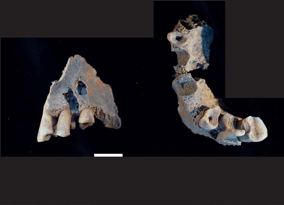
The burial chambers in the funerary monuments contained generally more than one individual. Burial 15, discovered in 2011, contained at least 11 individuals, mostly adults (Fig. 16.3). But it included also the left vertebral arch of the 2nd cervical vertebra of a late foetus or a neonate. The burial in funerary monument 7, excavated in 2014 contained also a minimum of 11 individuals: at least six adults, one child and three late foetuses or neonates and fragments of a cremated individual. From an archaeological point of view, the foetuses or neonates could not be linked to any of the females buried in the same tomb. Therefore, it remains unclear, if they were the remains of foetal/neonatal burials or of one or more pregnant females. Their skeletal remains are of particular interest. There is one secure case from the South-East Necropolis and a questionable case from the North Cemetery: in grave monument 6 a 25–35-year-old female skeleton (PE07 So4 006) was excavated. During the examination of the bones the left tibia of a foetus was found between the pelvis and the lumbar vertebrae. Age determination of foetuses is possible using their long bone length. The tibia belonged to a foetus in the seventh gestational month (Teegen 2009). In the Byzantine North Cemetery, a foetus of the same age can probably also be linked to a young female. Unfortunately, this burial was plundered by grave robbers. The context is, therefore, not totally clear. Nevertheless, these cases clearly show the dangers of pregnancy and giving birth in Antiquity and beyond. They can also explain the early mortality of adult females.
Fig. 16.2. Pergamon. Localization of the cemeteries mentioned in text. 1. Roman South-East Necropolis; 2. Early Byzantine warrior burial; 3. Late Byzantine North Cemetery. Modified after Pirson 2012.
Fig. 16.3. Pergamon. Roman South-East Necropolis. Multiple burial (PE11-So-11-Gr15) with at least 11 individuals.
PALAEOPATHOLOGY
Traces of several diseases could be found analysing the bone finds. This included diseases of the teeth and jaws, the skull, the respiratory system, joints (vertebrae and postcranial bones), trauma, tumours, and malformation.
The dental diseases present in the samples from Pergamon are dental caries, abscesses, dental calculus, parodontopathies and intra vitam tooth loss. They are discussed in the paper by Propstmeier et al.(this volume). Furthermore, teeth were probably used as tools.
Enamel hypoplasia
Unspecific stress markers like linear transverse enamel hypoplasias are quite common (Fig. 16.4). They were the result of a growth disruption during tooth development (Hillson 2014 with extensive bibliography). Severe cases also cause growth disruption on the underlying dentine tissue. This could sometimes be observed in cremations from Pergamon, where the enamel of the tooth crown was not preserved (Fig. 16.5).
The onset of enamel hypoplasias can be determined by their distance to the dento-enamel junction, using either metrics (cf. Rose et al. 1985, 294 Fig. 9.4; Hillson 2014, 179–81) or counting the number of pericymata (Hillson 2014, 182–4). The latter is more precise, but requires, however, the application of microscopic techniques. This is quite difficult in the field.
Enamel hypoplasia of the deciduous dentition develops between the late foetal stages and approximately two years of age. In the permanent dentition it develops mainly between the first and the eleventh year of age. Further information can be derived by observing the hypoplastic defects in the root dentine (Teegen 2004; cf. Fig. 16.4). By analyzing the third molar we can also collect information about tooth growth disruption between 12 and approximately18 years of age, when the root is fully grown.
Not only in Pergamon, but also in the other sites studied, enamel hypoplasias were rarely found in the crowns of deciduous teeth. In one case from Pergamonan enamel defect was found at the crown tips of a deciduous molar (Fig. 16.6). Simon Hillson (2014, Fig. 3.13) has charted the development of the deciduous teeth around birth. This means that the enamel defect on Fig. 16.6 was caused by a growth disturbance during the late phase of pregnancy or around birth.
Fig. 16.4. Pergamon. Roman South-East Necropolis. Canine with enamel and root hypoplasias (PE11-So-11-044-Gr24). Scale 1 cm.
Fig. 16.5. Pergamon. Roman South-East Necropolis. Dentine hypoplasia in a cremated premolar (PE11-So-11-041-1s). Scale 1 cm.
In the samples studied, enamel hypoplasias were primarily observed in the permanent teeth. In Pergamon they mainly developed between two and seven years, with a peak between three and four years. There were only a few observations below two years of age. Tooth abrasion in later life has to be taken into consideration.
Fig. 16.6. Pergamon. Roman South-East Necropolis. ‘Birth line’ in a deciduous molar of a small infant (PE07-So04-017, KF 147). Scale 1 cm.
Fig. 16.7. Pergamon. Roman South-East Necropolis. Teeth as tools (?) in the upper jaw of an adult individual (PE11-So-11-031_#1). Scale 1 cm.
Teeth as tools
The last point regarding the teeth is the observation of unusual abrasion. The individual PE11-So-11-031_#1 exhibits a groove with a diameter of approximately 5 mm on the occlusal surface of an incisive tooth, running from the buccal to the palatinal side of the tooth (Fig. 16.7). Normal chewing cannot produce this kind of abrasion. This individual probably held something between his teeth, regularly and over a long period. The traces are quite characteristic of using teeth as tools (cf. Alt and Pichler 1998).
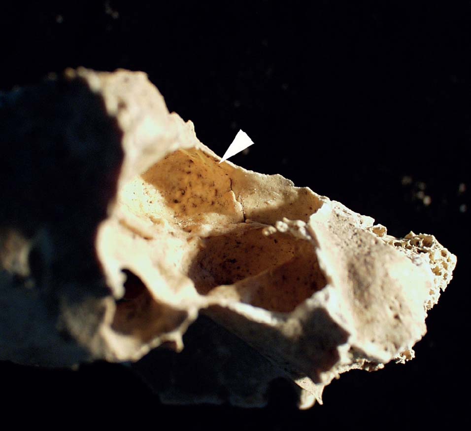
In the Roman population from the South-East Necropolis diseases of the internal lamina of the skull (Schultz 1993; 2001) are rare. This is true for infants and adults alike and stands in contrast to Byzantine burials from Pergamon itself (Schultz 1989a; Schultz and Schmidt-Schultz 1994; Teegen 2014) or Byzantine Kyme (see below). One of these rare cases is a skull fragment of a young adult female. Here, several tree-shaped areas of new bone formation and serpent-like vessel impressions are present (Fig. 16.8). They are very likely the result of an organized epidural haematoma (Schultz 1993; 2001; 2003). This could have been caused by an accident or interpersonal violence.
Fig. 16.8. Pergamon. Roman South-East Necropolis. Organised epidural hematoma on the internal lamina of the skull of a young female (PE14-Ar-02-084).
Fig. 16.9. Pergamon. Roman South-East Necropolis. Maxillary sinusitis (PE07-So04-003).
Respiratory diseases
The most common infection in the osteoarchaeological record is sinusitis, the inflammation of the pneumatic cavities in the skull (cf. Schultz 1993; Aufderheide and Rodriguez-Martín 1998, 257). They are quite common in all cemetery groups and therefore in all social classes. The frequency of maxillary sinusitis (Fig. 16.9) is generally higher than all other forms due to dental abscesses in the upper jaw. Sinusitis at Pergamon was probably at least partially caused by rainy winters, with storms and poor heating systems as well as allergic reactions to the fumes. It is interesting to note that even people of rank, like the old man buried in the monumental İlyastepe tumulus near Pergamon, suffered from this type of infection (Teegen 2011a).
Other diseases of the skull were rarely observed in adults. However, in one case, a traumatic injury of the external lamina of the skull was present, which will be presented later (cf. Fig. 16.13).
Joint diseases
Degenerative joint diseases (cf. Schultz 1988, 481–7; Aufderheide and Rodriguez-Martín 1998, 93–7) were mainly observed in the spinal column rather than in the body joints. The femoral head of an adult from the cremation burial PE11-So-10-047-Gr26 (Fig. 16.10) shows the characteristic new bone formation at the rim and the fungus-like structure. In general, the degree of degenerative joint diseases is quite low in the Roman population studied. This probably correlates with the middle class appearance of the South-East Necropolis.
Fig. 16.10. Pergamon. Roman South-East Necropolis. Degenerative joint disease of the femoral head of a cremated adult (PE11-So-10- 047-Gr26). Scale 1 cm.
Due to poor preservation, the degree of joint diseases (cf. Schultz 1988, figs. 170–1) in the individuals from the Byzantine North Cemetery could rarely be determined.
Degenerative changes of the vertebral joints are more common. Sometimes there are also Schmorl’s nodes present. In one adult individual (PE11-So-11-053-Gr27) at least three thoracic vertebrae are ankylosed due to ossification of the anterior ligaments (Fig. 16.11). Here, an ankylosing spondylitis seems likely (cf. Aufderheide and Rodriguez-Martín 1998, Fig. 6.14).
Fig. 16.11. Pergamon. Roman South-East Necropolis. Ankylosis of the anterior ligament of three thoracic vertebrae (PE11-So-11-053-Gr27). Scale 1 cm.
Fig. 16.12. Pergamon. Late Byzantine North Cemetery. Burial 4 with osteomyelitic process of both tibiae. Fine plaque-like structures indicating a process which was still on-going at the time of death. Scale 1 cm.
Unspecific infections
Remarkable is a case of osteomyelitis from the Byzantine North Cemetery (Fig. 16.12). It is very likely that it was caused by an infectious disease. Both tibia shafts are enlarged and exhibit an anomalous surface structure. Unfortunately, both tibiae are quite badly preserved and broken. This makes it easily possible to see the internal structure of the shafts. They show several layers of newly built bone. This is a quite typical reaction to an inflammatory process.
Histological analyses of a bone sample were carried out by Michael Schultz from Göttingen University; the results of which will be published elsewhere. The macroscopic image of the outer layer of the tibia also shows some fine plaque-like structures, indicating a process which was still ongoing at the time of death (Fig. 16.11). Probably, the spread of infection was also the cause of death, for example, by a general sepsis.
Trauma
Traumas both of the skull and the postcranial skeleton are rare in the Romans from Pergamon. There are two cases of cranial trauma (cf. Fig. 16.13) and several rib fractures. A well-healed fracture of a tibia probably indicates some kind of medical intervention.
Tumours
Today, tumours are quite a common cause of death. In Antiquity, the contrary was the case, mainly due to the lower life expectancy. But nevertheless, tumours can be regularly found in the archaeological record. From Roman Pergamon, at least three probable cases are known. One case seems to be a fibro-osseous tumour in the frontal sinus (Fig. 16.14). This kind of benign tumour often develops after chronic inflammatory processes of the sinus.
Fig. 16.13. Pergamon. Roman South-East Necropolis. Sharp trauma on the skull of an adult male (PE11-So-11-044-Gr24).
The other case is a little more problematic. In the left metatus acusticus externus of an adult male from the multiple burial 15 (cf. Fig. 16.3) (PE11-So-11-Gr15) an external auditory exostosis (EAE) was discovered (Fig. 16.15). The form of the alteration is clear and it can easily be described as EAE. Here, however, it is not clear if it is a benign tumour or the result of taking cold baths or an epigenetic trait (Torus auditivus). There is also one case of a slight torus auditivus, observed in the burials from the 2007 rescue excavation in the same South-East Cemetery. When we consider the outcome of this phenomenon, it is clear that these two people could have had difficulties in hearing.
Fig. 16.14. Pergamon. Roman South-East Necropolis. Probable fibro-osseous tumour (Ø ca. 5 mm) in the left frontal sinus (PE11-So-11-053-Gr27).
As D. Campillo and J. Baxarias (2008) have shown, a cholesteatoma is a quite likely interpretation for the case of the adult male PE11-So-11-Gr15 (Fig. 16.15). Computer tomography has, however, still to be performed. The presence of EAE on only one side makes it not very likely that this alteration was the result of cold baths.
Sometimes tumours can be found in cremated bones. This is the case in the adult individual PE11-So-10-044-4 (Teegen 2013, 139, fig. 63).
Malformations
Studying the last skeletons from the 2007 rescue excavation in summer 2013, a quite interesting case was discovered. Burial PE07-So-09-008 contained the skeleton of a young female. Her skull is deformed (Fig. 16.16) and the mastoid processes are of different dimensions. The latter could indicate the presence of a torticollis. Analyzing the sutures of the skull, a premature craniosynostosis seems likely. Both alterations can be found together (Kutterer and Alt 2008). Nearby, another deformed skull was found, belonging to a middle-aged individual (PE07-So-09-009). Deformation by soil pressure seems not very likely. Due to its more advanced age, the cause for the deformation could not clearly be ascertained.
People with premature craniosynostosis and torticollis have a quite different physical appearance. There is one unusual Roman helmet from the Xanten area in the province of Germania inferior (von Prittwitz und Gaffron et al. 1991), which was especially designed for an officer with torticollis. People with skull deformation due to premature craniosynostosis have a normal IQ, but sometimes learning disorders (Magge et al. 2002).
Fig. 16.15. Pergamon. Roman South-East Necropolis. External auditory exostosis (PE11-So-11-Gr15_Skull).
Fig. 16.16. Pergamon. Roman South-East Necropolis. Torticollis and skull deformation (Female, 21-25 years; PE07-So09-008). Scale 1 cm.
Priene
The ancient city Priene (Turkey; Fig. 16.1) was excavated by German archaeologists between 1895 and 1898 (Wiegand and Schrader 1904). Since 1998, teams from the Universities of Frankfurt and Bonn, with support from the German Archaeological Institute at Istanbul, have continued the excavations (Raeck 2008).
During the excavations by the University of Frankfurt (2005–10) in the former sanctuary of the Egyptian Gods (AEG) (Hennemeyer 2005; Raeck 2008; 2009), a late Byzantine cemetery was discovered. It probably dates to the 13th/14th century AD. Radiocarbon dates are still missing. Furthermore, some small grave groups were excavated throughout the ancient city (Fig. 16.17); several late Byzantine burials were discovered: in the so-called Agora chapel (AK) three burials with at least six individuals were excavated. Nearby, in the so-called south complex (SK), two other burials were found and excavated. Outside the city near the street to the acropolis (Teloneia) burials of a previously unknown Byzantine cemetery were discovered. In the Hellenistic to Roman cemetery, by the main gate, some dispersed human bones were also found.
Results
The anthropological and palaeopathological analysis of the late Byzantine skeletons from Priene revealed a minimal number of 50 individuals from the AEG, AK and SK sites. The inhumations from Priene are similarly preserved as those from Pergamon.
Fig. 16.17. Priene. Localization of the Late Byzantine cemeteries: 1. Former sanctuary of the Egyptian Gods (AEG); 2. Agora (AK); 3. Teloneia. Modified after Wiegand and Schrader 1904.
SPONDYLOLYSIS
Degenerative joint diseases of the spine and of the large body joints are common in late Byzantine Priene. Furthermore, there are three cases of spondylolysis (Fig. 16.18). Two cases of spondylolysis of the 5th lumbar vertebra (L5) from the AEG area and one from the SK area were discovered. The frequency of three out of 12 individuals (25%) with a preserved 5th lumbar vertebra is quite high. This approximates the frequency found in Inuit populations (Ortner 2003, 147). In modern populations of the Old World the frequency lies between 2.3% and 10%, predominantly males (Walker 2012, 116). In Priene only males between 20 and 50 years of age are affected. In only one case is the vertebral arch still preserved (Fig. 16.18). In one individual the L5 has slipped approximately 5 mm ventrally. This indicates a slight spondylolisthesis.
All individuals with spondylolysis show severe degenerative joint disease (DJD) of the lumbar vertebral bodies, indicating heavy physical loading and probably lower back pain. It is striking that the individuals buried inside the Agora chapel, who probably belonged to a higher social class, show neither signs of spondylolysis nor severe DJD.
Due to severe DJD in the present cases, it seems more likely that spondylolysis was due to physical stress of the lumbar and sacral vertebrae, probably at a young age. A genetic origin, can, however, not fully be excluded.
Fig. 16.18. Priene. Late Byzantine Cemetery in the former sanctuary of the Egyptian Gods (AEG). Spondylolysis of the 5th lumbar vertebra (AEG 5.15/2). Scale 1 cm.
CONGENITAL ALTERATIONS
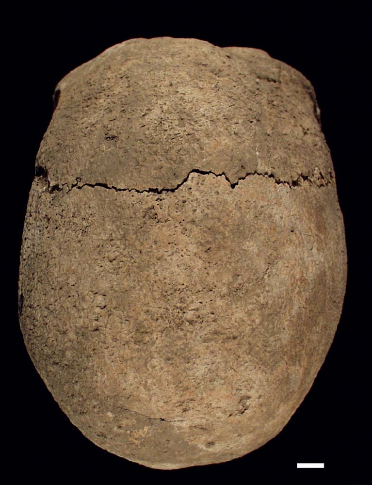
The most unusual case is a young female with syndromic sagittal craniosynostosis and twins. During the 2010 campaign at Priene, a somewhat disturbed burial was excavated at the periphery of the late Byzantine Cemetery in the former sanctuary of the Egyptian Gods (Fig. 16.19). The skeleton was nearly complete, only the lower leg and foot bones are not preserved due to a disturbance of the burial (Fig. 16.19).
The analysis revealed a young adult female of about 20–25 years of age with a calculated body height of 148±3 cm according to Pearson’s (1899) formulae, corrected by Rösing (1988). She died after a miscarriage of at least twins approximately 8 to 9 lunar months after conception. Her robust skull with a clear scaphocephalic appearance shows premature sagittal craniosynostosis (Fig. 16.20). The cranial index of 74.4 is in the dolicocephalic range. Furthermore, the skull is slightly deformed due to a closure of the right occipito-mastoid suture. There are also indications of a slight torticollis. The CT scan shows thickening of the left cranial vault of up to 10 mm. The right temporal and sphenoid bones are also heavily thickened and deformed. Their structure indicates a temporal lobe malformation. The internal lamina of the occipital bone shows an abnormal structure without marked areas of the occipital poles and the posterior cranial fossae. This is a strong indication of modifications of the outer form of the brain and the cerebellum. The frontal teeth of the upper and lower jaw are protruding, the lower jaw shows a trema of 15 mm width, giving her a singular appearance. Multiple linear enamel hypoplasias are present. Furthermore, there is a shortening of the limbs, but only on the left side. Due to this association of malformations the present case should be called a syndromic sagittal craniosynostosis. It is the only case of its kind in the late Byzantine population of Priene. Torticollis and cranial deformation was, however, also recorded in two burials nearby.
Kyme
During recent years the Italian archaeological excavation at Kyme (MIKE) has focused its work on the agora of the ancient city (La Marca and Mancuso 2012). Above the ancient structures, an early Christian basilica and other ecclesiastical buildings were found. Around the basilica is a Byzantine cemetery with 44 burials. Thirty of them contained bones of at least 49 humans.
Fig. 16.19. Priene. Late Byzantine Cemetery in the former sanctuary of the Egyptian Gods, burial 39. Young female with cranial deformation and premature closure of the sagittal suture.
Fig. 16.20. Priene. Late Byzantine Cemetery in the former sanctuary of the Egyptian Gods, burial 39. Premature closure of the sagittal suture. Scale 1 cm.
Results
The skeletal material from Kyme is very well preserved – much better than in Pergamon and Priene. This is due to the location of the tombs on the ancient agora with its structures made of marble.
The age distribution is quite different from all other sites. In the Agora Cemetery burials of neonates, infants, and children are most common. They have a much higher frequency than in both Pergamon and Priene. Furthermore, due to the excellent state of preservation, slight pathological changes can also be discovered.
MORTALITY
The age distribution of the Kyme sample is striking (Fig. 16.21: 48% of the individuals were below 12 months of age, and the major part were neonates (and a foetus = 30%). Only 26% of the sample belonged to adults. Similar patterns were observed by Arzu Demirel (this volume) for the Byzantine infants around the lower city church at Amorium.
The Byzantine Cemetery at Kyme offers a rare insight into infant mortality patterns. As we have seen before in the burials of pregnant females, pregnancy and giving birth were high risk periods for mother and child. Between four and six months of age the probability of dying was less. The death risk increases at the end of the first year of life. This probably corresponds with an early weaning period.
PALAEOPATHOLOGY
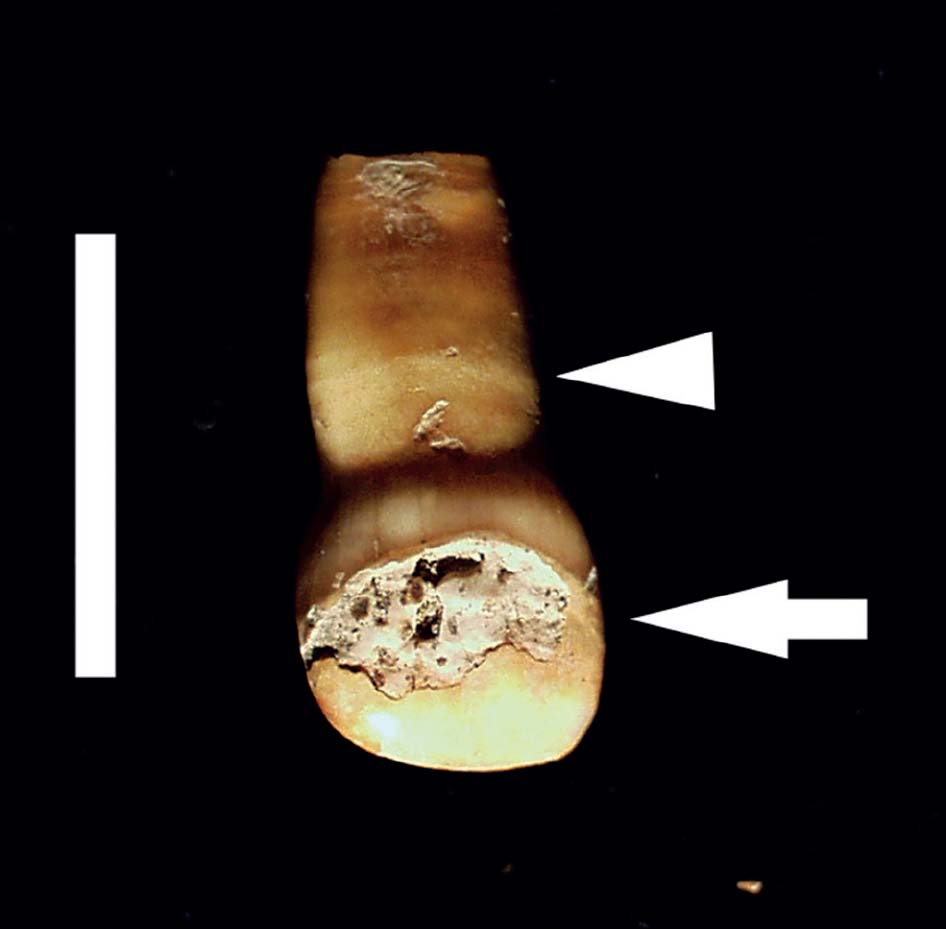
Unfortunately, the risky time of the first year of life is not reflected in the enamel hypoplasia ratios. As we have seen for Pergamon, individuals with enamel hypoplasia below two years of age were rarely found. One of them was found in burial 41. This small infant shows a severe enamel defect of the upper right deciduous first incisor (tooth 51) and root hypoplasia (Fig 16.22). The enamel defect developed in utero, the root hypoplasias around nine and 12 months post partum (Ferembachet al. 1980; Ubelaker 1989).
The same individual from burial 41 shows new bone formation on the internal lamina of the parietal bone (Fig. 16.23). This was the outcome of an inflammatory process, which was, however, not survived. The root hypoplasias possibly represent a growth disturbance caused by the inflammatory process of the internal lamina of the skull.
INFLAMMATORY PROCESSES OF THE INTERNAL LAMINA OF THE SKULL
Another small infant from burial 40 suffered from an inflammatory process in the skull base. Here, the ala minor ossis sphenoidalis shows clear signs of an inflammation (Fig. 16.24).
The 12–24-month-old infant from burial 29 showed severe pathological alterations on the internal lamina of the skull vault (Teegen 2012b, 18 figs. 4–7). Here, an inflammation of the sinus sagittalis superior and adjacent areas is present. Both occipital poles show signs of increased intracranial pressure. Furthermore, signs of an inflammatory process were also present in their impressions digitatae.
We can conclude that the infants from burials 29, 40, and 41 suffered from meningitis. This disease caused high fever, severe headaches, opisthotonus and was very likely the cause of death. Meningitis was not uncommon during the Byzantine period and was also observed by Michael Schultz (1989a), for example, at Pergamon or in Bogazköy (Schultz 1989b).
A neonate from burial 2 has a haematoma on the external lamina of the skull vault (Fig. 16.25). These are the remnants of a so-called kephal haematoma, which often develops during birth. As recent medico-legal findings show, they can reach considerable dimensions.
CRIBRA ORBITALIA
In the burial population from the Agora Cemetery at Kyme there is only one case of severe cribra orbitalia (grade III according to Hengen 1971). It belongs to a child aged between nine and 13 years. Traditionally it is believed that cribra orbitalia is a sign of anaemia (Steinbock 1976; Martin et al. 1985). Several diseases and alterations can cause a porotic or spongy orbita, e.g. tumours, inflammatory processes, anaemia, but also post-mortem erosion (Schultz 1986; Götz 1989). An exact diagnosis is, however, only possible by microscopic analysis of thin sections (Schultz 1993; 2001; 2003).
Fig. 16.21. Kyme. Byzantine Agora Cemetery. Age distribution of the buried individuals (N=49).
Fig. 16.22. Kyme. Byzantine Agora Cemetery, burial 41. Small infant with enamel defect of the upper right deciduous first incisor (tooth 51; arrow) and root hypoplasia (arrow head). Scale 1 cm.
Fig. 16.23. Kyme. Byzantine Agora Cemetery, burial 41. Small infant with new bone formation following an inflammatory process on the internal lamina of the parietal bone. Scale 1 cm.
SPONDYLOLYSIS
Also one of the few adult burials showed a spondylolysis of the 5th lumbar vertebra. Together with the degenerative joint diseases of the large joints and the vertebral joints this is an indication of heavy physical loading since childhood (see above). Furthermore, there is one juvenile with Morbus Scheuermannt, exhibiting Schmorl’s nodes in early years.
Fig. 16.24. Kyme. Byzantine Agora Cemetery, burial 40. Small infant with inflammatory process on the Ala minor ossissphenoidalis. Scale 1 cm.
Fig. 16.25. Kyme. Byzantine Agora Cemetery, burial 2. Small infant with hematoma on the external lamina of the skull vault. Scale 1 cm.
Short discussion
The people buried in the Roman South-East Necropolis at Pergamon can be interpreted as being some kind of ‘middle class’. This is indicated by the funerary monuments and also some luxurious grave goods, such as a funerary bed (Mania 2008). The more or less good health of these people correlates with their status.
In contrast, it is quite difficult to determine the social setting of the Byzantine burials, either from Pergamon, Kyme or Priene. Due to its remote location on the northern slopes of Pergamon’s acropolis hill, the North Cemetery seems to be relatively poor. For Priene and Kyme it is likely that they were also some kind of Byzantine ‘middle class’, like shopkeepers. They were neither poor peasants nor rich aristocrats. In contrast to their Roman counterparts, the Byzantine families exhibited signs of heavy physical loading.
It is difficult to determine when people died. The only reliable sources are grave inscriptions and other epigraphic sources. Walter Scheidel (1996) and others have shown that, in the Mediterranean and Egypt, mortality peaked in the summer months. The same can be presumed for Asia Minor and the cities mentioned in this paper. The graves themselves only rarely reveal the season of burial.
Studying the Roman and Byzantine populations from Pergamon, Kyme and Priene, differences between both time periods can be recognized (see also Kiesewetter, this volume). In particular, the degree of degenerative joint disease is quite low in the Roman sample and higher in the Byzantines. Furthermore it has to be noted that the cases of spondylolysis – mentioned above – were only observed in Byzantine Priene and Kyme and not in the sample from Roman Pergamon. The latter is not only larger but also contained more lumbar vertebrae than the populations mentioned before.
Also the presence of Schmorl’s nodes, protruding of the intervertebral disk in the adjacent vertebral bodies (Walker 2012, 179–80, figs. 284–5), in juveniles indicates heavy physical loading. Schmorl’s nodes in juveniles can be an indication of Scheuermann’s disease (Ortner 2003, 464). They have, until now, only been observed in Byzantine juvenile skeletons from Priene and recently also from Pergamon (Teegen 2014). The onset of Schmorl’s nodes in Romans from the South-East Necropolis begins generally only in the age range from the late 30s or early 40s. This is quite normal for prehistoric or historic populations (Aufderheide and Rodríguez-Martín 1998, 97).
Both, the presence or absence of spondylolysis and Morbus Scheuermann probably hints at different lifestyles and working conditions in Roman and Byzantine times in the three sites from Asia Minor.
In all three populations enamel (and root) hypoplasias are quite frequent. This indicates a lot of stress mostly between (one) two and seven years of age. In some cases (cf. Figs. 16.6 and 16.22) stress began before or during birth. This seems, however, to be the exception. In part, the enamel hypoplasis were probably caused during weaning and/or by (infectious) diseases in childhood; in some cases from Kyme, a connection between inflammatory processes on the internal lamina of the skull and the onset of enamel and root hypoplasias seems quite probable. There are, however, a wide range of causes for enamel hypoplasias (Jälevik and Norén 2000).
It is quite interesting to observe Galen’s notes regarding episodes of famine in the rural populations of the Roman Empire during the second half of the 2nd century AD (Schlange-Schöningen 2003). He mentioned poor peasants eating acorns and non-edible plant stuff. In times of famine the main efforts of the public administration was concentrated on procuring wheat for the urban population thus preventing riots. This could also result in different frequencies of enamel hypoplasia in urban and rural populations. In fact, in a recent paper, Saskia Hin (2014) argued for a lower rate of enamel hypoplasia in the urbs Rome, in confrontation with the rural parts of the campagna or rural Italy in general. For Pergamon and its hinterland the rural populations are still missing. In the city itself, enamel defects are, however, quite common.
In the palaeopathological literature we can often find the statement that enamel hypoplasia is an indicator for the lower social classes (Sweeny et al. 1971; Schultz 1982). This is, however, not true. High frequencies of enamel hypoplasia can regularly be observed in high rank burials – at least from the Iron Age onwards (cf. Little et al. 1992; Teegen and Schultz 2009, 19) – also in Hellenistic Pergamon (Teegen 2011a, 157). In this context it has to be considered that children of status were often brought up by a wet nurse. This could also lead to infections during breast-feeding and in consequence also to enamel hypoplasias. Due to these findings, enamel hypoplasia or other stress markers cannot be used as an indicator of higher or lower social status.
‘Food is a total social fact’ – with these words the eminent French sociologist and anthropologist Marcel Mauss concluded his main work Le don (Mauss 1923–4) in the English translation (Mauss 1954). This also includes the cultural sphere of diet and its social status. The consumption of meat (Purcell 2003) and fish (Wilkins 1993) is also a good status indicator in Antiquity. The composition of animal bone assemblages can give important clues on the social status of its consumers. Piglets, lambs, and calves together with domestic fowl will preferably be found in contexts of higher social status. Meat consumption can be determined by analysing stable isotopes of carbon and nitrogen (for details see the contributions by Propstmeier et al. and Wong et al., both this volume). Analysing skeletons of known status, as for example, the late Medieval and Renaissance family of the Medici in Florence (Italy), using stable isotopes, showed this very clearly (Fornaciari 2008).
Roman and Byzantine skeletons from Pergamon were analyzed for stable isotopes by G. H. Müldner (in Otten et al. 2011) and J. Propstmeier (2012; 2013; Propstmeier et al., this volume). These analyses reveal individuals with different diets (more plant or more meat oriented). Further studies are, however, needed to analyze whether this image also reflects different social status.
Medical history and palaeopathology
There is a strong bias in the epigraphical evidence regarding ancient medicine. Ancient medicine is generally seen through the eyes of its principal leaders (eminent doctors), who were dependent on their rich benefactors or patients, respectively (Nutton 2012). Furthermore, there are interested laymen, mostly belonging to the upper class, who could afford to spend time on such a hobby. Therefore, we know quite a lot about medicine available for those who could afford it. In contrast, we know very little about the medical problems of the middle classes and nearly nothing about those of the poor.
Galen himself was quite typical (Schlange-Schöningen 2003). He came from a wealthy family from Pergamon and belonged to the municipal aristocracy. Later he was surgeon to the gladiators, an extremely well-paid and acclaimed occupation – similar to some sports doctors today. Being surgeon to gladiators means also gaining spectacular insight into the anatomy and pathology of the human body and the skeleton. In Rome he was, at least at times, the personal doctor of five emperors, and an acclaimed author in the fields of medicine and philosophy. His writing influenced western medicine for more than one and a half millennia.
Palaeopathology gives us an invaluable insight into everyday life and diseases which often – or mostly – are not mentioned in the medical texts. They were, however, quite important for these ancient and Medieval populations. Furthermore, palaeopathology can evaluate health status in different social settings (Pitts and Griffin 2012), where often no textual evidence is present. Our investigations also contribute to the history of diseases itself as do also ancient images of diseased people, a collection of which can be found in the much-acclaimed book by Mrko Grmek and Danielle Gourevitch (1998).
Acknowledgments
I am most grateful to the Pergamon excavation of the German Archaeological Institute (director Prof. Felix Pirson), the Priene excavation (Prof. Wulf Raeck), the Kyme excavation (Prof. Antonio La Marca) and their teams, my collaborators Johanna Propstmeier MSc, Saskia Wunsch BA, and Sabrina Kutscher BA, the Gerda Henkel-Foundation, the Deutsche Forschungsgemeinschaft and last but not least the Ludwig-Maximilians-Universität München for support. The Turkish Ministry of Culture and Tourism (Ankara) kindly granted all necessary permits. I am further grateful to Dr Henrike Kiesewetter for reading my paper. The helpful comments of an anonymous reviewer are gratefully acknowledged. All remaining errors are of course my own. All figures are made by myself and are published here with courtesy by respectively the German Archaeological Institute at Istanbul (Pergamon excavations) (Figs. 16.1–16), the University of Frankfurt (Priene excavations) (Figs. 16.17–20), and the Italian archaeological excavation at Kyme (MIKE) (Figs. 16.21–25).
Bibliography
Alt, K. W. (1997) Odontologische Verwandtschaftsanalyse. Individuelle Charakteristika der Zähne in ihrer Bedeutung für Anthropologie, Archäologie und Rechtsmedizin. Stuttgart and Jena, Gustav Fischer Verlag.
Alt, K. W. and Pichler, S. L. (1998) Artificial modifications of human teeth. In K. W. Alt, F. W. Rösing, and M. Teschler-Nicola (eds.) Dental anthropology. Fundamentals, limits, and prospects, 387–415. Vienna, Springer.
Ameen, S., Staub, L., Ulrich, S., Vock, P., Ballmer, F., and Anderson, S. E. (2005) Harris lines of the tibia across centuries: A comparison of two populations, medieval and contemporary in Central Europe. Skeletal Radiology 34, 279–84.
Aufderheide, A. C. and Rodriguez-Martín, C. (1998) The Cambridge encyclopedia of human paleopathology. Cambridge, Cambridge University Press.
Bräuer, G. (1988) Osteometrie. In Knußmann (ed.), 160–232.
Campillo, D. and Baxarias, J. (eds.) (2008) Quaranta anys de paleopatologia en el Museu d’Arqueologia de Catalunya (Museu d’arqueologia de Catalunya Barcelona, Monografies 12). Barcelona, Museu d’arqueologia de Catalunya.
Demirel, F. A. (this volume) Infant and child skeletons from the Lower City Church at Byzantine Amorium, 306–17.
Ferembach, D., Schwidetzky, I., and Stloukal, M. (1980) Recommendations for age and sex diagnosis of skeletons. Journal of Human Evolution 9, 517–49.
Fornaciari, G. (2008) Food and disease at the Renaissance courts of Naples and Florence: A paleonutritional study. Appetite 51.1, 10–14.
Fornaciari, G. and Giuffra, V. (2009) Lezioni di paleopatologia, Genova, ed. ECIG.
Gilbert, R. I. and Mielke, J. H. (eds.) (1985) The analysis of prehistoric diets (Studies in Archaeology). Orlando, San Diego, and New York, Academic Press.
Götz, W. (1989) Histologische Untersuchungen an Cribra orbitalia, ein Beitrag zur Paläopathologie des Orbitadaches. Unpublished Master’s thesis, University of Göttingen.
Grmek, M. D. and Gourevitch, D. (1998) Les maladies dans l’art antique. Paris, Broccard.
Hauser, G. and De Stefano, G. F. (1989) Epigenetic variants of the human skull. Stuttgart, Schweizerbartsche Verlagsbuchhandlung.
Hengen, O. P. (1971) Cribra orbitalia: Pathogenesis and probable etiology. Homo 22, 57–75.
Hennemeyer, A. (2005) Das Heiligtum der Ägyptischen Götter in Priene. In A. Hoffmann (ed.), Ägyptische Kulte und ihre Heiligtümer im Osten des Römischen Reichs (Byzas 1), 139–53. Istanbul, Ege Yayınları.
Hillson, S. (2014) Tooth development in human evolution and bioarchaeology. Cambridge and New York, Cambridge University Press.
Hin, S. (2014) The first healthy metropolis in Europe’s history? Urban-rural differences in health status in ancient Rome. http://iussp.org/sites/default/files/event_call_for_papers/ Hin_HealthyMetropolis.pdf (visited 07.01.2015).
Jälevik, B. and Norén, J. G. (2000) Enamel hypomineralization of permanent first molars: A morphological study and survey of possible aetiological factors. International Journal of Paediatric Dentistry 10, 278–89.
Kiesewetter, H. (this volume) Toothache, back pain, and fatal injuries: What skeletons reveal about life and death at Roman and Byzantine Hierapolis, 268–85.
Knußmann, R. (ed.) (1988) Anthropologie (Handbuch der vergleichenden Biologie des Menschen 1,1), 160–232. Stuttgart and New York, Gustav Fischer Verlag.
Kósa, F. (1978) Identifikation der Feten durch Skelettuntersuchungen. In H. Hunger and D. Leopold (eds.), Identifikation, 211–41. Leipzig, Barth.
Kutterer, A., and Alt, K. W. (2008) Cranial deformations in an Iron Age population from Münsingen-Rain, Switzerland. International Journal of Osteoarchaeology 18, 392–406.
La Marca, A., and Mancuso, S. (eds.) (2012) Catalogo della Mostra Fotografica ‘Scavi archeologici a Kyme d’Eolide (Turchia)’ (Centro Direzionale BCC Mediocrati, maggio 2012). Arcavacata di Rende, Centro Editoriale e Librario dell’Università della Calabria.
Lewis, M. E. (2009) The bioarchaeology of children. Perspectives from biological and forensic anthropology (Cambridge Studies in Biological and Evolutionary Anthropology 50). Cambridge and New York, Cambridge University Press.
Little, B. J., Lanphear, K. M., and Owsley, D. W. (1992) Mortuary display and status in a 19th-century Anglo-American cemetery in Manassas, Virginia. American Antiquity 57, 397–418.
Magge, S. N., Westerveld, M., Pruzinsky, T., and Persing, J. A. (2002) Long-term neuropsychological effects of sagittal craniosynostosis on child development. Journal of Craniofacial Surgery 13.1, 99–104.
Mania, U. (2008) Die Südostnekropole. ArchäologischerAnzeiger 2008.2, 112–8.
Mann, R. W., Jantz, R. L., Bass, W. M., and Willey, P. S. (1991) Maxillary suture obliteration: A visual method for estimating skeletal age. Journal of Forensic Science 36.3, 781–91.
Martin, D. L., Goodman, A. H., and Armelagos, G. J. (1985) Skeletal pathologies as indicators of quality and quantity of diet. In Gilbert and Mielke (eds.), 227–79.
Martin, R. (1928) Lehrbuch der Anthropologie in systematischer Darstellung. Bd. 2: Kraniologie, Osteologie. Jena, Gustav Fischer Verlag.
Mauss, M. (1923–4) Essai sur le don. L’Année Sociologique N.S. 1, 30–186.
Mauss, M. (1954) The gift: Forms and functions of exchange in archiac societies. New York, Norton.
Nutton, V. (2012) Ancient medicine. Second ed. (Sciences of Antiquity Series). London and New York, Routledge.
Ortner, D. J. (2003) Identification of pathological conditions in human skeletal remains (2nd ed.). San Diego, Academic Press.
Otten, T., Evans, J., Lamb, A., Müldner, G., Pirson, A., and Teegen, W.-R. (2011) Ein frühbyzantinisches Waffengrab aus Pergamon. Interpretationsmöglichkeiten aus archäologischer und naturwissenschaftlicher Sicht. Istanbuler Mitteilungen 61, 347–422.
Pearson, K. (1899) Mathematical contributions to the theory of evolution. V. On the reconstruction of the stature of prehistoric races. Philosophical Transactions of the Royal Society of London A 192, 169–244.
Pirson, F. (2008) Pergamon – Bericht Über die Arbeiten in der Kampagne 2007. Archäologischer Anzeiger 2008.2, 83–155.
Pirson, F. (2012) Pergamon – Bericht Über die Arbeiten in der Kampagne 2011. Archäologischer Anzeiger 2012.2, 175–274.
Pirson, F. (2013) Pergamon – Bericht Über die Arbeiten in der Kampagne 2012. Archäologischer Anzeiger 2013.2, 79–164.
Pirson, F. (2014) Pergamon – Bericht Über die Arbeiten in der Kampagne 2013. Archäologischer Anzeiger 2014.2, 101–76.
Pirson, F., Japp, S., Kelp, U., Nováček, J., Schultz, M., Stappmanns, V., Teegen, W.-R., and Wirsching, A. (2011) Der Tumulus auf dem İlyastepe und die pergamenischen Grabhügel. Istanbuler Mitteilungen 61, 117–203.
Pitts, M. and Griffin, R. (2012) Exploring health and social well-being in Late Roman Britain: An intercemetery approach. American Journal of Archaeology 116.2, 253–76.
Plicht, J. van der (2015) Unpublished Reports CIO/433-2015/PWL and CIO/582-2015/PWL. 07.04.2015 and 21.09.2015.
Prittwitz und Gaffron, H.-H. von, Spiering, B., and Eggert, G. (1991) Der Reiterhelm des Tortikollis. Bonner Jahrbücher 191, 225–46.
Propstmeier, J. (2012) Die Lebensbedingungen in Pergamon: Nahrungsrekonstruktion mit Hilfe stabiler Stickstoff- und Kohlenstoffisotope einer römischen und spätbyzantinischen Nekropole. Unpublished Master’s thesis, University of Munich.
Propstmeier, J. (2013) Analyse stabiler Isotope zur Ernährungsrekonstruktion. Archäologischer Anzeiger 2013.2, 141–2.
Propstmeier, J., Nehlich, O., Richards, M. P., Grupe, G., Müldner, G. H., and Teegen, W.-R. (this volume) Diet in Roman Pergamon: Preliminary results using stable isotope (C, N, S), osteoarchaeological and historical data, 237–49.
Purcell, N. (2003) The way we used to eat: Diet, community, and history at Rome. American Journal of Philology 124.3, 329–58.
Raeck, W. (2008) 2007 Yılı Priene Çaslismalari/Die Arbeiten in Priene im Jahre 2007. In 30. Kazı Sonuçları Toplantısı. 1. Cilt. 26–30 Mayis 2008 Ankara, 33–52. Ankara, T. C. Kültür Bakanliği.
Raeck, W. (2009) Urbanistische Veränderung und archäologischerBefund in Priene. In A. Matthaei and M. Zimmermann (eds.), Stadtbilder im Hellenismus, 307–21. Berlin, Verlag Antike.
Rösing, F. W. (1988) Körperhöhenrekonstruktion aus Skelettmaßen. In Knußmann (ed.), 586–600.
Rösing, F. W., Graw, M., Marré, B., Ritz-Timme, S., Rothschild, M. A., Rötzscher, K., Schmeling, A., Schröder, I., and Geserick, G. (2007) Recommendations for the forensic diagnosis of sex and age from skeletons. Homo 58, 75–89.
Rose, J. C., Condon, K. W., and Goodman, A. H. (1985) Diet and dentition: Developmental disturbances. In Gilbert and Mielke (eds.), 281–305.
Schaefer, M., Scheuer, L. and Black, S. M. (2009) Juvenile osteology. A laboratory and field manual. Amsterdam, Elsevier.
Scheidel, W. (1996) Seasonal mortality in the Roman Empire. In W. Scheidel, Measuring sex, age and death in the Roman Empire. Explorations in ancient demography (Journal of Roman Archaeology, Supplementary Series 21), 139–63. Ann Arbor, Journal of Roman Archaeology.
Scheuer, L. and Black, S. (2000) Developmental juvenile osteology. San Diego, Academic Press.
Schlange-Schöningen, H. (2003) Die römische Gesellschaft bei Galen: Biographie und Sozialgeschichte (Untersuchungen zur antiken Literatur und Geschichte 65). Berlin and New York, Walter de Gruyter.
Schmid, F. and Künle, A. (1958) Das Längenwachstum der langen Röhrenknochen in bezug auf Körperlänge und Lebensalter. Fortschritte Röntgenstrahlen [Röfo] 89.3, 350–6.
Schultz, M. (1982) Umwelt und Krankheit des vor- und frühgeschichtlichen Menschen. In Kindlers Enzyklopädie. Der Mensch Bd. 2, 259–312. Munich, Kindler.
Schultz, M. (1986) Die mikroskopische Untersuchung prähistorischer Skeletfunde. Anwendung und Aussagemöglichkeiten der differentialdiagnostischen Untersuchung in der Paläopathologie (Archäologie und Museum 6). Liestal, Amt für Museen und Archäologie des Kantons Basel-Land.
Schultz, M. (1988) Paläopathologische Diagnostik. In Knußmann (ed.), 480–96.
Schultz, M. (1989a) Osteologische Untersuchungen an den spätmittelalterlichen Skeleten von Pergamon – Ein vorläufiger Bericht. In IV. Arkeometri Sonuçları Toplantısı (Ankara 23–7 Mayis 1988), 111–8. Ankara, T. C. Kültür Bakanliği.
Schultz, M. (1989b) Nachweis äußerer Lebensbedingungen an den Skeleten der frühmittelalterlichen Bevölkerung von Bogazkale/Hattussa. In IV. Arkeometri Sonuçları Toplantısı (Ankara 23–7 Mayis 1988), 119–20. Ankara, T. C. Kültür Bakanliği.
Schultz, M. (1993) Vestiges of non-specific inflammations of the skull in prehistoric and historic populations. A contribution to palaeopathology (Anthropologische Beiträge 4A/B). Aesch BL, Anthropologisches Forschungsinstitut Aesch and Anthropologische Gesellschaft Basel.
Schultz, M. (2001) Paleohistopathology of bone: A new approach to the study of ancient diseases. Yearbook of Physical Anthropology 44, 106–47.
Schultz, M. (2003) Light microscopic analysis in skeletal paleopathology. In D. Ortner, 73–108.
Schultz, M. and Schmidt-Schultz, T. H. (1994) Krankheiten des Kindesalters in der mittelalterlichen Population von Pergamon. Ergebnisse einer paläopathologischen Untersuchung. Istanbuler Mitteilungen 44, 181–201.
Schultz, M. and Schmidt-Schultz, T. H. (2004) ‘Der Bogenschütze von Pergamon’. Die paläopathologisch-biographische Rekonstruktion einer interessanten spätbyzantinischen Bestattung. Istanbuler Mitteilungen 54, 243–56.
Schutkowski, H. (1990) Zur Geschlechtsdiagnose von Kinderskeletten. Morphognostische, metrische und diskriminanzanalytische Untersuchungen. Unpublished PhD thesis, University of Göttingen.
Schutkowski, H. (1993) Sex determination of infant and juvenile skeletons: I. Morphognostic features. American Journal of Physical Anthropology 90, 199–205.
Steinbock, T. R. (1976) Paleopathological diagnosis and interpretation. Bone diseases in ancient human populations. Springfield (Ill.), Charles C. Thomas.
Stloukal, M. and Hanáková, H. (1978) Die Länge der Langknochen altslawischer Bevölkerungen – Unter besonderer Berücksichtigung von Wachstumsfragen. Homo 29, 53–69.
Sweeney, E. A., Saffir, J. A., and De Leon, R. (1971) Linear hypoplasia of deciduous incisor teeth in malnourished children. American Journal of Clinical Nutrition 24, 29–31.
Teegen, W.-R. (2004) Hypoplasia of the tooth root: A new unspecific stress marker in human and animal paleopathology [abstract]. American Journal of Physical Anthropology, Supplement 38, 193.
Teegen, W.-R. (2009) Burials of a pregnant woman and neonates from the Roman South-East Necropolis of Pergamon (Bergama, Prov. Izmir, Turkey): A preliminary report. Poster, Réncontres autour de la mort des tout-petits. Mortalité foetale et infantile. 3–4 décembre 2009, Musée d’Archéologie Nationale Saint-Germain-en-Laye. http://gaaf.emonsite.com/pages/rencontres/rencontre-autour-des-tout-petits-2009/ pre-actes.html (visited 03.08.2013).
Teegen, W.-R. (2010) Spondylolysis in Late Byzantine Priene (Turkey) [abstract]. American Journal of Physical Anthropology, Supplement 50, 228–9.
Teegen, W.-R. (2011a) Die menschlichen Skelettreste. In Pirson et al., 146–65.
Teegen, W.-R. (2011b) Der anthropologisch-paläopathologische Befund. In Otten et al., 369–93.
Teegen, W.-R. (2011c) A female skeleton with syndromic sagittal craniosynostosis from Late Byzantine Priene (Turkey) – a CT investigation [abstract]. American Journal of Physical Anthropology, Supplement 52, 292.
Teegen, W.-R. (2011d) Die anthropologisch-paläopathologischen Untersuchungen. Archäologischer Anzeiger 2011.2, 186–8.
Teegen, W.-R. (2012a) Die anthropologisch-paläopathologischen Untersuchungen 2011. Archäologischer Anzeiger 2012.2, 255–8.
Teegen, W.-R. (2012b) La Tomba 29 nell’area dell’agorà: un caso studio paleopatologico. In La Marca and Mancuso (eds.), 18.
Teegen, W.-R. (2013) Die anthropologisch-paläopathologischen Untersuchungen 2012. Archäologischer Anzeiger 2013.2, 138–43.
Teegen, W.-R. (2014) Die anthropologisch-paläopathologischen Untersuchungen in Pergamon 2013. Archäologischer Anzeiger 2014.2, 152–56.
Teegen, W.-R. (2015) Die anthropologisch-paläopathologischen Untersuchungen in Pergamon 2014. Archäologischer Anzeiger 2015.2, 158–63.
Teegen, W.-R (forthcoming) Gli uomini della necropolis bizantina sull’agora di Kyme Rapporto preliminare su gli studi antropologici e paleopatologici. In La Marca (ed.), Kyme Eolica VI (submitted, not yet published).
Teegen, W.-R. and Schultz, M. (2009) Eine slawische Burg und ihre ‘fürstlichen’ Bewohner: Starigard/Oldenburg 10. Jh. In L. Clemens and S. Schmidt (eds.) Sozialgeschichte der mittelalterlichen Burg (Interdisziplinärer Dialog zwischen Archäologie und Geschichte 1), 13–24. Trier, Kliomedia.
Ubelaker, D. H. (1989) Human skeletal remains: Excavation, analysis, interpretation (2nd ed.) (Manuals on Archaeology 2). Washington D.C., Taraxum.
Waldron, T. (2001) Palaeopathology (Manuals in Archaeology). Cambridge, Cambridge University Press.
Walker, D. (2012) Disease in London, 1st–19th centuries. An illustrated guide to diagnosis (MOLA Monograph 56). London, Museum of London.
Wiegand, T. and Schrader, H. (1904) Priene. Ergebnisse der Ausgrabungen und Untersuchungen in den Jahren 1895–8. Berlin, Georg Reimer.
Wilkins, J. (1993) Social status and fish in Greece and Rome. Food, Culture and History 1, 191–203.
Wiltschke-Schrotta, K. (1988) Das frühbronzezeitliche Gräberfeld von Franzhausen I. Analyse der morphologischen Merkmale mit besonderer Berücksichtigung der epigenetischen Varianten. Unpublished PhD thesis, University of Vienna.
Wong, M., Naumann, E., Jaouen, K., and Richards, M. (this volume) Isotopic investigations of human diet and mobility at the site of Hierapolis, Turkey, 306–17.