Adult elbow
Normal Anatomy
AP view
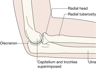
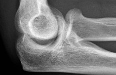
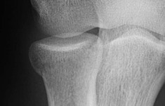
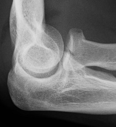
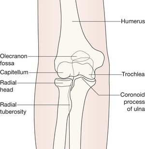
The olecranon is obscured by the humerus. The capitellum is lateral and articulates with the radial head. The trochlea is medial and articulates with the ulna.
Radiocapitellar line
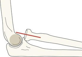
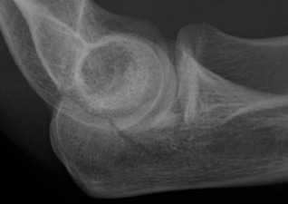
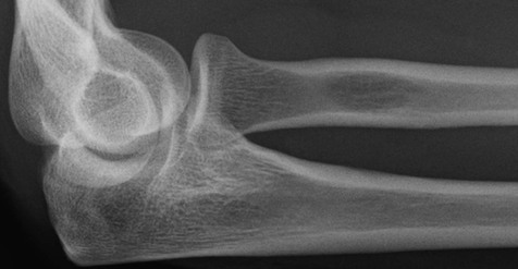
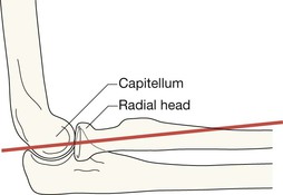
On the lateral view: the line to draw is along the long axis of the proximal 2–3 cm of the shaft of the radius. This line should pass through the capitellum.
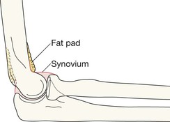
Elbow fat pads
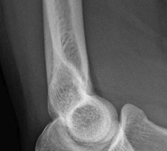
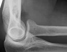
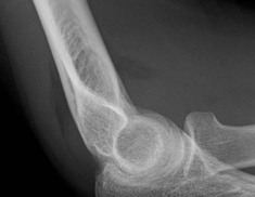
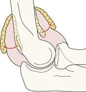
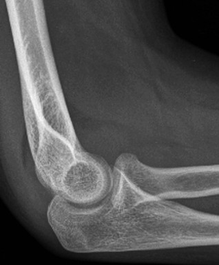
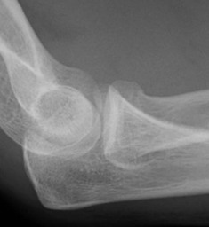
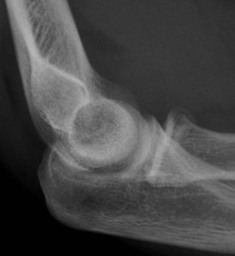
Two pads of fat are situated close to the cortex of the distal humerus. Referred to as the anterior and posterior fat pads, these are external to the synovial lining of the joint. The fat pads are never visualised on the AP projection. Look for them on the lateral view. The fat is seen as a dark streak in the surrounding grey soft tissues.
▪ The anterior fat pad will be seen in most (but not all) normal elbows and is closely applied to the humerus.
▪ The posterior fat pad is not visible on the radiograph of a normal elbow because it is situated deep within the olecranon fossa. Because the elbow is radiographed in the flexed position this fat shadow is hidden by the overlying bone.
Analysis: three questions to answer
If the AP and lateral radiographs appear normal it is important that the images are again checked in a methodical manner. Ask yourself three questions.
Question 1—Are the fat pads normal on the lateral view?
The same principles apply as for the paediatric elbow (p. 102). Recap:
The common injuries
Fracture of the head or neck of the radius
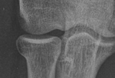
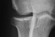
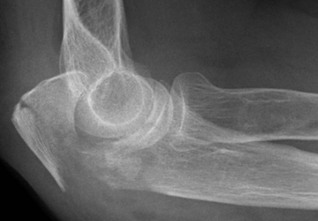
In the adult, these fractures represent 50% of all fractures at the elbow.
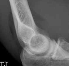
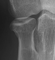
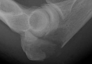
An additional radiograph. Some Emergency Departments routinely add a third (an angled) projection to the standard AP and lateral views. This is termed the radial head-capitellum view3. The patient is positioned as for the lateral view but the tube is angled 45° to the joint. This is an excellent projection for evaluation of the radial head.
A rare but important injury
The Monteggia injury1,2,5
This combination injury represents less than 5% of all elbow fractures or dislocations4–6. Often referred to as a Monteggia fracture–dislocation or a Monteggia lesion. It comprises a fracture of the ulna and dislocation of the head of the radius.
Background: the forearm bones can be regarded as acting as a single functional unit as they are bound together by the strong interosseous membrane and ligaments. Consequently, a displaced or angulated fracture of just one of these two bones will be accompanied by an injury affecting the other bone—invariably a dislocation. Whenever there is a displaced fracture of the ulna, but an intact radius it is important to assess the proximal radio-ulnar joint for a dislocation of the head of the radius utilising the radiocapitellar line. This assessment will show whether a Monteggia injury is present.
Clinical impact guideline: early diagnosis is clinically very important… “the key to a good result following a Monteggia fracture–dislocation is prompt recognition of the injury pattern”2.
