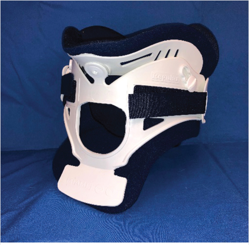
Fig. 19.1 Miami J orthosis. (This image is provided courtesy of Stephen R. Dsida, LP, CPO.)
Summary
Spine bracing has undergone extensive updates since the introduction of bracing in the 1930s and are now commonly recommended for patients undergoing rehabilitation from surgery or spine injury. Bracing is commonly employed after vertebral compression fractures but its effectiveness is still in question. The goal of bracing is to limit spine motion as much as possible and various types of braces exist for each anatomic segment of the spine. There are different types of cervical braces which are most commonly employed after a traumatic injury when nonsurgical management is the preferred method of treatment. Thoracic and lumbar braces are commonly used for compression fracture bracing. They can be either rigid or soft and provide varying degrees of spinal stability. Generally the spinal braces used for osteoporotic vertebral compression fractures (VCFs) have been shown to provide limited benefits in regard to pain and functional improvement or no benefit. Despite literature evidence supporting the use of bracing, it remains a common initial treatment for those patients with painful VCFs. There are also some potential complications of bracing that clinicians should be aware of including an increased risk of skin breakdown in the locations of the brace’s pressure points. As opposed to osteoporotic VCFs, bracing is generally recommended in cases of traumatic VCFs where surgery is performed or as an adjunct to surgical fixation. While bracing may confer mild benefit in regard to patient comfort or slightly reduced healing time, additional investigation will need to be done to determine the effectiveness of spinal bracing in the treatment of patients with painful VCFs.
Keywords: bracing, spine brace, lumbosacral orthosis, TLSO, Jewett, lumbosacral orthosis
Before the year 1931, spine braces were not patented and nor were they sold for use. Physicians would make them for specific patients. These early braces tended to be painful and not very functional and most of the bracing at that time was done to treat adolescent scoliosis. The Milwaukee brace, a removable cervicothoracolumbosacral orthosis (CTLSO) was invented in 1946.1 The brace was cumbersome and the metal bars went from the neck down the lower lumbar spine. The result was that a person wearing the brace could not look down for balance and the brace could not be fitted under clothing. In 1972, an adolescent female in Boston diagnosed with idiopathic scoliosis refused to wear the Milwaukee brace because of the neck strap. Her father made some adjustments by cutting off the top part of the brace, changing the metal to plastic and adding padding at the pressure points. This was subsequently dubbed the Boston brace and since then, numerous adjustments and alterations have improved the comfort and functionality of the brace.2
Spine braces are routinely recommended for patients during rehabilitation from back injuries and surgeries either as the primary treatment method for a less severe spine fracture or after a surgical repair. For more serious fractures, bracing is felt to be an important adjunct to surgical management. Despite its acceptance, bracing is not without controversy; it is has not been shown that any back brace actually improves outcomes. This chapter will explore the main uses of braces and some of the published evidence regarding their use.
The goal of any brace, spine or otherwise, is immobilization. Immobilization allows for bone healing to occur. Some braces are better at immobilizing spines than others and custom-fitted “clamshell” braces that encompass your entire trunk in rigid plastic are more likely to reduce spinal movement, especially bending and twisting, than loose-fitting metal braces. Even the most aggressive braces, however, can probably only do so much to stabilize the spine and mitigate further vertebral compression or collapse of the vertebral body.
Most fractures in the cervical spine are related to trauma and when neurological compromise or frank instability is present, surgery is usually the treatment of choice. When nonsurgical management (NSM) is the optimal course of treatment, a cervical orthosis is usually utilized. Johnson et al described four types of cervical braces: cervical collars, poster braces, cervical-thoracic braces, and the halo vest.3 Each brace is utilized in specific situations as described below.
Most stable cervical spine fractures are placed in rigid or semirigid collars. The most common of these is the Philadelphia collar. The Philadelphia collar is a two-piece semirigid orthosis made of Plastazote and reinforced with anterior and posterior plastic struts. The Philadelphia collar restricts motion much better than a soft collar but is less comfortable. Other similar collars are the Miami J collar (▶Fig. 19.1) and the Aspen collar. Despite their stiffness, the rigid collars lack the ability to effectively immobilize the lower cervical spine.4
The SOMI (Sterno-Occipito-Mandibular Immobilizer) orthosis provides better restriction of motion of the low-cervical spine and cervicothoracic junction compared to the collar option. The SOMI brace provides good restriction to flexion (93% restriction of motion), but is less beneficial for control of neck extension (42% restriction of motion), lateral bending (66% restriction of motion), and axial rotation (66% restriction of motion).5,6 This brace is good for providing additional support to stable mid and lower cervical spine fractures.
For unstable upper cervical spine fractures, the orthosis most often utilized is the halo orthosis (▶Fig. 19.2). The use of a halo vest is usually confined to injuries with limited displacement. The duration of treatment varies between 6 weeks and 4 months.6 A halo vest is the most rigid external immobilizer, especially for the upper cervical spine. It restricts up to 75% of flexion–extension at C1–C2, and offers superior control of lateral bending and rotation compared to other cervical braces.7 As effective as the halo vest is at immobilizing the cervical spine, it can limit pulmonary function. Taitsman et al found that the halo vest leads to increased risk of pulmonary complications including pneumonia.8
Fig. 19.3 Cruciform anterior spinal hyperextension (CASH) thoracolumbar orthosis. (This image is provided courtesy of Stephen R. Dsida, LP, CPO.)
Bracing treatment of lower cervical and upper thoracic spine fractures may also be done by using the Minerva brace, a cervical collar jacket.9 Sharpe et al found that for cervical fractures, except for above C1, the Minerva provided “improvement in control of flexion/extension of the upper cervical spine and in control of rotation.”9
Spinal orthoses have been widely used in the management of thoracolumbar fractures. A large variety of braces are available as either prefabricated from numerous manufacturers or custom-fabricated units created with the assistance of a prosthetist/orthotist. The following is a review of the types of braces available and evidence regarding their efficacy in traumatic and osteoporotic fractures.
TLSOs can be either rigid or soft. Both prefabricated and custom-fabricated options are available. The rigid-type design controls extension, flexion, lateral bending, and rotation. The TLSOs are primarily used in the management of VCFs with significant deformity or stable burst fractures from T6 to L4 that do not require surgical intervention.10 Examples of TLSOs in order of increasing immobilization include the dorsal lumbar corset, the Jewett hyperextension brace, the cruciform anterior sternal hyperextension (CASH) brace (▶Fig. 19.3), and the custom-molded thermoplastic TLSO (▶Fig. 19.4).
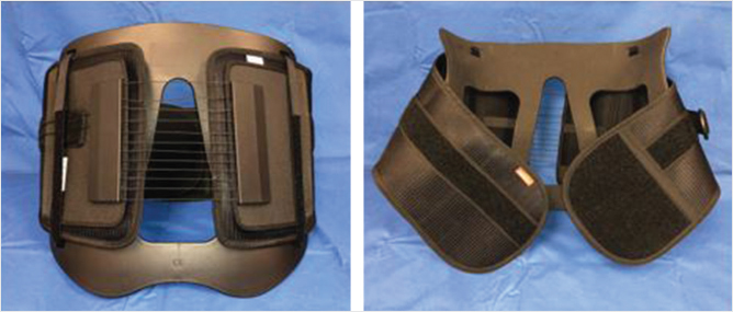
Lumbosacral-type orthoses can also be either rigid or soft. Both prefabricated and custom-fabricated options are available. A rigid lumbosacral orthosis (LSO) provides the most restriction of motion and is typically used to immobilize the lumbar spine at the L3 segment and below. If there is a need to provide significantly more immobilization to the L4–L5 and L5–S1 segments, a unilateral thigh extension can be added.11 The lumbosacral corset is a typically made from cloth and is most often used as an adjunctive support for individuals with low back pain (▶Fig. 19.5). The lumbosacral chairback orthoses (▶Fig. 19.6) are designed with a circumferential band that typically starts at the lower thoracic levels and extends down midway between the iliac crest and the greater trochanter. Chairback orthoses are also often used to provide support for those with low back pain or postoperatively following lumbar fusion surgery.
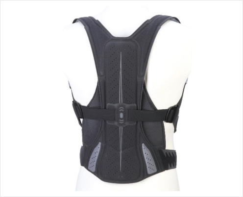
Garment-type orthoses are semirigid or flexible, prefabricated braces designed to fit closely to the body with shoulder straps and a pelvic band to prevent migration.12 Examples include the SpinoMed (▶Fig. 19.7) and OsteoMed orthoses. They have been used primarily in women with osteoporosis for long-term management of osteoporotic compression fractures. In general, they have been shown to provide small and limited benefits by reducing pain, improving strength, and improving posture or kyphosis.13–15
Kypho-orthoses are semirigid or flexible, prefabricated braces that limit or counteract flexion. An example of this type of brace is the Posturex, a semirigid, backpack-style brace with adjustable paravertebral bars. This is a weighted kypho-orthosis that has a soft, backpack design with light weights suspended on the posterior aspect for the brace to encourage extension. Kypho-orthoses have primarily been used in women with subacute osteoporotic VCFs or in the long-term management of osteoporosis. A few small studies have evaluated their efficacy and found limited improvements primarily in balance and gait.16–18
Orthoses have been employed in the management of osteoporotic VCFs with the goal to provide stability to the fracture, reduce pain, and allow for earlier mobility. However, their overall effectiveness in these areas is uncertain and there are no compelling data to support their use as an effective method of adjunctive treatment in patients with osteoporotic VCFs.19,20 A number of studies have examined the role of bracing in osteoporotic compression fractures or for osteoporosis with and without compression fractures. These efforts have varied widely in the types of orthoses evaluated, time after injury that bracing was applied, the duration of treatment, and the outcomes accessed. The heterogeneity and overall paucity of these data have resulted in a confusing picture of what role bracing should have in the management of osteoporotic VCFs. Some current guidelines provide conflicting recommendations with some recommending for the use of back braces in patients with painful VCFs and some guidelines recommending against their use.21,22 Despite evidence supporting spinal bracing, it remains a common first-line treatment in the NSM of VCFs in most cases. A brief review of recent data may provide some guidance as to the use of bracing in patients with painful VCFs.
Fig. 19.4 Custom-fabricated thoracolumbar sacral orthosis (TLSO). (This image is provided courtesy of Stephen R. Dsida, LP, CPO.)
Fig. 19.6 Lumbosacral chairback orthosis. (This image is provided courtesy of Stephen R. Dsida, LP, CPO.)
One of the best studies evaluating the outcomes of patients with acute osteoporotic compression fractures without neurologic injury is a randomized controlled trial comparing types of braces and no bracing. These patients were either treated using a rigid TLSO, a soft brace or no brace within 3 days after injury. After 12 weeks, the baseline-adjusted Oswestry Disability Index for the no brace group was not inferior to the rigid TLSO or soft brace groups. All groups had statistically significant improved adjusted Oswestry Disability Index, visual analog scores, and improvement in anterior vertebral body compression.20 The lack of difference between the braced groups and the group without the brace along with the overall improvement in all groups is important to consider when evaluating studies comparing one brace to another due to the fact that any overall improvement of the patient discomfort of functional capacity may be due to the improvement of their condition apart from their use of a brace.
The rates of radiographic fracture union with use of a TLSO following osteoporotic compression fracture were investigated by Murata et al with approximately 54% of fractures achieving union after 2 months, 80% at 3 months, and 89% at 6 months.23 In comparison, the authors report approximately 60% of osteoporotic compression fractures treated with a soft corset exhibited dynamic radiographic union at an average of 20 months. The authors conclude that a plastic TLSO plays a biomechanical role in the healing of osteoporotic VCFs but acknowledge that when comparing the two groups clinically there was no significant difference between the groups at 6 months.
Several studies specifically investigated the effectiveness of the garment-type SpinoMed orthosis in female subjects. When treatment was initiated 1 week following the fracture for a total treatment time of 3 weeks, the SpinoMed was no better in reducing pain or improving function compared with a soft lumbar orthosis.13 However, wearing the orthosis for 6 months following acute fracture was associated with increased back extensor strength, increased abdominal flexor strength, decreased kyphotic angle, increased vital capacity, less pain, and improved function when compared with no brace controls (A14, A23). Further, in a study designed to evaluate the long-term effect of using the SpinoMed orthosis in the setting of osteoporosis and prior compression fractures of indeterminate age, females who used the orthosis for at least 2 hours a day for 6 months had significantly decreased back pain and increased isometric trunk muscle strength.24 Finally, a study that compared the SpinoMed brace with a three-point orthosis in both males and females found a statistically significant improvement in pain, disability, and forced expiratory volume in the SpinoMed group.25 These small studies point to favorable outcomes following long-term use of the SpinoMed orthoses but have looked at fractures of indeterminate age, have compared one type of brace to another, and have compared bracing to treatments with calcium and bisphosphonates, none of which is optimal NSM for patients with acute or subacute VCFs.
The treatment of patients with bracing is also not without potential complications. A single small trial found 11% of patients immobilized with plaster corset for an average of 2.3 months had skin breakdown requiring plaster corset removal compared with no skin changes seen in patients treated with other orthoses for an average of 2.9 months.26
Plaster corsets have been out of favor for quite some time and thoracic and lumbar fractures are currently treated with the Jewett brace or the CASH brace because of their hyperextension properties. The Jewett brace is a three-point pressure system with two anterior pads that place pressure over the sternum and pubic symphysis, and one posterior pad to produce an opposing pressure in the midthoracic region. Although the Jewett and CASH braces provide excellent sagittal hyperextension and limit flexion–extension motion, they are not able to prominently reduce the motion in the coronal and transverse planes. A new design of brace known as the Knight–Taylor brace is less effective for preventing flexion and extension but is much better in preventing lateral bending. Similar to the Jewett and CASH braces, the Knight–Taylor brace is ineffective in restricting axial rotation.27 The movement that is allowed in the Knight–Taylor brace, however, has been shown to allow patients to maintain static and dynamic motor balance.28
A traumatic burst fracture is a spinal fracture where the vertebra fractures from immediate and severe axial loading. Acute trauma such as a car accident or a fall from a height is the leading cause for burst fractures. Often there are pieces of the vertebra that are displaced into the surrounding tissues and sometimes the spinal canal. For fractures that are unstable (three-column fractures) or associated with neurological injury, surgical intervention is typically the treatment of choice with TLSO bracing used postoperatively as an adjunctive treatment. For relatively stable fractures as defined by less than 25- to 30-degree kyphosis, less than 50% loss of vertebral body height, and less than 50% retropulsion of bone into the spinal canal, bracing is recommend.29 A TLSO is typically worn for 8 to 12 weeks after the injury and the patient is monitored with serial radiographs. Wood et al reported that while early analysis (4 years) revealed few significant differences between the two groups, the long-term follow-up (between 16 and 22 years), those with relatively stable burst fracture who were treated nonoperatively reported less pain, no worse kyphosis, and had better function compared with those who were treated surgically.30
Another group of studies showed that while surgical stabilization and possible decompression may result in earlier mobilization, reduced time to hospital discharge, and faster return to work, surgical intervention may also expose patients to more complications and increased risk for subsequent revision surgery and may end up as a more expensive treatment associated with greater overall health care costs.31,32 The conclusion of one major study was that: “Non-operative management including symptomatic pain control, early mobilization, and perhaps a brace may be an acceptable alternative in properly selected patients.”33
Compared with evidence of bracing in traumatic fractures, the available evidence regarding bracing for patient with osteoporotic VCFs is limited.34 There are not many high-quality studies in this patient population that have been performed or published. The origin of brace use after osteoporotic spinal fracture is largely empiric and extrapolated from the use of bracing in higher velocity traumatic fractures. The theory is immobilization and reduction of compressive forces that would cause additional compression of the vertebrae. The success of bracing to prevent further deformity and allow healing relies on the supposition that the spinal braces actually immobilize the spine.
One prospective randomized study used TLSO to manage osteoporotic VCFs and found no difference in outcome whether a brace was used or not.20 Another study looked at 62 patients with such fractures. When comparing one brace to another, there was increased trunk muscle strength, improved posture, and improved vertebral height in the braced group using a brace that allowed more movement.14
A second randomized control trial evaluated the outcomes of patients with acute osteoporotic compression fractures without neurologic injury. These patients were treated using either a rigid brace or no brace within 3 days after injury. The primary outcome was the patient function as measured by the Roland–Morris Disability Questionnaire at 3 months postinjury and secondary outcomes were assessed until 2 years after the injury. They found no significant difference between the treatment groups for any outcome measured at any time point throughout the entire study.35 The authors concluded that treatment without bracing was equivalent to treatment with a TLSO brace for severely comminuted type A3 fractures and the influence of the brace on early pain control and late function remains yet to be determined. They also concluded that even comminuted burst fractures, in the absence of an associated posterior ligamentous complex injury, are a very stable injury and may not require a brace.
A separate question when the treating clinician elects to use a brace is what the best orthosis is for thoracic and lumbar osteoporotic VCFs. One study found that plaster corsets gave better stability and patients had more mobility but were less compliant. Another study found that compared to three-point orthoses, patients treated with a dynamic orthosis had a greater reduction in pain and a greater improvement in quality of life and respiratory function, with equal effectiveness in stabilizing the fracture, and fewer complications.25 An additional study found that a lower profile LSO was less effective than a full TLSO for restricting motion not only in the upper lumbar but in the lower lumbar spine as well.12,35,36
In summary, despite the widespread acceptance of spinal bracing in treating osteoporotic VCFs, evidence proving the effectiveness of these orthoses in treating patients with VCFs is lacking and much of the better quality studies show no differences in patient outcomes whether a brace is used or not. There may be a benefit of increased patient comfort or reduced healing time when a TLSO is used, but this has not been reliably shown and further investigations are needed. In patients with subacute fractures, there is some evidence that garment-type and kyphosis-limiting orthoses may be of benefit by decreasing pain and improving core strength, but it is unknown if this increase in strength has any clinical significance.20,37 The efficacy for bracing in osteoporotic compression fracture remains inconclusive. In the future, additional studies are needed with standardized outcome measures to demonstrate the benefit of bracing in the setting of vertebral fracture.
What is conspicuously absent is a study comparing vertebral augmentation to bracing, which is a decision commonly encountered by clinicians on a daily basis throughout the country. Based on current literature information, bracing would likely be far inferior to vertebral augmentation, but this study has yet to be done.
[1] Fayssoux RS, Cho RH, Herman MJ. A history of bracing for idiopathic scoliosis in North America. Clin Orthop Relat Res 2010;468(3):654–664
[2] Périé D, Aubin CE, Petit Y, Beauséjour M, Dansereau J, Labelle H. Boston brace correction in idiopathic scoliosis: a biomechanical study. Spine 2003;28(15): 1672–1677
[3] Johnson RM, Hart DL, Simmons EF, Ramsby GR, Southwick WO. Cervical orthoses. A study comparing their effectiveness in restricting cervical motion in normal subjects. J Bone Joint Surg Am 1977;59(3):332–339
[4] Lauweryns P. Role of conservative treatment of cervical spine injuries. Eur Spine J 2010;19(Suppl 1):S23–S26
[5] Basu S, Chatterjee S, Bhattacharya MK, Seal K. Injuries of the upper cervical spine: A series of 28 cases. Indian J Orthop 2007;41(4):305–311
[6] Fisher SV. Proper fitting of the cervical orthosis. Arch Phys Med Rehabil 1978;59(11):505–507
[7] Richter D, Latta LL, Milne EL, et al. The stabilizing effects of different orthoses in the intact and unstable upper cervical spine: a cadaver study. J Trauma 2001;50(5):848–854
[8] Taitsman LA, Altman DT, Hecht AC, Pedlow FX. Complications of cervical halo-vest orthoses in elderly patients. Orthopedics 2008;31(5):446
[9] Sharpe KP, Rao S, Ziogas A. Evaluation of the effectiveness of the Minerva cervicothoracic orthosis. Spine 1995;20(13):1475–1479
[10] Agabegi SS, Asghar FA, Herkowitz HN. Spinal orthoses. J Am Acad Orthop Surg 2010;18(11):657–667
[11] Fidler MW, Plasmans CM. The effect of four types of support on the segmental mobility of the lumbosacral spine. J Bone Joint Surg Am 1983;65(7):943–947
[12] Newman M, Minns Lowe C, Barker K. Spinal orthoses for vertebral osteoporosis and osteoporotic vertebral fracture: a systematic review. Arch Phys Med Rehabil 2016;97(6):1013–1025
[13] Li M, Law SW, Cheng J, Kee HM, Wong MS. A comparison study on the efficacy of SpinoMed and soft lumbar orthosis for osteoporotic vertebral fracture. Prosthet Orthot Int 2015;39(4):270–276
[14] Pfeifer M, Kohlwey L, Begerow B, Minne HW. Effects of two newly developed spinal orthoses on trunk muscle strength, posture, and quality-of-life in women with postmenopausal osteoporosis: a randomized trial. Am J Phys Med Rehabil 2011;90(10):805–815
[15] Vogt L, Hübscher M, Brettmann K, Banzer W, Fink M. Postural correction by osteoporosis orthosis (Osteo-med): a randomized, placebo-controlled trial. Prosthet Orthot Int 2008;32(1):103–110
[16] Gündoğdu M, Oncel S, Sahin E, Baydar M, Dilek B. The effect of posture support corset on balance, quality of life, dorsal kyphosis in patients with kyphosis due to osteoporosis. Turk Geriatri Derg 2013;16:253–259
[17] Sinaki M, Brey RH, Hughes CA, Larson DR, Kaufman KR. Significant reduction in risk of falls and back pain in osteoporotic-kyphotic women through a Spinal Proprioceptive Extension Exercise Dynamic (SPEED) program. Mayo Clin Proc 2005;80(7):849–855
[18] Raeissadat SA, Sedighipour L, Pournajaf S, Vahab Kashani R, Sadeghi S. Effect of posture training with weighted kypho-orthosis (WKO) on improving balance in women with osteoporosis. J Aging Res 2014;2014:427–903
[19] Dionyssiotis Y, Trovas G, Thoma S, Lyritis G, Papaioannou N. Prospective study of spinal orthoses in women. Prosthet Orthot Int 2015;39(6):487–495
[20] Kim HJ, Yi JM, Cho HG, et al. Comparative study of the treatment outcomes of osteoporotic compression fractures without neurologic injury using a rigid brace, a soft brace, and no brace: a prospective randomized controlled noninferiority trial. J Bone Joint Surg Am 2014;96(23):1959–1966
[21] Faciszewski T, McKiernan F. Calling all vertebral fractures classification of vertebral compression fractures: a consensus for comparison of treatment and outcome. J Bone Miner Res 2002;17(2):185–191
[22] Esses SI, McGuire R, Jenkins J, et al. American Academy of Orthopaedic Surgeons clinical practice guideline on: the treatment of osteoporotic spinal compression fractures. J Bone Joint Surg Am 2011;93(20):1934–1936
[23] Murata K, Watanabe G, Kawaguchi S, et al. Union rates and prognostic variables of osteoporotic vertebral fractures treated with a rigid external support. J Neurosurg Spine 2012;17(5):469–475
[24] Pfeifer M, Begerow B, Minne HW. Effects of a new spinal orthosis on posture, trunk strength, and quality of life in women with postmenopausal osteoporosis: a randomized trial. Am J Phys Med Rehabil 2004;83(3):177–186
[25] Meccariello L, Muzii VF, Falzarano G, et al. Dynamic corset versus three-point brace in the treatment of osteoporotic compression fractures of the thoracic and lumbar spine: a prospective, comparative study. Aging Clin Exp Res 2017;29(3):443–449
[26] Talic A, Kapetanovic J, Dizdar A. Effects of conservative treatment for osteoporotic thoracolumbal spine fractures. Mater Sociomed 2012;24(1):16–20
[27] Patwardhan AG, Li SP, Gavin T, Lorenz M, Meade KP, Zindrick M. Orthotic stabilization of thoracolumbar injuries. A biomechanical analysis of the Jewett hyperextension orthosis. Spine 1990;15(7):654–661
[28] Liaw MY, Chen CL, Chen JF, Tang FT, Wong AM, Ho HH. Effects of Knight-Taylor brace on balance performance in osteoporotic patients with vertebral compression fracture. J Back Musculoskeletal Rehabil 2009;22(2):75–81
[29] Rajasekaran S. Thoracolumbar burst fractures without neurological deficit: the role for conservative treatment. Eur Spine J 2010;19(Suppl 1):S40–S47
[30] Wood KB, Butterman GR, Phukan R, et al. Operative compared with nonoperative treatment of a thoracolumbar burst fracture without neurological deficit a prospective randomized study with follow-up at sixteen to twenty-two years. J Bone Joint Surg Am 2015;97(1):3–9
[31] Thomas KC, Bailey CS, Dvorak MF, Kwon B, Fisher C. Comparison of operative and nonoperative treatment for thoracolumbar burst fractures in patients without neurological deficit: a systematic review. J Neurosurg Spine 2006; 4(5):351–358
[32] Aleem IS, Nassr A. Cochrane in CORR: surgical versus non-surgical treatment for thoracolumbar burst fractures without neurological deficit. Clin Orthop Relat Res 2016;474(3):619–624
[33] Abudou M, Chen X, Kong X, Wu T. Surgical versus non-surgical treatment for thoracolumbar burst fractures without neurological deficit. Cochrane Database Syst Rev 2013(6):CD005079
[34] Longo UG, Loppini M, Denaro L, Brandi ML, Maffulli N, Denaro V. The effectiveness and safety of vertebroplasty for osteoporotic vertebral compression fractures. A double blind, prospective, randomized, controlled study. Clinical Cases in Mineral and Bone Metabolism 2010;7(2):109–113
[35] Bailey CS, Dvorak MF, Thomas KC, et al. Comparison of thoracolumbosacral orthosis and no orthosis for the treatment of thoracolumbar burst fractures: interim analysis of a multicenter randomized clinical equivalence trial. J Neurosurg Spine 2009;11(3):295–303
[36] Tuong NH, Dansereau J, Maurais G, Herrera R. Three-dimensional evaluation of lumbar orthosis effects on spinal behavior. J Rehabil Res Dev 1998;35(1):34–42
[37] Chang V, Holly LT. Bracing for thoracolumbar fractures. Neurosurg Focus 2014;37(1):E3