Fig. 4.1 Arteries of the back
The structures of the back are supplied by branches of the aa. intercostales posteriores, which arise from the pars thoracica aortae (aorta thoracica) or from the a. subclavia.
Fig. 4.1 Arteries of the back
The structures of the back are supplied by branches of the aa. intercostales posteriores, which arise from the pars thoracica aortae (aorta thoracica) or from the a. subclavia.
Fig. 4.2 Veins of the back
The veins of the back drain into the v. azygos via the vv. intercostales posteriores, v. hemiazygos, and vv. lumbales ascendentes. The interior of the columna vertebralis is drained by the plexus venosus vertebralis that runs the length of the spine.
 The back receives its innervation from branches of the nn. spinales. The rr. posteriores (dorsales) of the nn. spinales supply most of the mm. dorsi proprii. The extrinsic muscles of the back are supplied by the rr. anteriores (ventrales) of the nn. spinales.
Fig. 4.3 Nerves of the back
Cross section of the columna vertebralis and medulla spinalis with surrounding musculature, superior view.
Fig. 4.4 Nerves of the nuchal region
Right side, posterior view. Like the back, the nuchal region receives most of its motor and sensory innervation from the rr. posteriores of the nn. spinales. The rr. posteriores of C1–C3 have specific names: n. suboccipitalis (C1), n. occipitalis major (C2), and n. occipitalis tertius (C3). The nn. occipitalis minor and auricularis magnus arise from the rr. anteriores of the nn. spinales C1–C4 (plexus cervicalis) and innervate the skin of the anterolateral head and neck. The rr. anteriores of C1–C4 also give rise to the ansa cervicalis, which innervates the mm. infrahyoidei (see p. 620).
Fig. 4.5 Cutaneous innervation of the back
Color denotes the skin areas innervated by (A) particular peripheral nerves or (B) particular pairs of segmental nn. spinales. Patterns of loss of cutaneous sensation can be helpful in diagnosis of nerve lesions.
 The dura mater of the cavitas cranii (dura mater cranialis) is composed of two layers, the periosteal and meningeal. Only the meningeal layer extends into the canalis vertebralis with the medulla spinalis. The periosteal layer of dura terminates at the foramen magnum and is replaced in the canalis vertebralis with the periosteum of the vertebral bone. Due to this structural difference in the two regions, the dural sac is not adherent to the bone of the canalis vertebralis as it is in the cavitas cranii.
Fig. 4.7 Medulla spinalis and its meningeal layers
Posterior view. The dura mater is opened and the arachnoidea mater is sectioned. The detailed anatomy of the medulla spinalis can be found on pp. 678–679.
Fig. 4.8 Pars cervicalis medullae spinalis in situ: Transverse section
Superior view. Medulla spinalis at level of vertebra C4.
Fig. 4.9 Cauda equina in the canalis vertebralis
Posterior view. The laminae arcuum vertebrarum and facies dorsalis of the os sacrum have been partially removed.
Fig. 4.10 Cauda equina in situ: Transverse section
Superior view. Cauda equina at level of vertebra L2.
Fig. 4.11 Medulla spinalis, dural sac, and columna vertebralis at different ages
Anterior view. Longitudinal growth of the medulla spinalis lags behind that of the columna vertebralis. At birth, the distal end of the medulla spinalis, the conus medullaris, is at the level of the corpus vertebrae L3, but in the average adult it extends to the level of L1/L2. The dural sac always extends into the upper os sacrum.
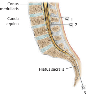
Lumbar puncture
A needle introduced into the dural sac (cisterna lumbalis) generally slips past the radices nervorum spinalium without injuring the medulla spinalis or nn. spinales. Liquor cerebrospinalis (cerebrospinal fluid, CSF) samples are therefore taken between the vertebrae L3 and L4 (2), once the patient has leaned forward to separate the procc. spinosi of the lumbar spine.

Anesthesia
Lumbar anesthesia may be administered in a similar fashion (2). Epidural anesthesia is administered by placing a catheter in the spatium epidurale without penetrating the dural sac (1). This may also be done by passing a needle through the hiatus sacralis (3).
Fig. 4.12 Segment of the medulla spinalis
The medulla spinalis (spinal cord) consists of 31 segments, each innervating a specific area of the skin (a dermatome) of the head, trunk, or limbs. Afferent (sensory) fila radicularia posteriora and efferent (motor) fila radicularia anteriora form the radices posterior and anterior of the n. spinalis for that segment. The two radices fuse to form a mixed (motor and sensory) n. spinalis that exits the foramen intervertebrale and immediately thereafter divides into a r. anterior and a r. posterior.
Fig. 4.13 Segments of the medulla spinalis, dermatomes, and effects of spinal cord lesions
The medulla spinalis is divided into four major regions: pars cervicalis, pars thoracica, pars lumbalis, and pars sacralis. The regions of the medulla spinalis are designated by colors: red, pars cervicalis; brown, pars thoracica; green, pars lumbalis; blue, pars sacralis.
 Like the medulla spinalis itself, the arteries and veins of the medulla spinalis consist of multiple horizontal systems (blood vessels of the seg menta medullae spinalis) that are integrated into a vertical system.
Fig. 4.15 Arteries of the medulla spinalis
The unpaired a. spinalis anterior and paired aa. spinales posteriores typically arise from the aa. vertebrales. As they descend within the canalis vertebralis, the aa. spinales are reinforced by anterior and posterior aa. medullares segmentales. Depending on the spinal level, these reinforcing branches may arise from the aa. vertebrales, cervicalis ascendens or profunda, intercostales posteriores, lumbales, or sacrales laterales.
Fig. 4.16 Veins of the medulla spinalis
The interior of the medulla spinalis drains via venous plexuses into a v. spinalis anterior and a v. spinalis posterior. The radicular and spinal veins connect the vv. medullae spinalis with the plexus venosus vertebralis internus. The vv. intervertebrales and basivertebrales connect the plexus venosi vertebrales interni and externi, which drain into the v. azygos system.
Fig. 4.17 Neurovasculature of the nuchal region
Posterior view. Removed: Mm. trapezius, sternocleidomastoideus, and semispinalis capitis. Revealed: Suboccipital region.
Fig. 4.18 Neurovasculature of the back
Posterior view. Removed: Muscle fascia (except lamina posterior of fascia thoracolumbalis); m. latissimus dorsi (right). Reflected: M. trapezius (right). Revealed: A. transversa colli in the deep scapular region. See p. 72 for the course of the intercostal vessels.