Fig. 13.1 Bony framework of the abdomen
Anterior view. These bones are the site of attachment for the muscles and ligaments of the anterolateral abdominal wall and form a bony cage that protects certain abdominal organs.
Fig. 13.1 Bony framework of the abdomen
Anterior view. These bones are the site of attachment for the muscles and ligaments of the anterolateral abdominal wall and form a bony cage that protects certain abdominal organs.
Fig. 13.3 Abdominal wall muscle attachment sites
Left hip bone. Muscle origins are in red, insertions in blue.
 The oblique muscles of the anterolateral abdominal wall consist of the mm. obliqui externus and internus abdominis and the m. transversus abdominis. The posterior or deep abdominal wall muscles (notably the m. psoas major) are functionally hip muscles (see p. 148).
Fig. 13.6 Diaphragma in situ
The diaphragma, which separates the thorax from the abdomen, has two asymmetric domes and three apertures (for the aorta, v. cava, and oesophagus).
Fig. 13.9 Posterior abdominal wall muscles
Anterior view. The mm. psoas major and iliacus are together known as the m. iliopsoas.
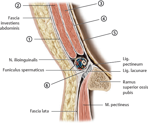
 The regio inguinalis (inguen) is the junction of the anterior abdominal wall and the anterior thigh. The canalis inguinalis is an important site for the passage of structures into and out of the cavitas abdominis (e.g., components of the funiculus spermaticus).

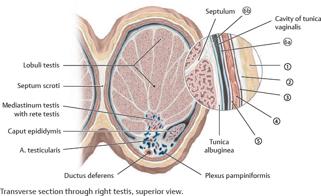
 The coverings of the scrotum, testis, and funiculus spermaticus are continuations of muscular and fascial layers of the anterior abdominal wall, as are those of the canalis inguinalis.
Fig. 13.14 Scrotum and funiculus spermaticus
Anterior view. Removed: Skin over the scrotum and funiculus spermaticus.

Table 13.4 Coverings of the testis
Covering layer |
Derived from |
|
|
Scrotal skin |
Abdominal skin |
|
Tunica dartos |
Stratum membranosum |
|
Fascia spermatica externa |
Aponeurosis musculi obliqui externi abdominis |
|
M. cremaster and/or fascia cremasterica |
M. obliquus internus abdominis |
|
Fascia spermatica interna |
Fascia transversalis |
|
Lamina parietalis tunicae vaginalis testis |
Peritoneum |
|
Lamina visceralis tunicae vaginalis testis |
|
* The m. transversus abdominis has no contribution to the funiculus spermaticus or covering of the testis. |
||
 The vagina musculi recti abdominis is created by fusion of the aponeuroses of the mm. transversus abdominis and obliqui abdominalis. The inferior edge of the lamina posterior of the vagina musculi recti abdominis is called the linea arcuata (vaginae musculi recti abdominis).
Fig. 13.18 Inferior anterior abdominal wall: Structure and fossae
Coronal section, posterior (internal) view of left inferior portion of the anterior abdominal wall.
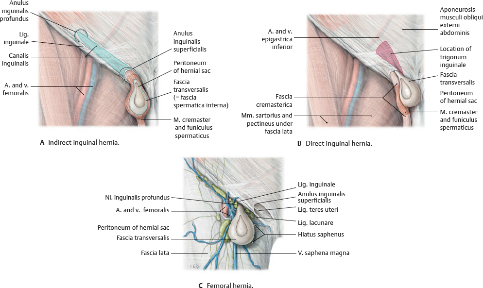
Inguinal and femoral hernias
Indirect inguinal hernias occur in younger males and may be congenital or acquired; direct inguinal hernias generally occur in older males and are always acquired. Femoral hernias are acquired and more common in females.
