19 Bones, Ligaments & Muscles
Cingulum Pelvicum
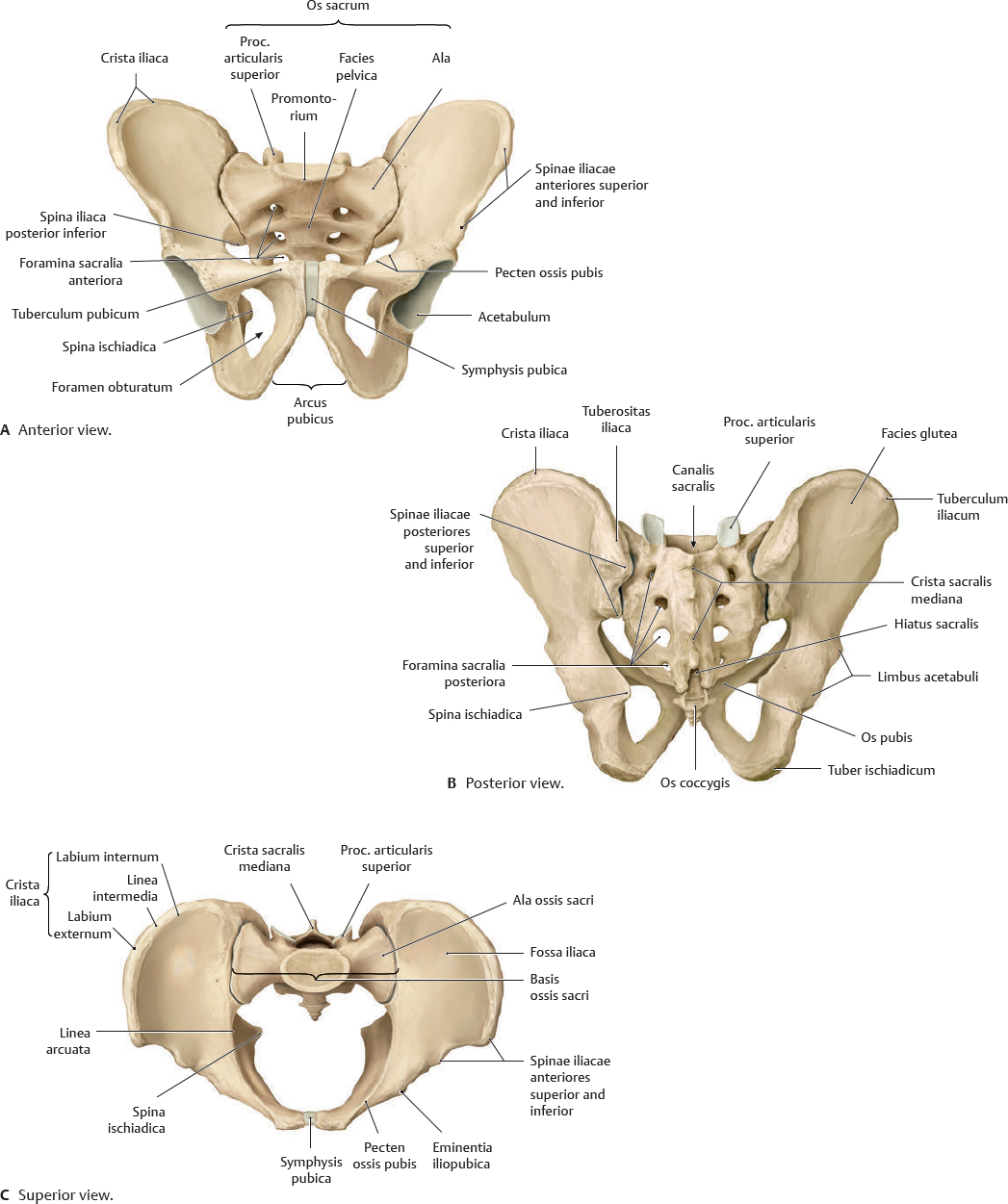
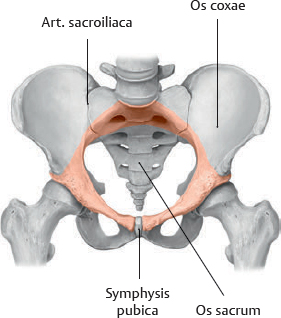
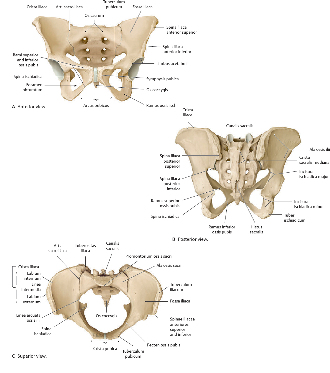
 The pelvis is the region of the body inferior to the abdomen and surrounded by the pelvic girdle (cingulum pelvicum), which is the two ossa coxarum and the os sacrum that connect the columna vertebralis to the femur. The two ossa coxarum are connected to each other at the cartilaginous symphysis pubica and to the os sacrum via the artt. sacroiliacae, creating the pelvic brim (red, Fig. 19.1). The stability of the cingulum pelvicum is necessary for the transfer of trunk loads to the lower limb, which occurs in normal gait.
Fig. 19.1 Cingulum pelvicum
Anterosuperior view. The cingulum pelvicum consists of the two ossa coxarum and the os sacrum.
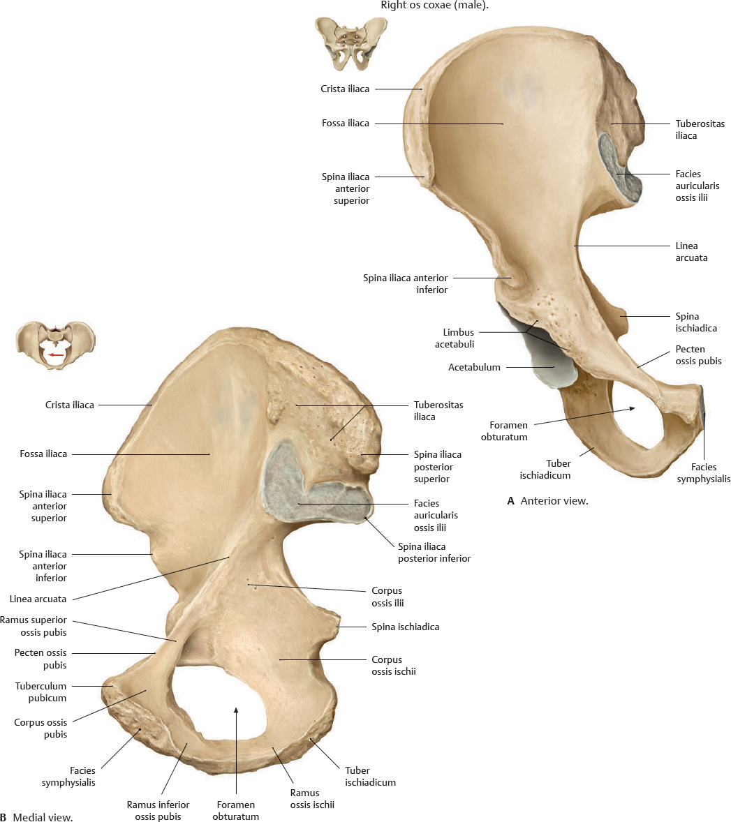
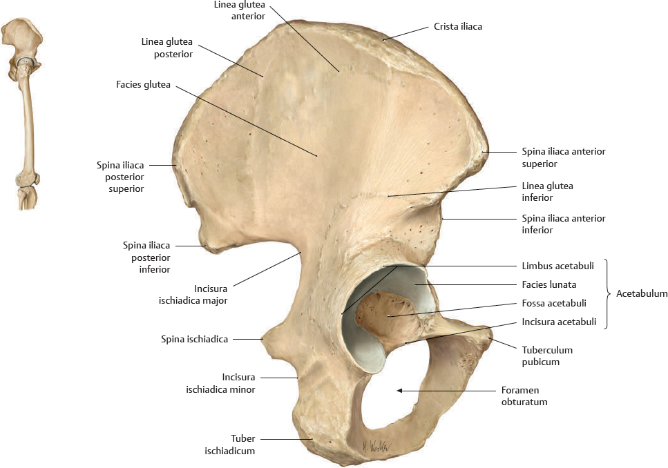
Fig. 19.2 Os coxae
Right os coxae (male).
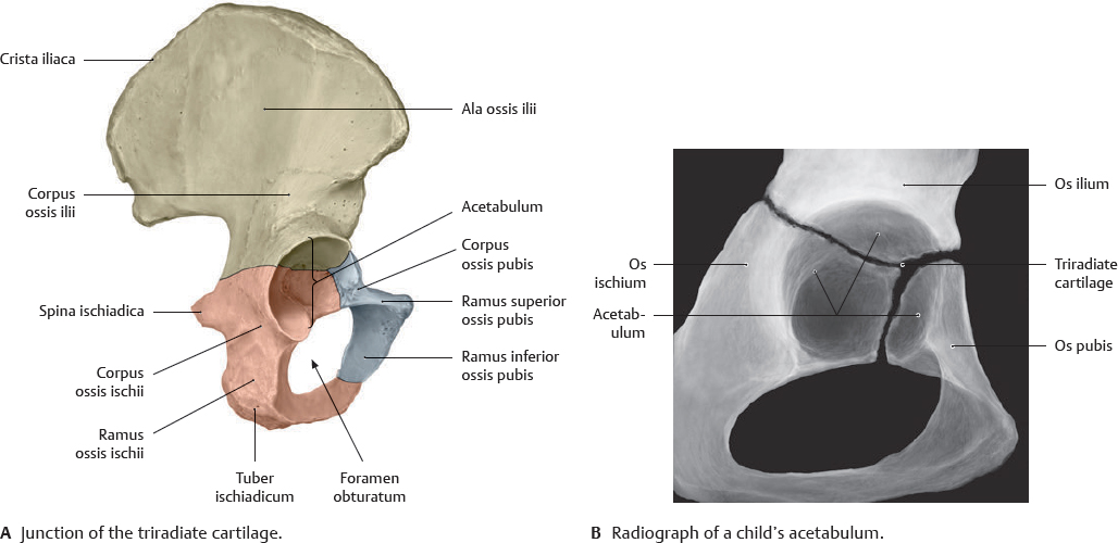
Fig. 19.3 Triradiate cartilage of the os coxae
Right os coxae, lateral view. The hip bone consists of the ossa ilium, ischium, and pubis.
Fig. 19.4 Os coxae: Lateral view
Right os coxae (male).
Female & Male Pelvis
 Clinical box 19.1
Childbirth
A non-optimal relation between the maternal pelvis and the fetal head may lead to complications during childbirth, potentially necessitating a caesarean section. Maternal causes include earlier pelvic trauma and innate malformations. Fetal causes include hydrocephalus (disturbed circulation of liquor cerebrospinalis, leading to brain dilation and cranial expansion).
Female & Male Pelvic Measurements
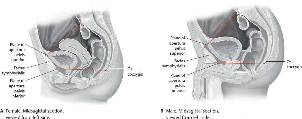
 The pelvic inlet, the apertura pelvis superior, is the boundary between the cavitates abdominis and pelvis. It is defined by the plane that passes through its edge, the pelvic brim, which is the prominence of the os sacrum, the linea arcuata and pecten ossis pubis, and the upper margin of the symphysis pubica. Occasionally, the terms apertura pelvis superior (pelvic inlet) and pelvic brim are used interchangeably. The pelvic outlet is the plane of the apertura pelvis inferior, passing through the arcus pubicus, the tubera ischiadica, the inferior margin of the lig. sacrotuberale, and the tip of the os coccygis.
Table 19.1 Gender-specific features of the pelvis
Structure |

|

|
Pelvis major |
Wide and shallow |
Narrow and deep |
Apertura pelvis superior |
Transversely oval |
Heart-shaped |
Apertura pelvis inferior |
Roomy and round |
Narrow and oblong |
Tubera ischiadica |
Everted |
Inverted |
Cavitas pelvis |
Roomy and shallow |
Narrow and deep |
Os sacrum |
Short, wide, and flat |
Long, narrow, and convex |
Angulus subpubicus |
90–100 degrees |
70 degrees |
Fig. 19.7 Pelves minor and pelvis major
The pelvis is the region of the body inferior to the abdomen, surrounded by the cingulum pelvicum. The pelvis major is immediately inferior to the cavitas abdominis, between the alae ossium iliacorum, and superior to the apertura pelvis superior. The pelvis minor is the bony-walled space between the aperturae pelvis superior and inferior. It is bounded inferiorly by the diaphragma pelvis, also called the pelvic floor.
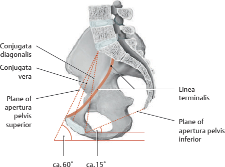
Fig. 19.8 Narrowest diameter of female pelvic canal
The conjugata vera, the distance between the promontorium ossis sacri and the most posterosuperior point of the symphysis pubica, is the narrowest AP (anteroposterior) diameter of the pelvic (birth) canal. This diameter is difficult to measure due to the viscera, so the conjugata diagonalis, the distance between the promontorium and the inferior border of the symphysis pubica, is used to estimate it. The linea terminalis is part of the border defining the apertura pelvis superior.
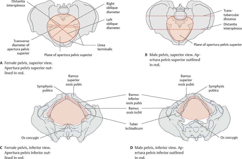
Fig. 19.9 Apertura pelvis superior and apertura pelvis inferior
The measurements shown are applicable to both male and female. The transverse and oblique diameters of the female apertura pelvis superior are obstetrically important, as they are the measure of the diameter of the pelvic (birth) canal. The distantia interspinosa is the narrowest diameter of the apertura pelvis inferior.
Pelvic Ligaments
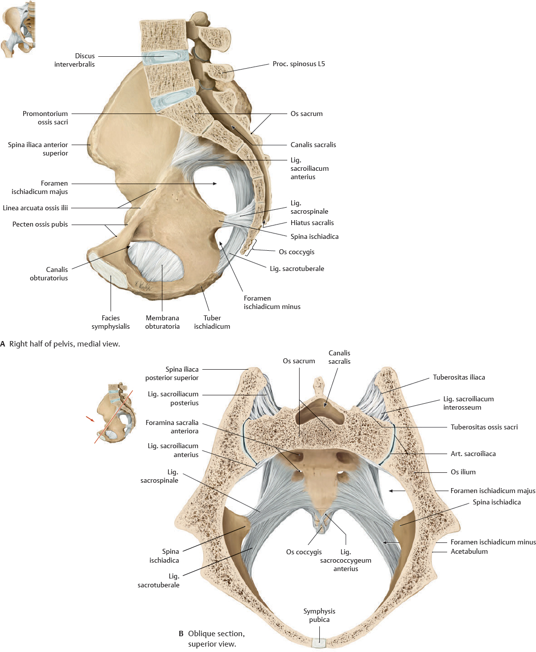
Fig. 19.10 Ligaments of the pelvis
Male pelvis.
Fig. 19.11 Ligaments of the articulatio sacroiliaca
Male pelvis.
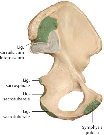
Fig. 19.12 Pelvic ligament attachment sites on os coxae
Left os coxae, medial view. Ligament attachments are shown in green.
Muscles of the Diaphragma Pelvis & Perineum
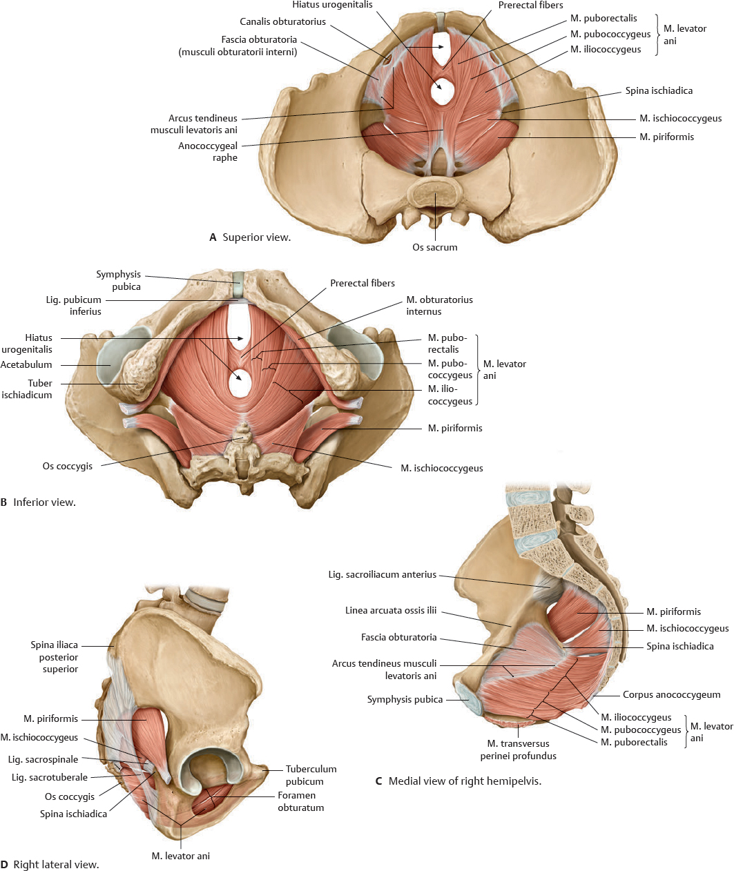
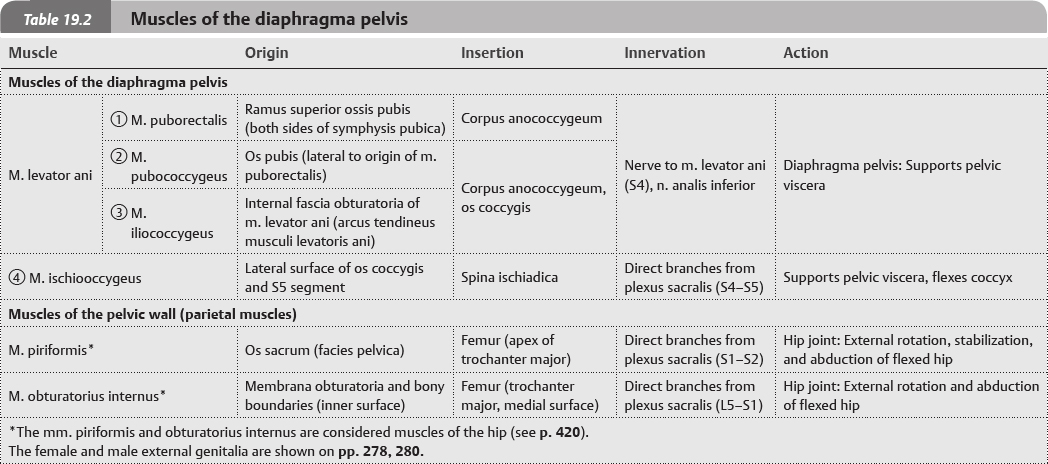
Fig. 19.13 Muscles of the diaphragma pelvis
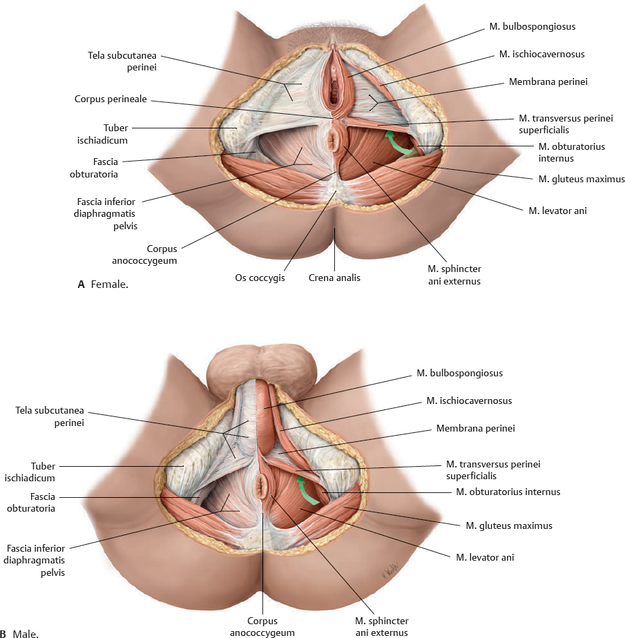
Fig. 19.14 Muscles and fascia of the diaphragma pelvis and perineum, in situ
Lithotomy position. Removed on left side: Tela subcutanea perinei, fascia inferior diaphragmatis pelvis, and fascia obturatoria. Note: The green arrows are pointing forward to the anterior recess of the fossa ischioanalis.
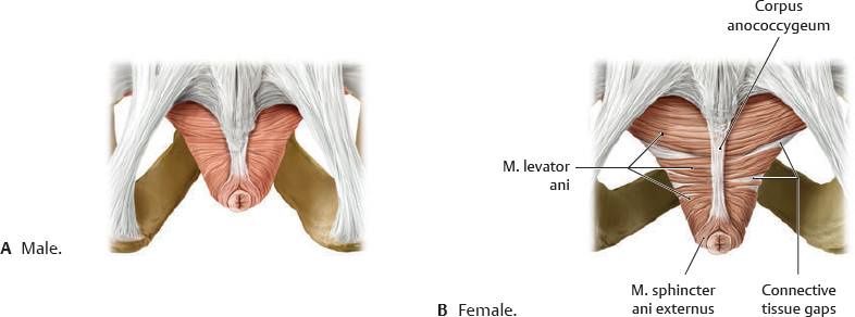
Fig. 19.15 Genderrelated differences in structure of the musculus levator ani
Posterior view. Note the connective tissue gaps between muscular parts of the m. levator ani in the female.
Diaphragma Pelvis & Perineal Muscle Facts
Fig. 19.16 Muscles of the diaphragma pelvis
Superior view.
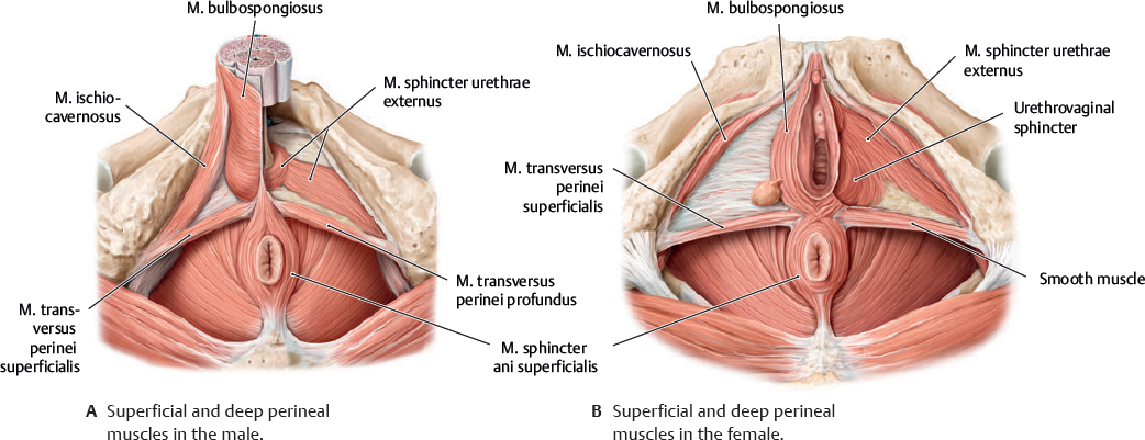
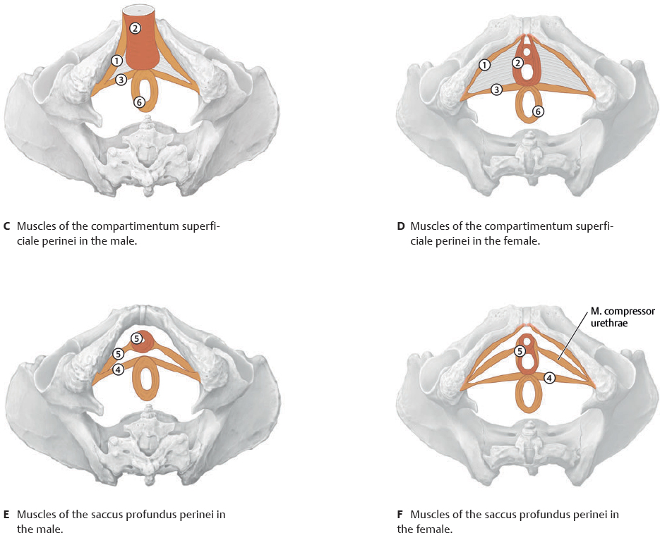
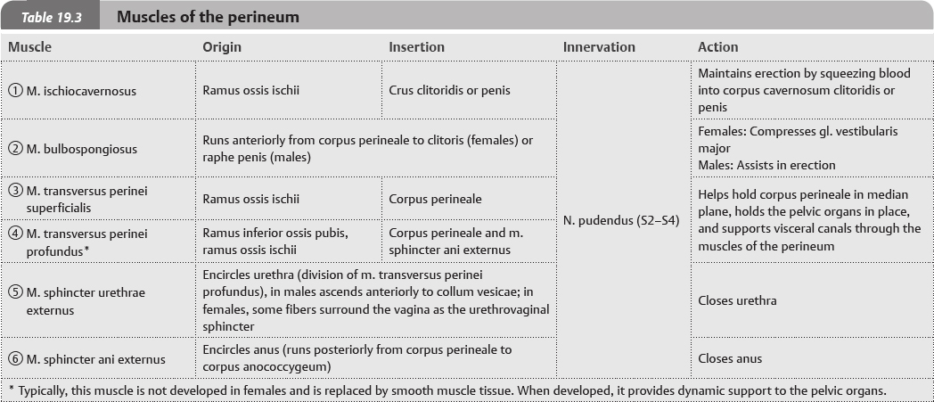
Fig. 19.17 Muscles of the perineum
Inferior view.
 The pelvis is the region of the body inferior to the abdomen and surrounded by the pelvic girdle (cingulum pelvicum), which is the two ossa coxarum and the os sacrum that connect the columna vertebralis to the femur. The two ossa coxarum are connected to each other at the cartilaginous symphysis pubica and to the os sacrum via the artt. sacroiliacae, creating the pelvic brim (red, Fig. 19.1). The stability of the cingulum pelvicum is necessary for the transfer of trunk loads to the lower limb, which occurs in normal gait.
 Clinical box 19.1