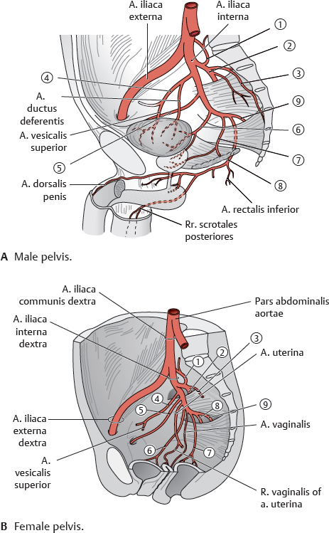

Table 22.1 Branches of the arteria iliaca interna
The a. iliaca interna gives off five parietal (pelvic wall) and four visceral (pelvic organs) branches.* Parietal branches are shown in italics. |
||
Branches |
||
|
A. iliolumbalis |
|
|
A. glutea superior |
|
|
Aa. sacrales laterales |
|
|
A. umbilicalis |
A. ductus deferentis |
Aa. vesicales superiores |
||
|
A. obturatoria |
|
|
A. vesicalis inferior |
|
|
A. rectalis media |
|
|
A. pudenda interna |
A. rectalis inferior |
A. dorsalis penis |
||
Rr. scrotales posteriores |
||
|
A. glutea inferior |
|
* In the female pelvis, the aa. uterina and vaginalis arise directly from the anterior division of the a. iliaca interna. |
||

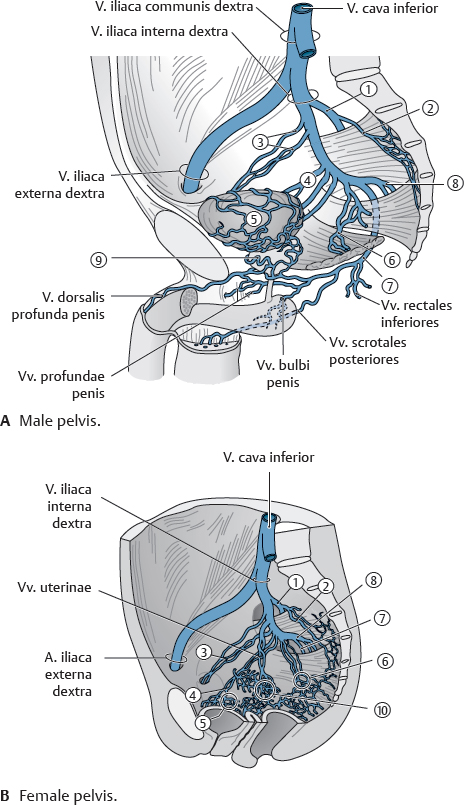
Table 22.2 Venous drainage of the pelvis
Tributaries |
|
|
Vv. gluteae superiores |
|
Vv. sacrales laterales |
|
Vv. obturatoriae |
|
Vv. vesicales |
|
Plexus venosus vesicalis |
|
Vv. rectales mediae (plexus venosus rectalis) (also vv. rectales superiores and inferiores, not shown) |
|
V. pudenda interna |
|
Vv. gluteae inferiores |
|
Plexus venosus prostaticus |
|
Plexus venosi uterinus and vaginalis |
The male pelvis also contains veins draining the penis and scrotum. |
|
Fig. 22.2 Blood vessels of the rectum
A posterior view. The main blood supply to the rectum is from the aa. rectales superiores; the aa. rectales mediae serve as an anastomosis between the aa. rectales superior and inferior.
Fig. 22.3 Arterial supply of the cavernous body of the rectum (hemorrhoidal plexus)
Caudal view with patient in lithotomy position. The hemorrhoidal plexus is a permanently distended cavernous body. It's supplied by three branches of the a. rectalis superior at the 3, 7, and 11 o'clock positions where they form three major cushions and four minor cushions in the area of the columnae anales. These circular cavernous structures filled with blood serve as an effective continence mechanism that ensures liquid- and gas-tight closure. The sustained contraction of the muscular sphincter apparatus inhibits venous drainage, and blood is allowed to drain from the cavernous body when the sphincters relax during defecation.
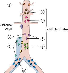
Fig. 22.5 Lymphatic drainage of the internal organs
Lymph draining from the abdomen, pelvis, and lower limb ultimately passes through the nll. lumbales sinistri and dextri. (See Table 22.3 for numbering.) The nll. lumbales consist of the nll. cavales and aortici laterales, the nll. preaortici, and the nll. retroaortici/retrocavales. Effer ent lymph vessels from the nll. aortici and cavales laterales and the nll. retroaortici and retrocavales form the trunci lumbales and those from the nll. preaortici form the trunci intestinales. The trunci lumbales and intestinales terminate in the cisterna chyli.

Table 22.3 Nodi lymphoidei of the abdomen
|
||
Nll. lumbales |
Nll. preaortici |
|
|
||
|
||
|
||
|
||
|
||
|
||

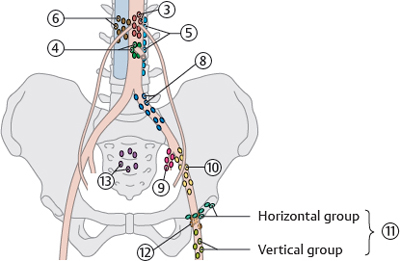
Table 22.4 Nodi lymphoidei of the pelvis
Numbers continued from Table 22.3. |
||
Nll. preaortici |
|
|
|
||
|
||
|
||
|
||
|
||
|
||
|
Horizontal group |
|
Vertical group |
||
|
||
|
||
Fig. 22.10 Nodi lymphoidei of the male genitalia
Anterior view. Removed: Gastrointestinal tract (except rectal stump) and peritoneum.
Fig. 22.11 Nodi lymphoidei of the female genitalia
Anterior view. Removed: Gastrointestinal tract (except rectal stump) and peritoneum. Retracted: Uterus.
Fig. 22.13 Autonomic plexuses in the pelvis
Anterior view of the male lower abdomen. Removed: Peritoneum and abdominopelvic organs except renes.
Fig. 22.14 Innervation of the urinary organs
Anterior view of the male lower abdomen and pelvis. Removed: Peritoneum and abdominopelvic organs except renes, gll. suprarenales, rectal stump, and vesica urinaria. See p. 276 for schematic of innervation of urinary organs.
Fig. 22.15 Innervation of the female pelvis
Right pelvis, left lateral view. Reflected: Uterus and rectum. See p. 277 for schematic of innervation of genitalia.
Fig. 22.16 Innervation of the male pelvis
Right pelvis, left lateral view. See p. 277 for schematic of innervation of genitalia.
Referred pain from the internal organs
The convergence of somatic and visceral neurofibrae afferentes to a common actual sites of pain, a phenomenon known as referred pain. Pain impulses from a particular organ are consistently projected to the same well-defined skin area.

Fig. 22.19 Innervation of the rectum
Left side of image shows somatic nerves (nn. spinales); right side of image shows nn. autonomici.
Fig. 22.22 Neurovasculature of the female perineum
Lithotomy position. Removed from left side: Mm. bulbospongiosus and ischiocavernosus.