31 Hip & Thigh
Bones of the Lower Limb
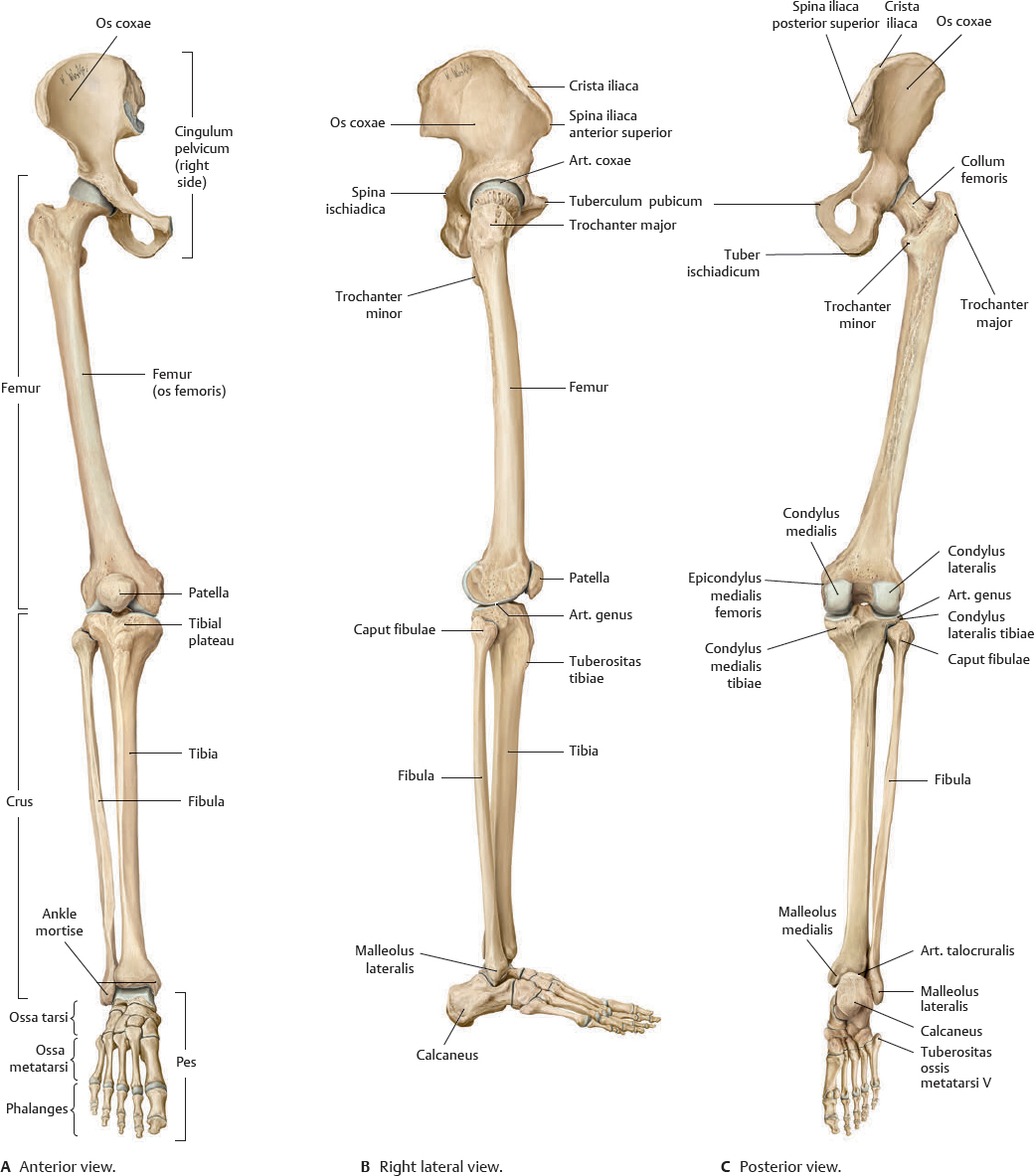
 The skeleton of the lower limb (membrum inferius) consists of an os coxae and a free part (pars libera). The paired ossa coxarum attach to the trunk at the art. sacroiliaca to form the cingulum pelvicum (see p. 228), and the pars libera, divided into a femur, crus, and pes, attaches to the cingulum pelvicum at the art. coxae. Stability of the cingulum pelvicum is important in the distribution of weight from the upper body to the lower limbs.
Fig. 31.1 Bones of the lower limb
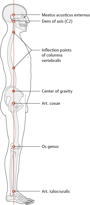
Fig. 31.2 Line of gravity
Right lateral view. The line of gravity runs vertically from the whole-body center of gravity to the ground with characteristic points of intersection.
Fig. 31.3 The ossa coxarum and their relation to bones of the trunk.
The paired ossa coxarum and os sacrum form the cingulum pelvicum (see p. 228).
Femur
 Clinical box 31.1
Fractures of the femur
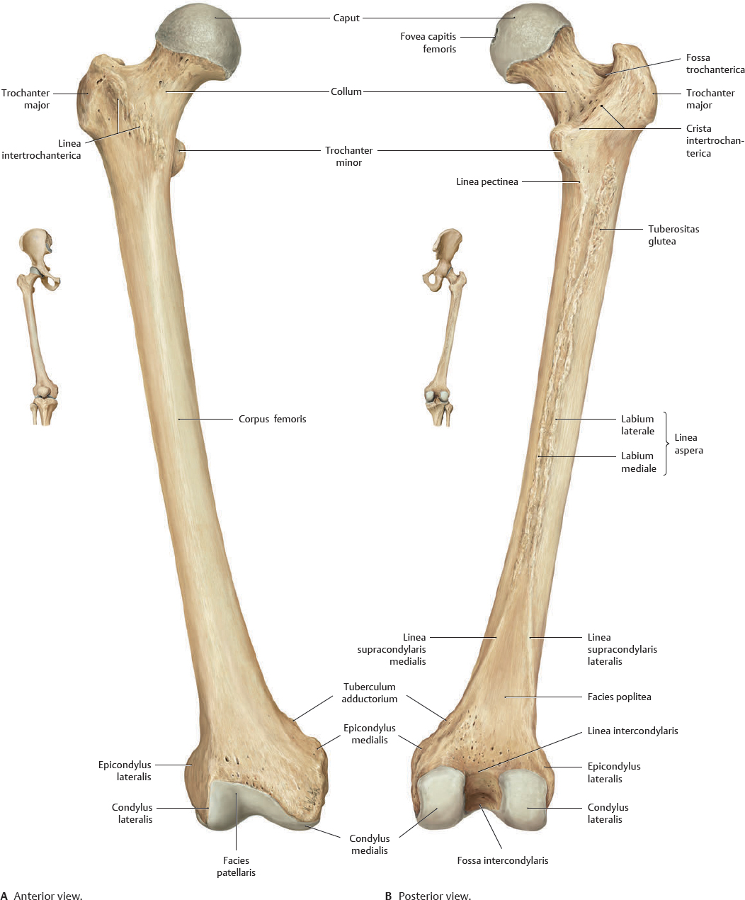
Femoral fractures caused by falls in patients with osteoporosis are most frequently located in the collum femoris. Corpus femoris fractures are less frequent and are usually caused by strong trauma (e.g., a car accident).
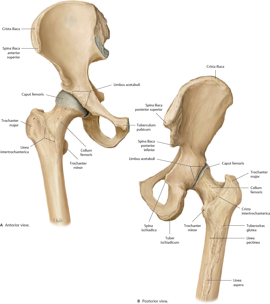
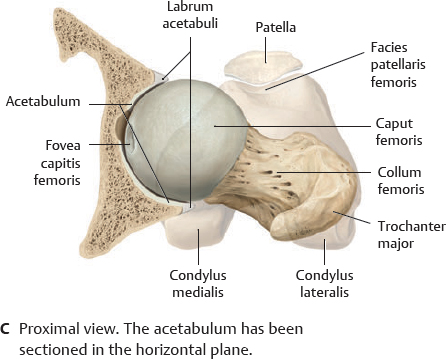
Fig. 31.5 Caput femoris in the articulatio coxae
Right art. coxae, superior view.
Articulatio Coxae: Overview
Fig. 31.6 Right articulatio coxae
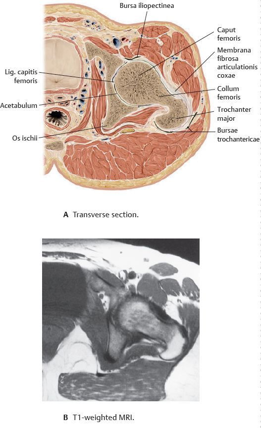
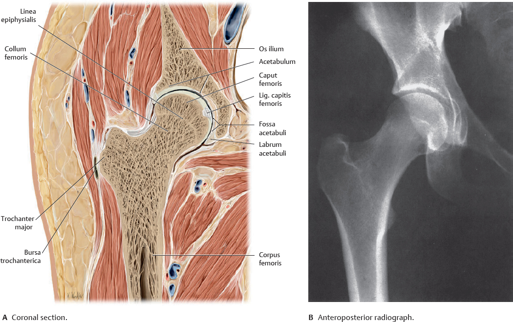
The caput femoris articulates with the acetabulum of the pelvis at the art. coxae, a special type of spheroidal (ball-and-socket) joint. The roughly spherical caput femoris (with an average radius of curvature of approximately 2.5 cm) is largely contained within the acetabulum.
Fig. 31.7 Articulatio coxae: Coronal section
Right art. coxae, anterior view.
 Clinical box 31.2
Diagnosing hip dysplasia and dislocation
Ultrasonography, the most important imaging method for screening the infant hip, is used to identify morphological changes such as hip dysplasia and dislocation. Clinically, hip dislocation presents with instability and limited abduction of the art. coxae, and leg shortening with asymmetry of the sulci gluteales.
Articulatio Coxae: Ligaments & Capsula Articularis
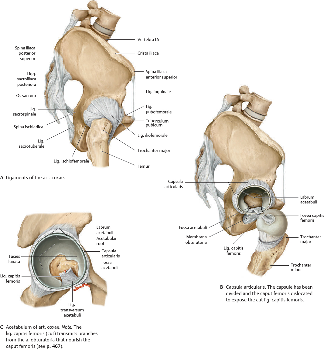
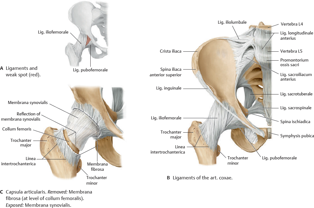
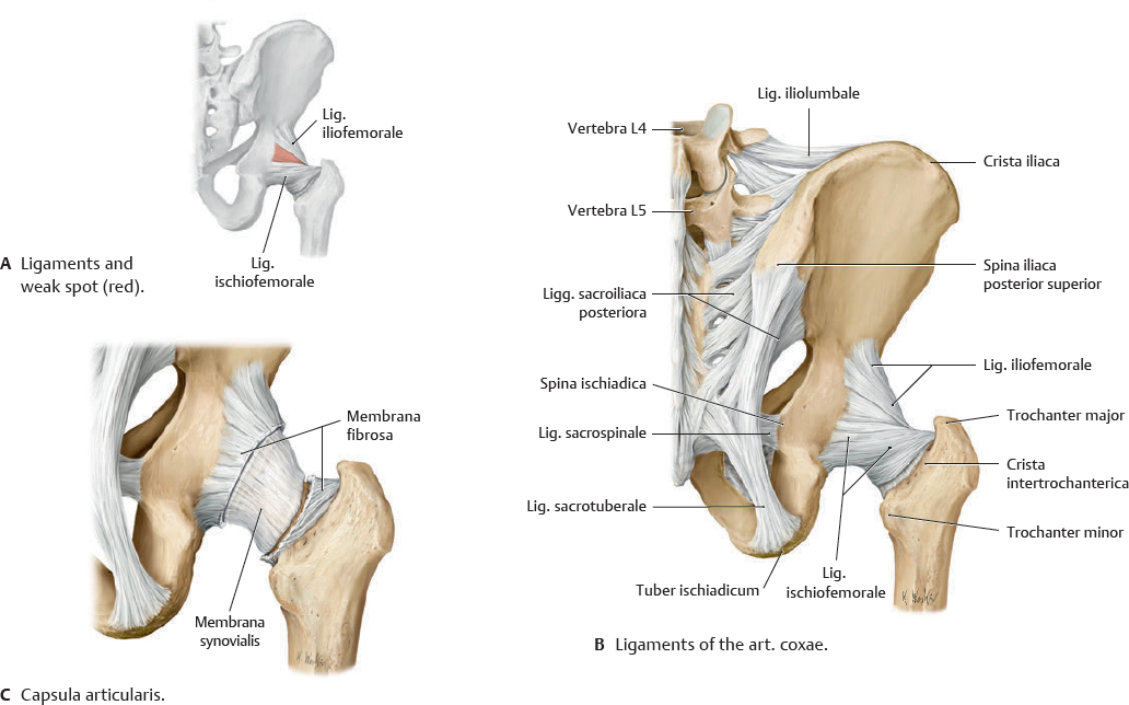
 The art. coxae has three major ligaments: ligg. iliofemorale, pubofemorale, and lig. ischiofemorale. The zona orbicularis (annular ligament) is not visible externally and encircles the collum femoris like a buttonhole.
Fig. 31.8 Articulatio coxae: Lateral view
Right art. coxae.
Fig. 31.9 Articulatio coxae: Anterior view
Right art. coxae.
Fig. 31.10 Articulatio coxae: Posterior view
Right art. coxae.
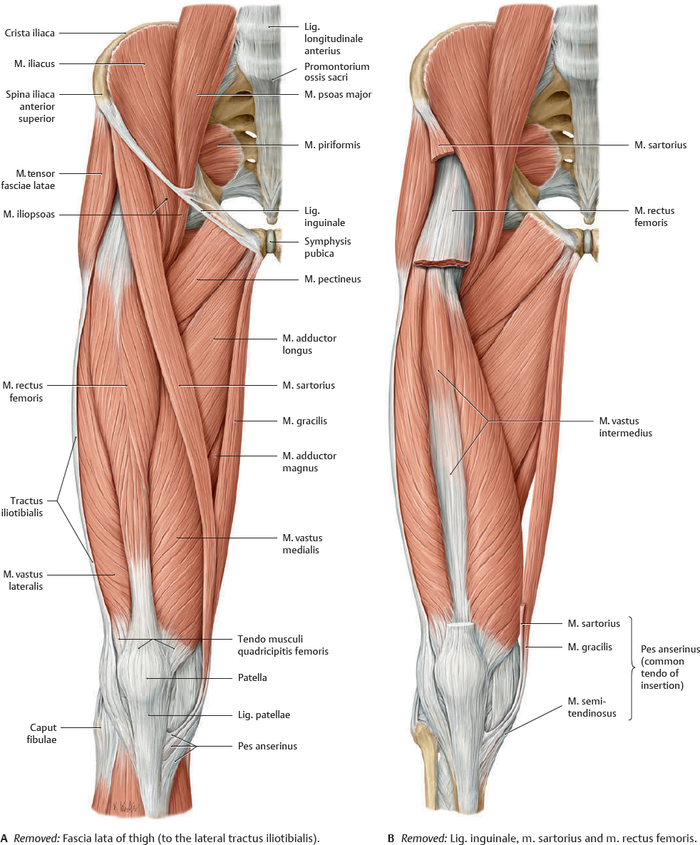
Anterior Muscles of the Regiones Coxae, Femoris & Glutealis (I)
Fig. 31.11 Anterior muscles of the coxa and femur (I)
Right limb. Muscle origins are shown in red, insertions in blue.
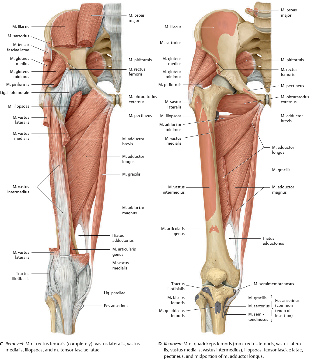
Anterior Muscles of the Regiones Coxae, Femoris & Glutealis (II)
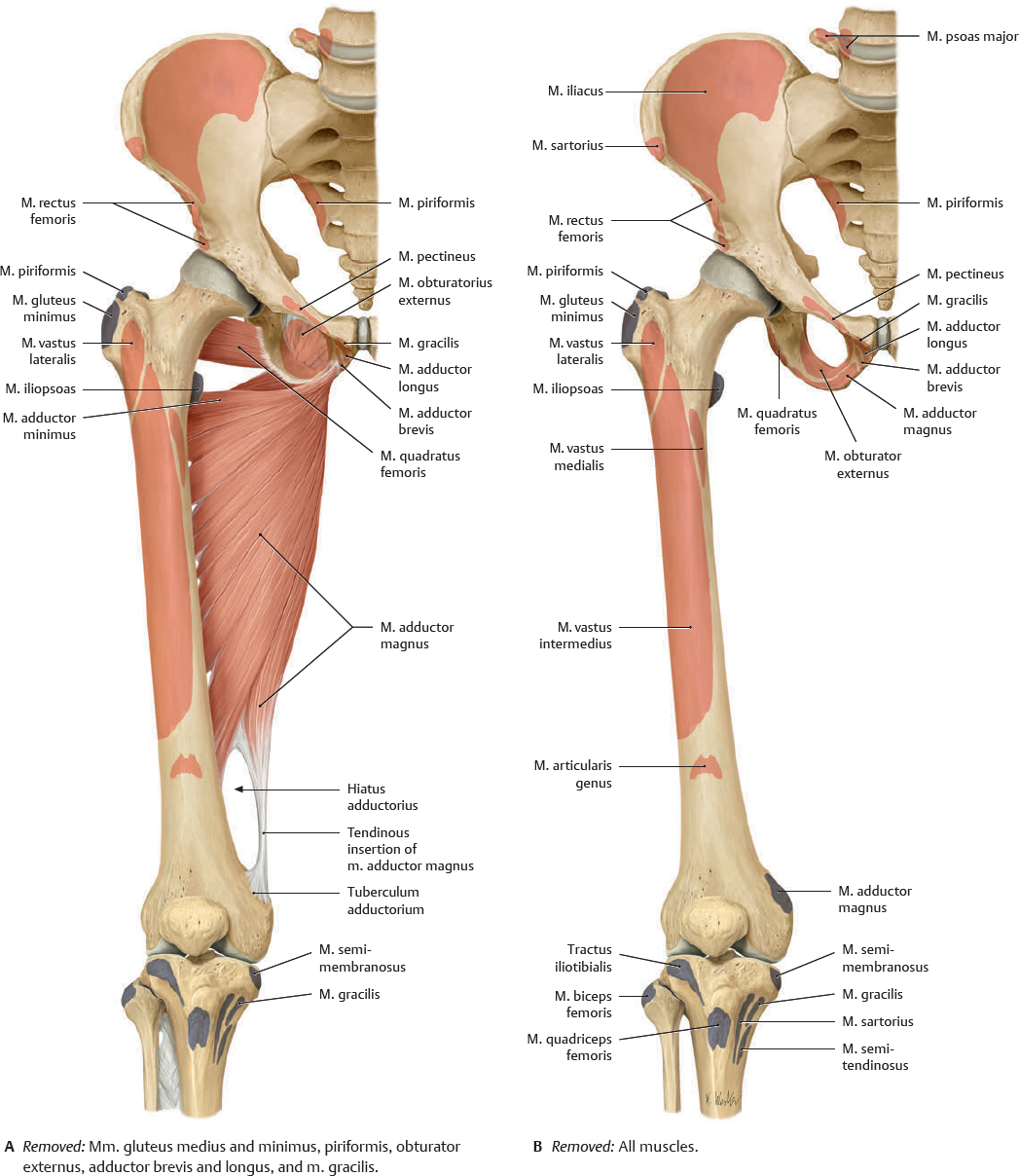
Fig. 31.12 Anterior muscles of the coxa and femur (II)
Right limb. Muscle origins are shown in red, insertions in blue.
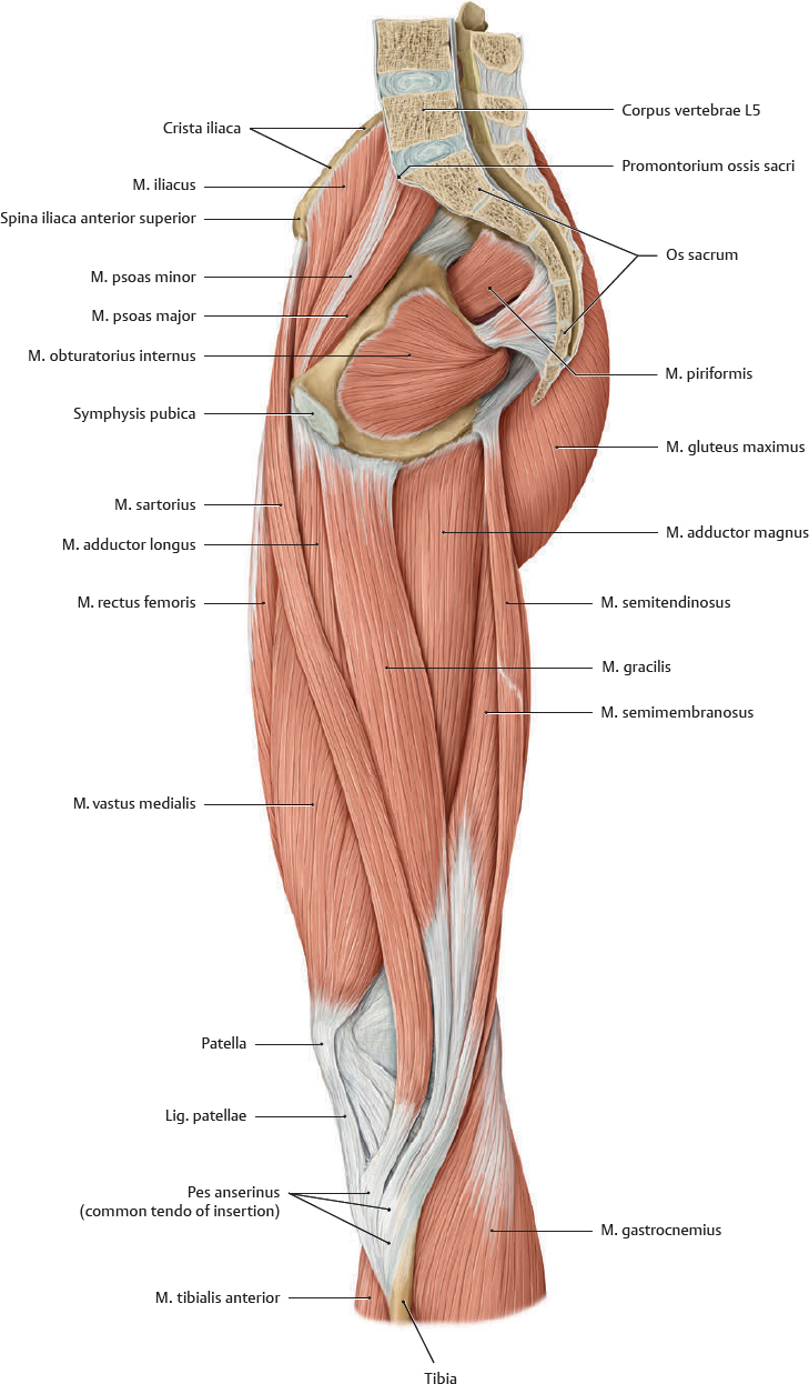
Fig. 31.13 Medial muscles of the regiones coxae, femoris, and glutealis
Midsagittal section.
Posterior Muscles of the Regiones Coxae, Femoris & Glutealis (I)
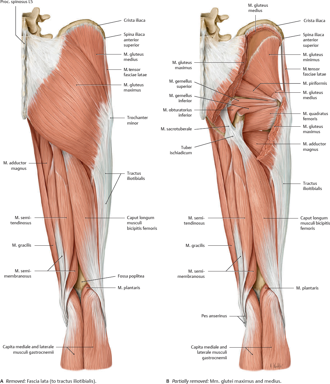
Fig. 31.14 Posterior muscles of the regiones coxae, femoris, and glutealis (I)
Right limb. Muscle origins are shown in red, insertions in blue.
Posterior Muscles of the Regiones Coxae, Femoris & Glutealis (II)
Fig. 31.15 Posterior muscles of the regiones coxae, femoris, and glutealis (II)
Right limb. Muscle origins are shown in red, insertions in blue.
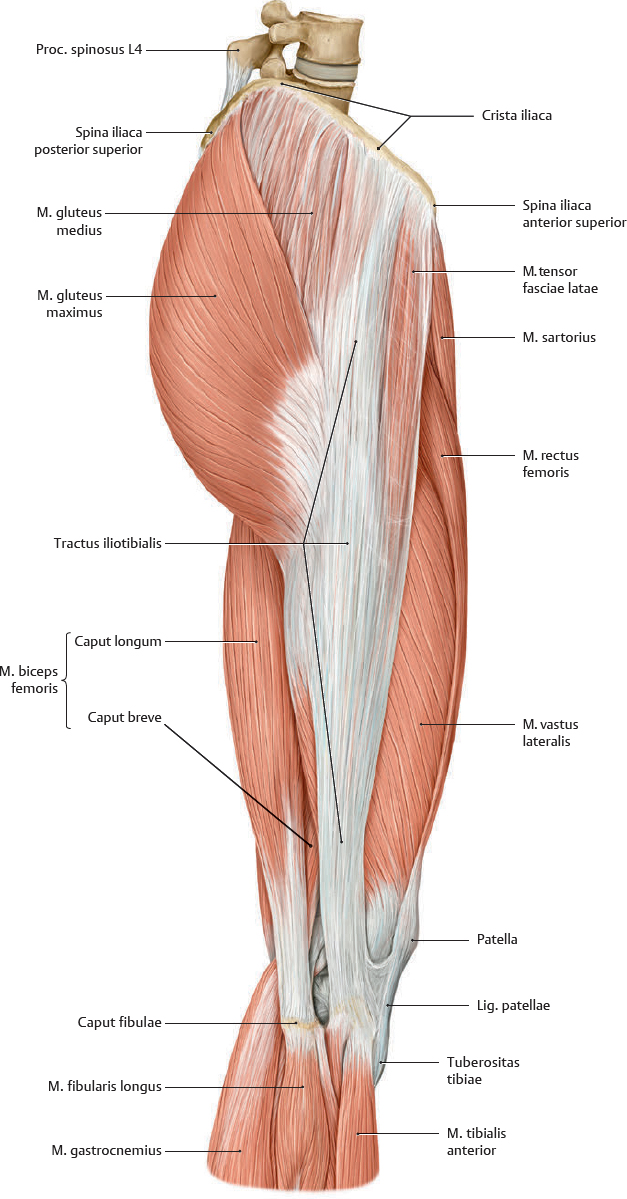
Fig. 31.16 Lateral muscles of the regiones coxae, femoris, and glutealis
Note: The tractus iliotibialis (the thickened band of fascia lata) functions as a tension band to reduce the bending loads on the proximal femur.
Muscle Facts (I)
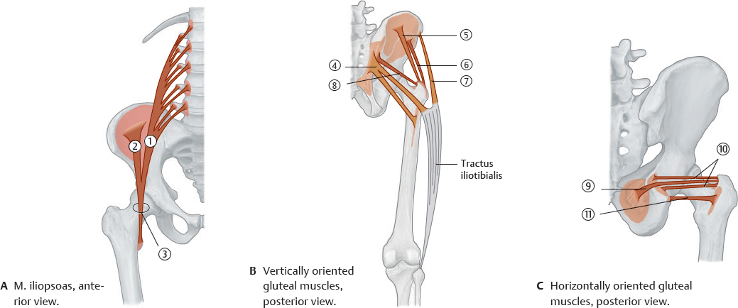
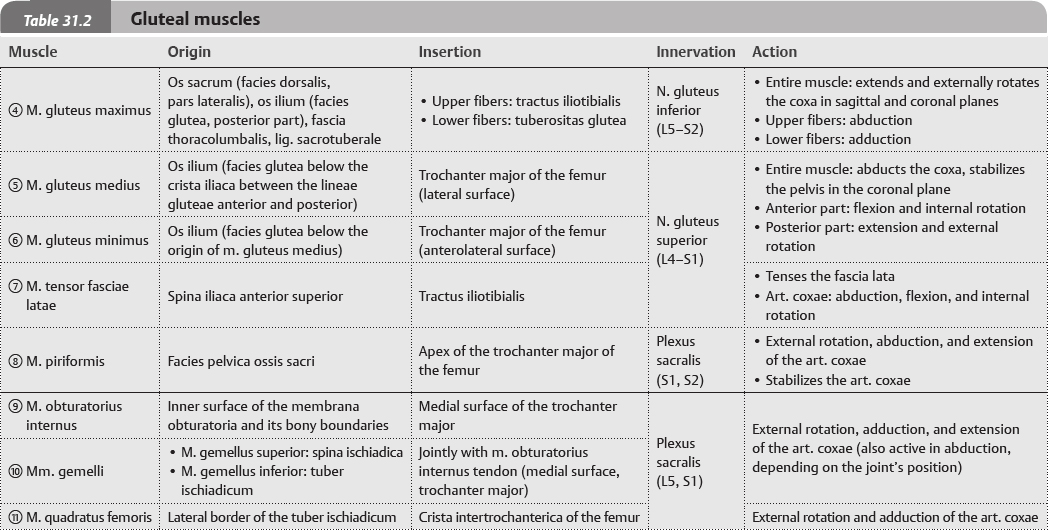
Fig. 31.17 Muscles of the coxa
Right side, schematic.
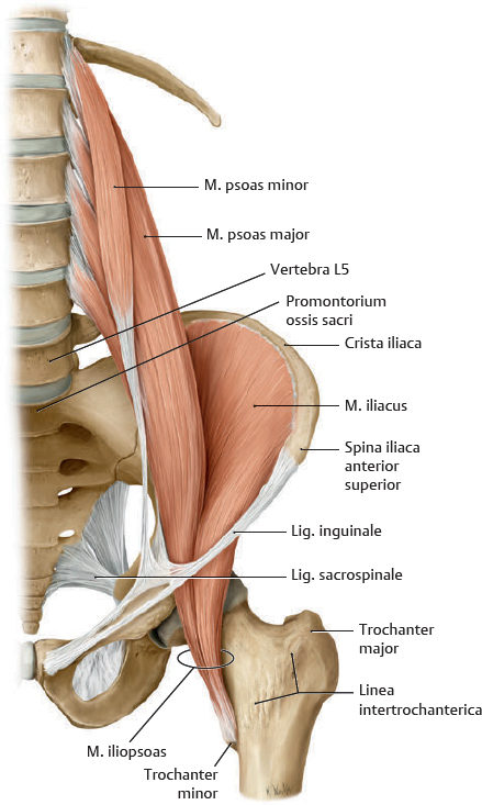
Fig. 31.18 Musculus psoas and musculus iliacus
Right side, anterior view.
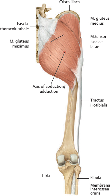
Fig. 31.19 Superficial muscles of the gluteal region
Right side, posterior view.
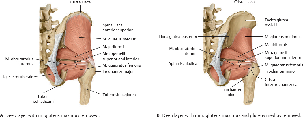
Fig. 31.20 Deep muscles of the gluteal region
Muscle Facts (II)
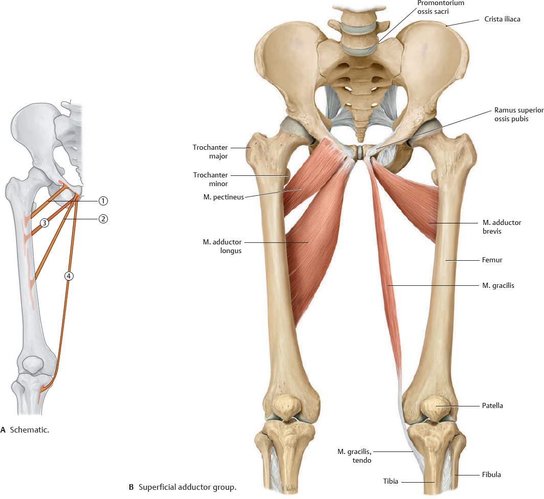
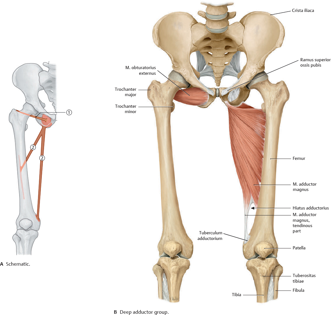
 Functionally, the medial thigh muscles are considered the adductors of the coxa.
Fig. 31.21 Medial thigh muscles: Superficial layer
Right side, anterior view.
Fig. 31.22 Medial thigh muscles: Deep layer
Right side, anterior view.
Muscle Facts (III)
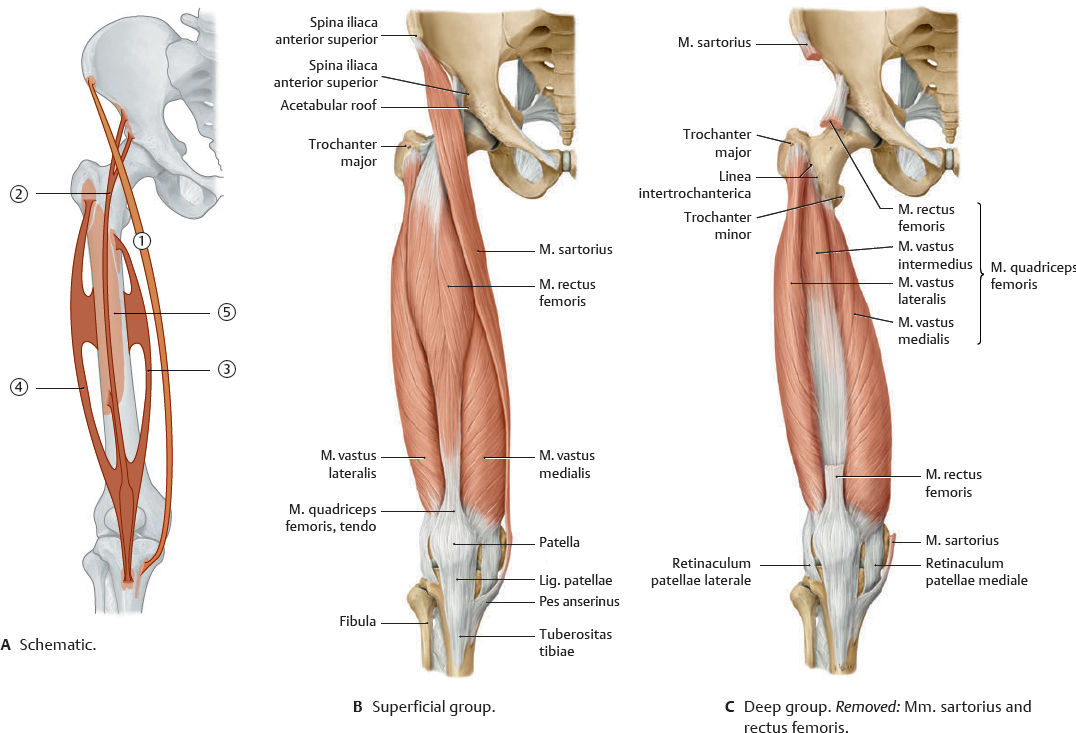
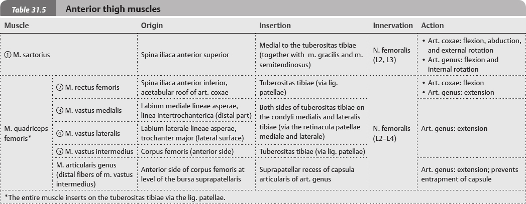
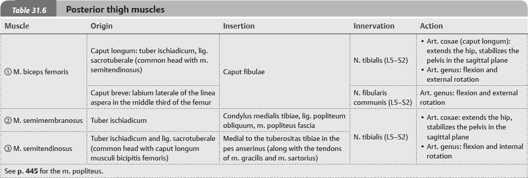
 The anterior and posterior muscles of the thigh can be classified as extensors and flexors, respectively, with regard to the art. genus.
Fig. 31.23 Anterior thigh muscles
Right side, anterior view.
Fig. 31.24 Posterior thigh muscles
Right side, posterior view.
 The skeleton of the lower limb (membrum inferius) consists of an os coxae and a free part (pars libera). The paired ossa coxarum attach to the trunk at the art. sacroiliaca to form the cingulum pelvicum (see p. 228), and the pars libera, divided into a femur, crus, and pes, attaches to the cingulum pelvicum at the art. coxae. Stability of the cingulum pelvicum is important in the distribution of weight from the upper body to the lower limbs.

 Clinical box 31.1