46 Sectional & Radiographic Anatomy
Sectional Anatomy of the Head & Neck (I)
Fig. 46.1 Coronal section through the orbital opening (aditus orbitalis)
Anterior view. This section shows four regions of the head: the cavitas oris, the cavitas nasi and sinus paranasales, the orbita, and the fossa cranii anterior. Muscles of the oral floor, the apex linguae, the palatum durum, the neurovascular structures in the canalis mandibulae, and the dens molaris primus are all seen in the region of the cavitas oris. This section reinforces the clinical implications of the relationship of the sinus maxillaris with the dentes maxillares and the paries inferior orbitae and with the n. maxillaris in the sulcus infraorbitalis. The paries medialis orbitae shares a thin bony wall (lamina orbitalis) with the cellulae ethmoidales. The section is enough anterior so that the parietes laterales of the orbitae are not included due to the lateral curvature of the cranium.
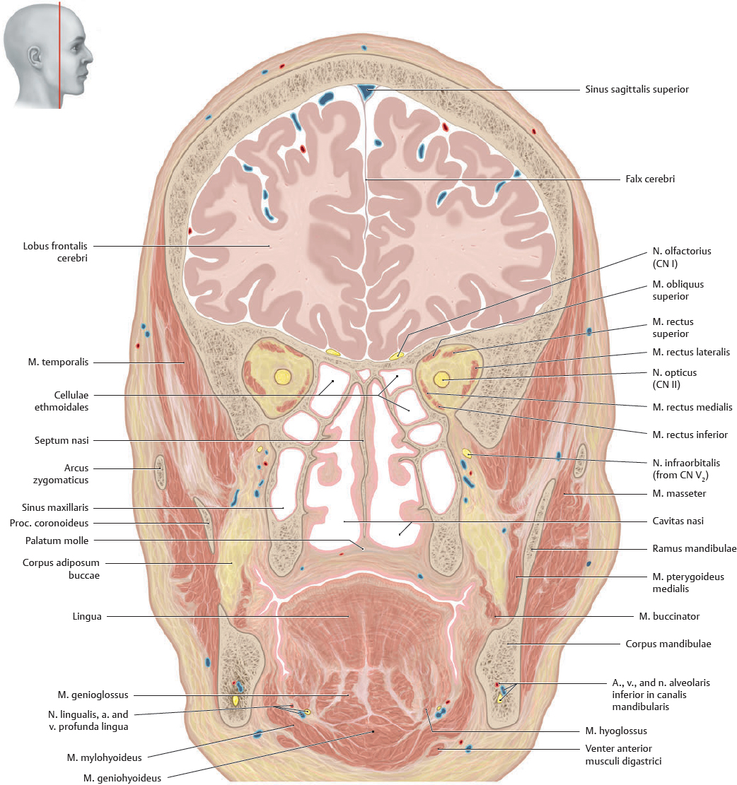
Fig. 46.2 Coronal section through the orbital apex
Anterior view. In this more posterior section than that of Fig. 46.1, the palatum molle now separates the cavitates oris and nasi. The corpus adiposum buccae is also visible. The section is slightly angled, producing an apparent discontinuity in the ramus mandibulae on the left side.
Sectional Anatomy of the Head & Neck (II)
Fig. 46.3 Coronal section through the hypophysis
Anterior view.
Fig. 46.4 Midsagittal section through the Septum nasi
Left lateral view.
Sectional Anatomy of the Head & Neck (III)
Fig. 46.5 Sagittal section through the paries medialis orbitae
Left lateral view. This section passes through the conchae nasi inferior and media of the lateral nasal wall. Three of the four sinus paranasales (cellulae ethmoidales, sinus sphenoidalis, and frontalis) are seen in this section and in relation to the cavitas nasi into which they drain. In the region of the cervical spine, the a. vertebralis is cut at multiple levels. The nn. spinales have been cut just prior to their lateral exit through the foramina intervertebralia.
Fig. 46.6 Sagittal section through the inner third of the orbita
Left lateral view. This section passes through the sinus maxillaris, frontalis, and sphenoidalis and a single cellula ethmoidalis. The mm. pharyngis and masticatorii are revealed grouped around the pars cartilaginea of the tuba auditiva. The tonsilla palatina of the cavitas oris and medial portion of the gl. submandibularis below the floor of the mouth are also seen in this section.
Sectional Anatomy of the Head & Neck (IV)
Fig. 46.7 Transverse section through the nervus opticus and hypophysis
Inferior view.
Fig. 46.8 Transverse section of head through the articulatio atlantoaxialis mediana
Superior view. This section passes through the palatum molle and mucoperiosteum of the palatum durum. The articulation of the odontoid process (dens axis) with the atlas (C1) at the art. atlantoaxialis mediana is shown, as well as the vagina carotica, containing the vertical neurovascular elements of the neck. The a. vertebralis is sectioned as it prepares to enter the foramen magnum and fuse with its opposite to form the a. basilaris.
Sectional Anatomy of the Head & Neck (V)
Fig. 46.9 Transverse section of the neck
Transverse section at the level of the corpus vertebrae C5. Inferior view. The vv. jugulares interna and externa are separated by the m. sternocleidomastoideus. The n. accessorius (CN XI) is just medial to this muscle as it prepares to innervate it from behind. The elongated proc. spinosus of the C7 vertebra (vertebra prominens) is also visible in the section due to the lordotic curvature of the collum.
Fig. 46.10 Transverse section at the level of the corpus vertebrae C6
Inferior view.
Fig. 46.11 Transverse section of the neck
Transverse section at the level of the symphysis intervertebralis C7/T1. Inferior view. This section reveals the roots of nn. spinales C6 to C8 of the plexus brachialis passing between the mm. scaleni anterior and medius. The n. phrenicus is on the anterior surface of the m. scalenus anterior and the components of the vagina carotica (v. jugularis interna, a. carotis communis, and n. vagus) lie in the interval between this muscle, the m. sternocleidomastoideus, and the gl. thyroidea.
Radiographic Anatomy of the Head & Neck (I)
Fig. 46.12 Radiograph of the cranium
Anteroposterior view.
Fig. 46.13 Coronal MRI through the bulbus oculi
Anterior view.
Fig. 46.14 Radiograph of the cranium
Left lateral view.
Fig. 46.15 Midsagittal MRI through the septum nasi
Left lateral view. Boxed area represents the location of the ventricular system, thalamus, and pons. A more detail labeled version of this area can be seen in Fig. 51.5, p.688.
Radiographic Anatomy of the Head & Neck (II)
Fig. 46.16 Radiograph of the cranium
Inferosuperior oblique view (Waters view).
Fig. 46.17 Radiograph of the mandibula
Left lateral view.
Fig. 46.18 Transverse MRI through the orbita and ductus nasolacrimalis
Inferior view.
Fig. 46.19 Transverse MRI through the neck
Inferior view
Radiographic Anatomy of the Head & Neck (III)
Fig. 46.20 Articulatio temporomandibularis (TMJ)
Coronal section.
Fig. 46.21 Articulatio temporomandibularis (TMJ)
Sagittal section, mouth closed.
Fig. 46.22 Cranial MR angiography
Cranial view. In this angiogram note that the right a. cerebri posterior arises from the a. carotis interna instead of the a. basilaris—a variant. The normal configuration is seen on the left side.
Fig. 46.23 Sinus durae matris system of the head
Right lateral view.