CHAPTER 1
Old Brain—New Brain
To understand how the brain creates intelligence, there are a few basics you need to know first.
Shortly after Charles Darwin published his theory of evolution, biologists realized that the human brain itself had evolved over time, and that its evolutionary history is evident from just looking at it. Unlike species which often disappear as new ones appear, the brain evolved by adding new parts on top of the older parts. For example, some of the oldest and simplest nervous systems are sets of neurons that run down the back of tiny worms. These neurons allow the worm to make simple movements, and they are the predecessor of our spinal cord, which is similarly responsible for many of our basic movements. Next to appear was a lump of neurons at one end of the body that controlled functions such as digestion and breathing. This lump is the predecessor of our brain stem, which similarly controls our digestion and breathing. The brain stem extended what was already there, but it did not replace it. Over time, the brain grew capable of increasingly complex behaviors by evolving new parts on top of the older parts. This method of growth by addition applies to the brains of most complex animals. It is easy to see why the old brain parts are still there. No matter how smart and sophisticated we are, breathing, eating, sex, and reflex reactions are still critical to our survival.
The newest part of our brain is the neocortex, which means “new outer layer.” All mammals, and only mammals, have a neocortex. The human neocortex is particularly large, occupying about 70 percent of the volume of our brain. If you could remove the neocortex from your head and iron it flat, it would be about the size of a large dinner napkin and twice as thick (about 2.5 millimeters). It wraps around the older parts of the brain such that when you look at a human brain, most of what you see is the neocortex (with its characteristic folds and creases), with bits of the old brain and the spinal cord sticking out the bottom.
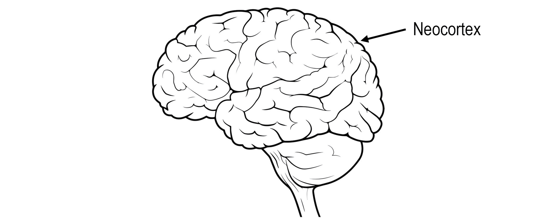
A human brain
The neocortex is the organ of intelligence. Almost all the capabilities we think of as intelligence—such as vision, language, music, math, science, and engineering—are created by the neocortex. When we think about something, it is mostly the neocortex doing the thinking. Your neocortex is reading or listening to this book, and my neocortex is writing this book. If we want to understand intelligence, then we have to understand what the neocortex does and how it does it.
An animal doesn’t need a neocortex to live a complex life. A crocodile’s brain is roughly equivalent to our brain, but without a proper neocortex. A crocodile has sophisticated behaviors, cares for its young, and knows how to navigate its environment. Most people would say a crocodile has some level of intelligence, but nothing close to human intelligence.
The neocortex and the older parts of the brain are connected via nerve fibers; therefore, we cannot think of them as completely separate organs. They are more like roommates, with separate agendas and personalities, but who need to cooperate to get anything done. The neocortex is in a decidedly unfair position, as it doesn’t control behavior directly. Unlike other parts of the brain, none of the cells in the neocortex connect directly to muscles, so it can’t, on its own, make any muscles move. When the neocortex wants to do something, it sends a signal to the old brain, in a sense asking the old brain to do its bidding. For example, breathing is a function of the brain stem, requiring no thought or input from the neocortex. The neocortex can temporarily control breathing, as when you consciously decide to hold your breath. But if the brain stem detects that your body needs more oxygen, it will ignore the neocortex and take back control. Similarly, the neocortex might think, “Don’t eat this piece of cake. It isn’t healthy.” But if older and more primitive parts of the brain say, “Looks good, smells good, eat it,” the cake can be hard to resist. This battle between the old and new brain is an underlying theme of this book. It will play an important role when we discuss the existential risks facing humanity.
The old brain contains dozens of separate organs, each with a specific function. They are visually distinct, and their shapes, sizes, and connections reflect what they do. For example, there are several pea-size organs in the amygdala, an older part of the brain, that are responsible for different types of aggression, such as premeditated and impulsive aggression.
The neocortex is surprisingly different. Although it occupies almost three-quarters of the brain’s volume and is responsible for a myriad of cognitive functions, it has no visually obvious divisions. The folds and creases are needed to fit the neocortex into the skull, similar to what you would see if you forced a napkin into a large wine glass. If you ignore the folds and creases, then the neocortex looks like one large sheet of cells, with no obvious divisions.
Nonetheless, the neocortex is still divided into several dozen areas, or regions, that perform different functions. Some of the regions are responsible for vision, some for hearing, and some for touch. There are regions responsible for language and planning. When the neocortex is damaged, the deficits that arise depend on what part of the neocortex is affected. Damage to the back of the head results in blindness, and damage to the left side could lead to loss of language.
The regions of the neocortex connect to each other via bundles of nerve fibers that travel under the neocortex, the so-called white matter of the brain. By carefully following these nerve fibers, scientists can determine how many regions there are and how they are connected. It is difficult to study human brains, so the first complex mammal that was analyzed this way was the macaque monkey. In 1991, two scientists, Daniel Felleman and David Van Essen, combined data from dozens of separate studies to create a famous illustration of the macaque monkey’s neocortex. Here is one of the images that they created (a map of a human’s neocortex would be different in detail, but similar in overall structure).
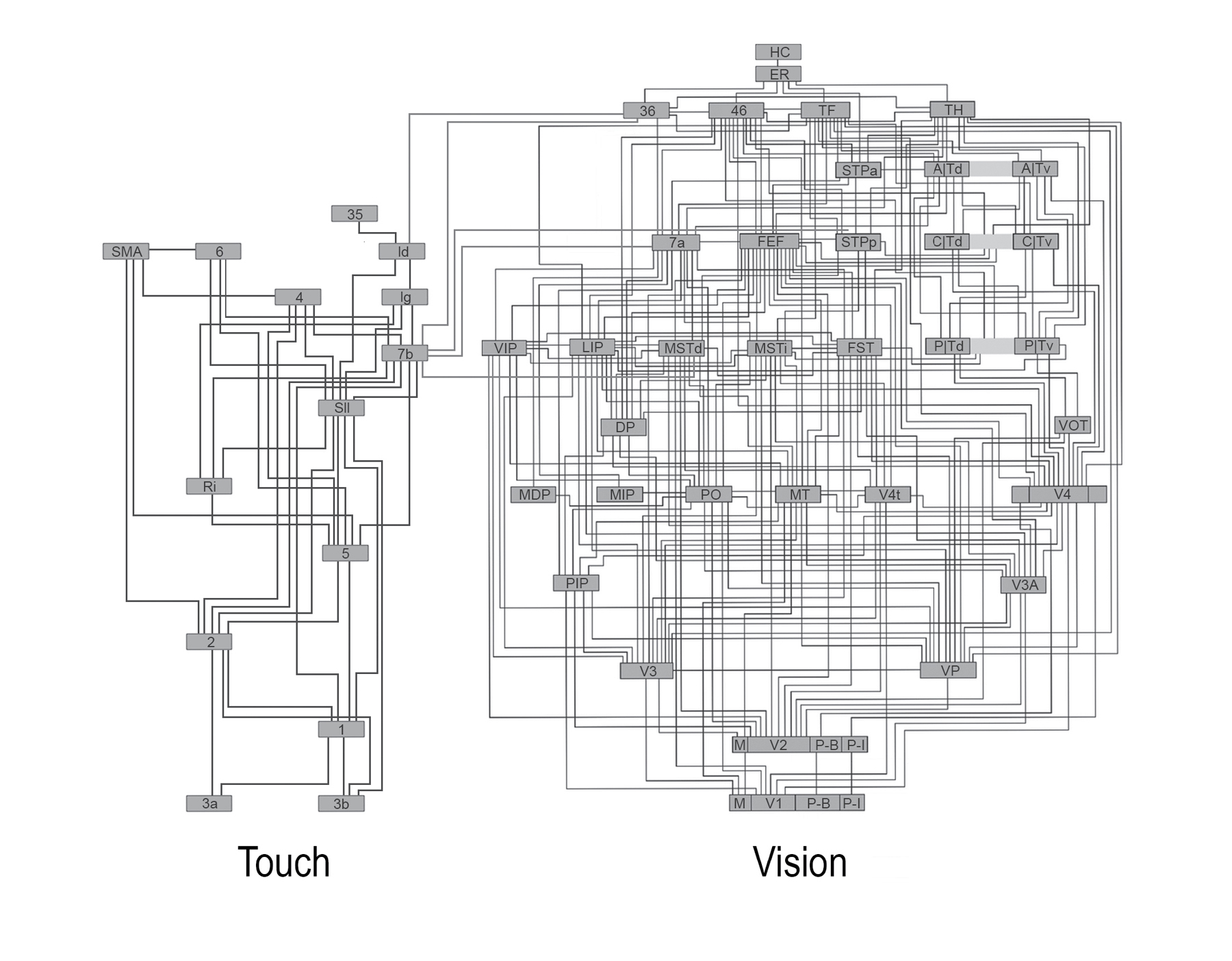
Connections in the neocortex
The dozens of small rectangles in this picture represent the different regions of the neocortex, and the lines represent how information flows from one region to another via the white matter.
A common interpretation of this image is that the neocortex is hierarchical, like a flowchart. Input from the senses enters at the bottom (in this diagram, input from the skin is on the left and input from the eyes is on the right). The input is processed in a series of steps, each of which extracts more and more complex features from the input. For example, the first region that gets input from the eyes might detect simple patterns such as lines or edges. This information is sent to the next region, which might detect more complex features such as corners or shapes. This stepwise process continues until some regions detect complete objects.
There is a lot of evidence supporting the flowchart hierarchy interpretation. For example, when scientists look at cells in regions at the bottom of the hierarchy, they see that they respond best to simple features, while cells in the next region respond to more complex features. And sometimes they find cells in higher regions that respond to complete objects. However, there is also a lot of evidence that suggests the neocortex is not like a flowchart. As you can see in the diagram, the regions aren’t arranged one on top of another as they would be in a flowchart. There are multiple regions at each level, and most regions connect to multiple levels of the hierarchy. In fact, the majority of connections between regions do not fit into a hierarchical scheme at all. In addition, only some of the cells in each region act like feature detectors; scientists have not been able to determine what the majority of cells in each region are doing.
We are left with a puzzle. The organ of intelligence, the neocortex, is divided into dozens of regions that do different things, but on the surface, they all look the same. The regions connect in a complex mishmash that is somewhat like a flowchart but mostly not. It is not immediately clear why the organ of intelligence looks this way.
The next obvious thing to do is to look inside the neocortex, to see the detailed circuitry within its 2.5 mm thickness. You might imagine that, even if different areas of the neocortex look the same on the outside, the detailed neural circuits that create vision, touch, and language would look different on the inside. But this is not the case.
The first person to look at the detailed circuitry inside the neocortex was Santiago Ramón y Cajal. In the late 1800s, staining techniques were discovered that allowed individual neurons in the brain to be seen with a microscope. Cajal used these stains to make pictures of every part of the brain. He created thousands of images that, for the first time, showed what brains look like at the cellular level. All of Cajal’s beautiful and intricate images of the brain were drawn by hand. He ultimately received the Nobel Prize for his work. Here are two drawings that Cajal made of the neocortex. The one on the left shows only the cell bodies of neurons. The one on the right includes the connections between cells. These images show a slice across the 2.5 mm thickness of the neocortex.
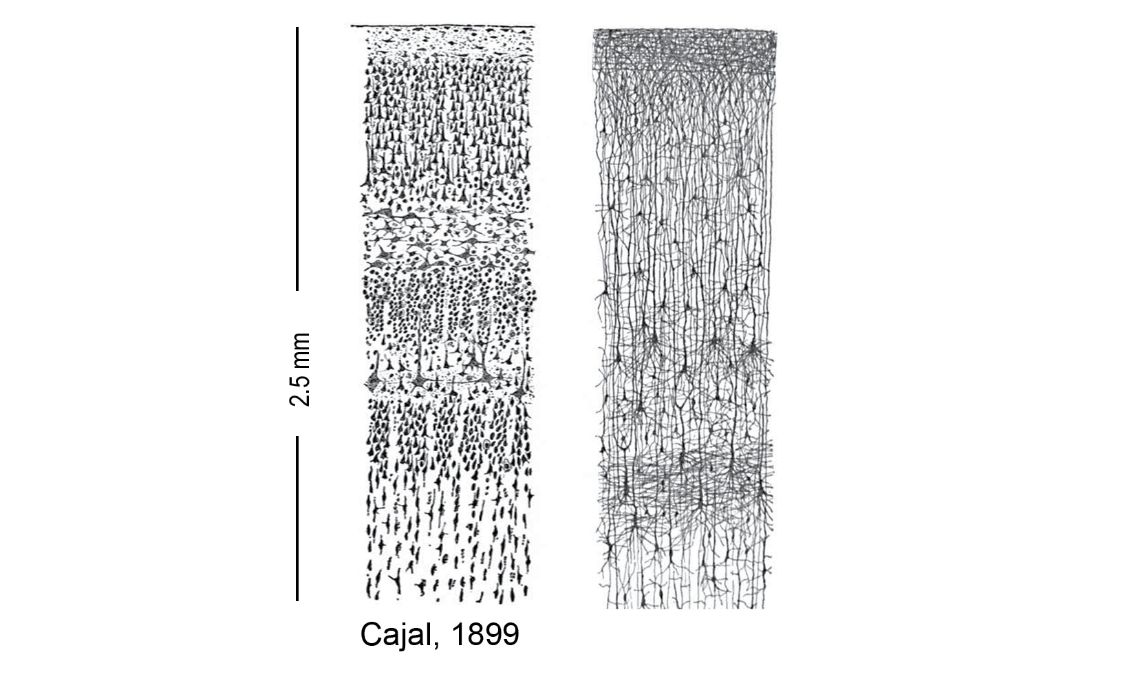
Neurons in a slice of neocortex
The stains used to make these images only color a small percentage of the cells. This is fortunate, because if every cell were stained then all we would see is black. Keep in mind that the actual number of neurons is much larger than what you see here.
The first thing Cajal and others observed was that the neurons in the neocortex appear to be arranged in layers. The layers, which run parallel to the surface of the neocortex (horizontal in the picture), are caused by differences in the size of the neurons and how closely they are packed. Imagine you had a glass tube and poured in an inch of peas, an inch of lentils, and an inch of soybeans. Looking at the tube from the side you would see three layers. You can see layers in the pictures above. How many layers there are depends on who is doing the counting and the criteria they use for distinguishing the layers. Cajal saw six layers. A simple interpretation is that each layer of neurons is doing something different.
Today we know that there are dozens of different types of neurons in the neocortex, not six. Scientists still use the six-layer terminology. For example, one type of cell might be found in Layer 3 and another in Layer 5. Layer 1 is on the outermost surface of the neocortex closest to the skull, at the top of Cajal’s drawing. Layer 6 is closest to the center of the brain, farthest from the skull. It is important to keep in mind that the layers are only a rough guide to where a particular type of neuron might be found. It matters more what a neuron connects to and how it behaves. When you classify neurons by their connectivity, there are dozens of types.
The second observation from these images was that most of the connections between neurons run vertically, between layers. Neurons have treelike appendages called axons and dendrites that allow them to send information to each other. Cajal saw that most of the axons ran between layers, perpendicular to the surface of the neocortex (up and down in the images here). Neurons in some layers make long-distance horizontal connections, but most of the connections are vertical. This means that information arriving in a region of the neocortex moves mostly up and down between the layers before being sent elsewhere.
In the 120 years since Cajal first imaged the brain, hundreds of scientists have studied the neocortex to discover as many details as possible about its neurons and circuits. There are thousands of scientific papers on this topic, far more than I can summarize. Instead, I want to highlight three general observations.
1. The Local Circuits in the Neocortex Are Complex
Under one square millimeter of neocortex (about 2.5 cubic millimeters), there are roughly one hundred thousand neurons, five hundred million connections between neurons (called synapses), and several kilometers of axons and dendrites. Imagine laying out several kilometers of wire along a road and then trying to squish it into two cubic millimeters, roughly the size of a grain of rice. There are dozens of different types of neurons under each square millimeter. Each type of neuron makes prototypical connections to other types of neurons. Scientists often describe regions of the neocortex as performing a simple function, such as detecting features. However, it only takes a handful of neurons to detect features. The precise and extremely complex neural circuits seen everywhere in the neocortex tell us that every region is doing something far more complex than feature detection.
2. The Neocortex Looks Similar Everywhere
The complex circuitry of the neocortex looks remarkably alike in visual regions, language regions, and touch regions. It even looks similar across species such as rats, cats, and humans. There are differences. For example, some regions of the neocortex have more of certain cells and less of others, and there are some regions that have an extra cell type not found elsewhere. Presumably, whatever these regions of the neocortex are doing benefits from these differences. But overall, the variations between regions are relatively small compared to the similarities.
3. Every Part of the Neocortex Generates Movement
For a long time, it was believed that information entered the neocortex via the “sensory regions,” went up and down the hierarchy of regions, and finally went down to the “motor region.” Cells in the motor region project to neurons in the spinal cord that move the muscles and limbs. We now know this description is misleading. In every region they have examined, scientists have found cells that project to some part of the old brain related to movement. For example, the visual regions that get input from the eyes send a signal down to the part of the old brain responsible for moving the eyes. Similarly, the auditory regions that get input from the ears project to the part of the old brain that moves the head. Moving your head changes what you hear, similar to how moving your eyes changes what you see. The evidence we have indicates that the complex circuitry seen everywhere in the neocortex performs a sensory-motor task. There are no pure motor regions and no pure sensory regions.
In summary, the neocortex is the organ of intelligence. It is a napkin-size sheet of neural tissue divided into dozens of regions. There are regions responsible for vision, hearing, touch, and language. There are regions that are not as easily labeled that are responsible for high-level thought and planning. The regions are connected to each other with bundles of nerve fibers. Some of the connections between the regions are hierarchical, suggesting that information flows from region to region in an orderly fashion like a flowchart. But there are other connections between the regions that seem to have little order, suggesting that information goes all over at once. All regions, no matter what function they perform, look similar in detail to all other regions.
We will meet the first person who made sense of these observations in the next chapter.

This is a good point to say a few words about the style of writing in this book. I am writing for an intellectually curious lay reader. My goal is to convey everything you need to know to understand the new theory, but not a lot more. I assume most readers will have limited prior knowledge of neuroscience. However, if you have a background in neuroscience, you will know where I am omitting details and simplifying complex topics. If that applies to you, I ask for your understanding. There is an annotated reading list at the back of the book where I discuss where to find more details for those who are interested.