One evening in 1953, a research scientist at the University of Cambridge in Britain walked into the Eagle, a well-known pub, and announced that he had “found the secret of life.”1 Turns out that he was right.
That man was Francis Crick, and he and his collaborator, James Watson, were about to publish one of the most significant and influential discoveries in modern science: a molecular model of deoxyribonucleic acid, or DNA. At last, the mysterious structure with its vital clues about the mechanism underlying heredity was revealed. The world would finally begin to understand the mechanism of inheritance—how living beings are brought into existence.
In the years since the Watson and Crick discovery, we’ve learned that DNA is an intricate and versatile molecule. Indeed, while the structure of DNA was discovered in 1953, it took another 47 years—and the invention of the supercomputer—to begin to decode the genome— the full set of genetic information represented inside of your DNA.
DNA is not only intricate, but delicate, and susceptible to damage— the type of damage that is believed to cause cancer and other serious diseases. Scientists, doctors, and researchers have long accepted that ionizing radiation—such as the ultraviolet rays that accompany sunlight, or the X-rays that you are exposed to in your doctor’s or dentist’s office—can harm and destroy DNA. It has been assumed, however, that non-ionizing radiation from power lines, television broadcasting, and cell phones did not harm DNA. For instance, in 2002, Dr. Robert L. Park of the American Physical Society, stated:
All known cancer-inducing agents . . . act by breaking chemical bonds, producing mutant strands of DNA. Not until the ultraviolet region of the electromagnetic spectrum is reached . . . do photons have sufficient energy to break chemical bonds. Microwave photons heat tissue, but they do not come close to the energy needed to break chemical bonds, no matter how intense the radiation.2
Powerful statements from prestigious organizations and respected academics, such as the one above, aim to assure the public that EMF is not a health hazard. You may well wonder how I can claim that dangers do exist. Well, the answer is that Dr. Park may know physics, but he is grossly uninformed about biology.
Biological science clearly demonstrates that all frequencies of EMF—including the non-ionizing radiation created by your cell phones, laptops, tablets, and the WiFi antennas in them—can react with and damage DNA. The DNA damage can then lead to cell death or remain as a mutation, which can lead directly to serious diseases, including cancer. This chapter will explain how this happens.
CELLS
All plants, animals, insects—all the organisms that you see around you that are alive—are made of cells. Their existence was first noted in 1665 by British philosopher and researcher Robert Hooke. Hooke, who was the curator of experiments (akin to the director of research and development) for the Royal Society in London, was an early pioneer of the use of microscopes in the field of biology. It was while using a microscope to examine a piece of cork that he made his great discovery, becoming the first person to see and publicly identify cells. (The microscope he used for this research is in the collection of the National Museum of Health and Medicine in Silver Spring, MD.)
It goes without saying that Hooke’s discovery of the cell was significant. At the time, however, Hooke did not appreciate what it was he had found, and it was not until over 170 years later, around 1839, when cell theory finally emerged.
Cell theory is based on a few core principles:
That last point on heredity proved to be a tricky one. While scientists learned much about cells and their function in the intervening century, it wasn’t until the 1950s that we understood what role DNA played in heredity. The three main aspects of DNA that will help us understand the harmful effect of EMF include the electrical properties of DNA, the replication process of DNA, and DNA’s role in coping with environmental stresses at the cellular level.
ELECTRICITY
DNA—with its two strands of genetic information, intertwined in a beautifully symmetrical twisting ladder—is among nature’s most impressive formations, a shape we call the double helix. Though the double-helix structure was noted in the original paper on the model of DNA, the term “double helix” was popularized much later, in the title of James Watson’s 1968 book, The Double Helix: A Personal Account of the Discovery of the Structure of DNA.
DNA is incredibly efficient, too. Inside of every cell in your body, you have a staggering six feet of DNA, coiled up in twists and folds to fit inside of the nucleus at the center of the cell—it’s an amazing feat of engineering with some very interesting electrical characteristics.
The intertwined strands of DNA are connected by rungs of molecules called nucleotides (sometimes also called bases). Each rung of your DNA is composed of two nucleotides, one from each strand, bonded in pairs. These nucleotides are held together by hydrogen bonds, where a single hydrogen atom, shared by two nucleotides, acts as the glue. The presence of so many nucleotides, connected with hydrogen bonds, results in a strong attraction between the two strands.
These hydrogen-bonded nucleotides are relatively flat molecules with electrons on both surfaces. Because the rungs of the DNA ladder are very close, the electrons form a continuous layer (often described as an electron cloud) that is able to conduct an electron current along the DNA chain. This makes it easy for electrons to be conducted (as in a wire) along the nucleotides that form your DNA rungs, a phenomenon called electron transfer. If an electron is released to the DNA rung by an oxidizing agent, that negative charge will flow through the nucleotides. Jacqueline K. Barton and her group at the California Institute of Technology have demonstrated long-range electron transfer in DNA and how this ability can vary with the composition of the base pairs.3
In other words, DNA conducts electricity.
DNA is such an efficient conductor of electricity, in fact, that it is a common building material in molecular electronics, or biological nanotechnology. Researchers who build extremely tiny machines out of living matter use DNA as one of their construction materials, precisely because it conducts electricity so well.
FRACTAL ANTENNA
Another fascinating electrical trait of DNA stems from its compact shape. One of the ways in which the six-foot-long DNA molecule is able to fit so efficiently in a space as tiny as the nucleus of a single cell is by packing itself into a tightly coiled fractal pattern. A fractal is a shape that displays self-similarity, where each part of the shape looks like the entire shape. No matter how far you zoom in or out, the shape looks the same. DNA is a fractal because the smaller coils are themselves coiled into larger coils, a shape known as a coiled coil.
It turns out that the coiled-coil structure and electrical conductivity seen in DNA are the two key characteristics of what we call fractal antennas. The coiled-coil structure of fractal antennas maximizes the length of the antenna, while minimizing its overall size. As a result, fractal antennas are both very long and exceedingly compact. This design can boost cell phone signal strength or radio reception, and amplify a wide range of electromagnetic frequencies. And DNA not only looks like a fractal antenna, but acts like one as well.
By definition, a fractal antenna can pick up and react to a wide range of frequencies of EMF, which means that many frequencies of EMF in the environment can and do react with your DNA. This is why DNA is very sensitive to electromagnetic radiation—notably more sensitive to EMF than other large molecules (such as proteins) in your body.
DNA REPLICATION
DNA is closely linked in people’s minds with inheritance—why you look like your parents or why dogs give birth only to puppies. But if that were the end of DNA’s importance, it would have little impact on your life after your birth.
Far more dynamically, DNA plays a vital and ongoing role throughout your life. Cells inside your body continually die, and new ones are constantly created to replace them. New cells are needed to sustain growth. This is how newborns grow into infants, then into children, and then into adults—by increasing the number of cells in their bodies and replacing those cells that die off. In children, this process is particularly dramatic. But even as adults, our bodies continuously produce new cells. For instance, on average we replace all the cells in our stomach lining every four days. Other multicellular structures and organs have slower cycles, but cell reproduction is going on in our bodies all the time.
For a new cell to be created, the DNA in a cell (referred to as a parent cell) has to be copied. This copying, or replication, process in DNA is truly amazing. Every time a human cell divides, its DNA replicates, copying and transmitting the exact same genetic data to the new cells. Given the trillions of cells in our bodies, the number of replications occurring on a daily basis is mind-boggling.
But cells do not do this perfectly.
MISTAKES
The objective of DNA replication is to create exact copies of the original DNA. However, given the immense scope of the DNA replication process, it’s to be expected that mistakes will happen—and they do. It is estimated that DNA makes replication mistakes 0.001% of the time. That may sound low (imagine a baseball pitcher who allowed only one hit for every 100,000 batters), but given the amount of DNA in each cell, there are approximately 120,000 mistakes in the DNA each time one of the cells in your body divides.
One of the most common types of error is termed a strand break, when a DNA chain breaks apart. When the break is in one strand of the DNA’s double helix, it is termed a single-strand break. When the break occurs in both strands, it is called a double-strand break.
FIXING MISTAKES
Fortunately, the cells in your body also contain tiny quality-assurance testers. These testing mechanisms validate the DNA copies, checking for mistakes. And in many cases, your cells are able to fix the mistakes. So while strand breaks occur all the time, the cell has repair mechanisms to correct many of these breaks. Indeed, studies show that the better your cells are at repairing this damage, the longer you’ll live.
Still, no matter how good your cells are at making repairs, there is a limit to what can be fixed. So when mistakes such as these strand breaks become too numerous (and that level will be different for each person), the cell’s repair mechanisms cannot cope and the damage remains. The DNA in the new cell has mutated from the original.
Often, mutated cells cannot function properly. When this occurs, the cell activates a process named apoptosis, or programmed cell death, to kill and remove the cell. This is an optimal outcome, because once the damaged cell is dead, it cannot harm the body or pass on its defective genes.
Sometimes, however, the damaged cell with the mutated DNA survives and replicates, becoming a permanent genetic mutation in the body. Sometimes such genetic mutations are harmless, or at least the damage is irrelevant to the cell’s operation. All of your DNA is in every cell of your body, but not all cells need all genes. For instance, the cells in your brain express different genes than the cells in your skin, and so on. Other times, however, the damage mutates the DNA (and its offspring) in a harmful way. This is the process through which diseases like cancer are believed to develop.
A variety of forces, both internal (your family’s genes) and external (such as exposure to pollutants), affects the rate at which DNA damage occurs. EMF is one of these forces.
EMF BREAKS DNA STRANDS
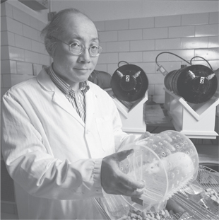
One of the most important series of studies on the question of DNA damage from exposure to non-ionizing electromagnetic radiation was performed by Drs. Henry Lai and Narendra Singh starting in 1994 and running through 1998. Lai and Singh, working at the University of Washington, wanted to answer a simple question: Does non-ionizing radiation damage DNA? To make the results more applicable to daily life, Lai and Singh decided to use levels of EMF radiation considered “safe” by government standards.
Professor Henry Lai, University of Washington Columns Magazine, March 2005
Their results showed that even exposures of only two hours increased the frequency of DNA strand breaks in the brain cells of living rats. Lai and Singh then performed similar experiments with lower frequencies of EMF. For instance, they exposed their subjects to EMF in the range you would find in your average desk lamp. Once again, they found increased occurrences of strand breaks. Lai and Singh’s research, demonstrating DNA strand breaks following exposure to non-ionizing EMF with field strengths of 0.25 or 0.5 millitesla (mT) has been replicated in other laboratories, and it clearly demonstrates that EMF can damage DNA even at low EMF-exposure levels.4 The levels of radiation at which Lai and Singh demonstrate this damage are well below the limits set by the current safety standards for technologies like cell phones, WiFi networks, and microwave ovens.
Even more disturbingly, Lai and Singh found that the DNA in the rat brains continued to break down for hours after exposure ended.5 This suggests that the exposure not only causes immediate damage, but also unleashes a chain of processes that continue to produce damage well after the exposure itself.
Many other studies have found similar genotoxic effects (effects that are poisonous to DNA) resulting from EMF exposure. As Dr. George Carlo and Martin Schram explain in their book Cell Phones: Invisible Hazards in the Wireless Age, multiple studies have demonstrated increased rates of micronuclei in the body following exposure to RF/ MW radiation (such as that emitted by cell phones). A micronucleus is a fragment of DNA with no known purpose, a by-product of errors that occur during cell division. The presence of micronuclei indicates a type of DNA damage so strongly associated with cancer that doctors test for them as a means of diagnosing cancer.
In 2009, Hugo W. Rüdiger, a professor at the Medical University of Vienna, released a study analyzing the results of 101 different published articles on the effects of low-frequency EMF on DNA. His review, published in the peer-reviewed journal Pathophysiology in August 2009, found that “of these, 49 report a genotoxic effect and 42 do not. In addition, 8 studies failed to detect an influence on the genetic material, but showed that RF-EMF enhanced the genotoxic action of other chemical or physical agents.” He concluded that “there is ample evidence that RF-EMF [low-energy radio frequency electromagnetic fields] can alter the genetic material of exposed cells.”6
PROTEIN SYNTHESIS
Another area in which low-frequency, non-ionizing radiation has been proven to harm DNA function is in your cell’s production of protein. Protein inside your cells is just like the protein your doctor tells you you should eat more of. In fact, it’s precisely because your cells need protein to function that you should be eating a healthy amount of the right types of protein. Protein is required for all the functions that your cells perform.
The science of proteins extends back to the 18th century, and since that time, an increasing number of proteins have been discovered and classified. Many of the body’s proteins deal with what might be considered basic housekeeping functions of life such as developing muscle to enable us to move and enzymes to enable us to digest food. Another class of proteins is antibodies, first identified in the 1890s, which your body’s cells create when you are under attack by foreign organisms (like a cold). The most recently discovered class of proteins is stress proteins, which are stimulated by potentially harmful environmental agents, including EMF.7
Fortunately, you don’t need to eat all of the exact proteins that your body needs. And even if you did, they would not be in the form that your body requires. That’s because your cells convert the proteins you eat into the types of proteins that it needs to function. When you eat protein, your body breaks that protein down into its constituent amino acids. Your cells then take these amino acids and build new proteins. Replacements are needed for damaged proteins, and new proteins are needed for new cells that are formed when cells divide. Because so many proteins have to be made, protein synthesis is happening in your body all the time.
DNA is central to protein synthesis. Human DNA has the ability to create about 25,000 different kinds of proteins; with those, your body can work to create an estimated 2,000,000 different types of protein that your body needs to function properly. Some proteins are always present (such as those that aid in the process of food digestion), and some proteins are created by your body on demand (such as antibodies that aid in the defense against viruses).
STRESS
While your body has a vast range of proteins that serves millions of functions, one type of protein is particularly relevant to the EMF issue: the proteins that help your cells cope with damage from environmental stress.
In the 1960s, Ferruccio Ritossa, an Italian scientist, made an unusual discovery in the chromosomes of a fruit fly. Over the course of an experiment, the flies were accidentally exposed to a temperature increase of a few degrees. Ritossa noticed that when this happened their chromosomes became enlarged at particular sites. It would be another 15 years before the significance of the finding was realized. When it was, the heat shock response, as it came to be termed, was found to occur in both animals and plants—including yeast. After extensive research in laboratories around the world, it was found that the heat shock response was the first stage in the synthesis of a special class of proteins called heat shock proteins.
Heat shock proteins repair other proteins. They serve a defensive role, defending cells against the ill effects of increases in temperature that could otherwise prove fatal. What’s more, they strengthen the cells to be more resilient to temperature increases in the future. Just like lifting weights today makes you stronger tomorrow, these heat shock proteins make your cells better able to cope with subsequent stresses of increased temperature. This greater resistance to the stress of heat shock is called thermotolerance.
Since the initial discovery of the heat shock response and heat shock proteins, we have discovered that cells produce similar proteins to cope with many different types of stress—not just temperature. And so, these proteins came to be collectively referred to as stress proteins, which are involved in the cellular stress response—the process by which individual cells cope with stress. (Because of the way these proteins were discovered, they are still designated with an “hsp” to show that it is a heat shock protein, and a number that is related to size.)
What stresses a cell? A variety of forces, as it turns out. The presence of heavy metals, changes in acidity, alcohol, viral infections, ultraviolet light, and low-oxygen conditions—all of these can damage your cells in the same way that an increase in temperature can. So, there are a variety of conditions that lead to the stress response—a rise in temperature, or thermal stress, is just one of many environmental factors. And on the whole, the cellular stress response has been very effective in helping cells cope with environmental stresses.
EMF AND THE STRESS RESPONSE
EMF—even at low-energy, non-ionizing frequencies—is among the environmental factors that stimulate the stress response in the cells of our bodies. Dr. Goodman and I demonstrated the relationship between EMF and the cellular stress response in research that we performed in 1994. We released a comprehensive review of the studies on EMF stimulation of stress protein synthesis in 1998, and an update in 2009 in the special EMF issue (volume 16) of the journal Pathophysiology.
Our initial studies found that when human cells are exposed to radiation in the extremely-low-frequency range (ELF similar to that from power lines), the stress response is triggered and cells begin to create stress proteins within five minutes. EMF appears to stimulate stress protein synthesis in much the same way that the natural electric fields that transmit signals in your nerves lead to the creation of proteins in your muscles.
We later repeated our experiment using EMF in the frequency range emitted by cell phones, and we found the same effect. These findings have been subsequently replicated in multiple experiments by researchers around the world.8
So we know that the presence of stress proteins is an indication that the cell has come into contact with something that it reacts to as harmful. And we know that EMF triggers the presence of stress proteins. If our bodies generate stress response proteins for EMF, doesn’t that mean that our bodies are coping with whatever damage electromagnetic radiation might otherwise cause?
Yes and no.
As we’ve seen, the cellular stress-response can be extraordinarily useful because it allows the organism to adapt to and overcome problems. The defensive value of the stress proteins is undeniable, and it is generally accepted that the short-term results from the generation of stress proteins in your cells is almost always beneficial. Stress proteins build your body’s defenses against damaging forces like increases in temperature and reductions in the oxygen supply that could otherwise prove life threatening. Because the body generates stress proteins to strengthen cellular resistance to EM radiation, you are well equipped to handle limited exposure to EMF-generating devices such as cell phones. However, there are limitations.
ELECTROMAGNETIC TOLERANCE
The long-term effects, however, are a different matter. Scientists have found that prolonged exposure to EMF (which, again, in the short term encourages the generation of stress proteins) has the opposite effect— extended exposure to EMF reduces the ability of your cells to produce stress proteins. Dr. Goodman and I first showed in 1996 that there was a decreased response when EMF stimuli are repeated.9 A reduced stress response is similar to the thermotolerance that results from prolonged exposure to heat shock. The evidence shows that extended exposure to EMF begins breaking down your DNA’s cellular stress response. Given the rise of wireless and other electronic technologies, more people are increasingly subject to prolonged EMF exposures and potentially developing an electromagnetic tolerance at the cellular level.
Drs. A. DiCarlo, J.M. Farrell and T. Litovitz at Catholic University of America in Washington, DC, observed similar results in an experiment performed on chicken embryos.10 In those eggs exposed to ELF radiation of 8 μT (such as that emitted by power lines) for 30 or 60 minutes at a time, twice a day for four days, production of hsp70 (heat shock protein 70, created in response to oxygen deprivation) declined. The same response was noted in eggs exposed to RF radiation (such as that emitted by cell phones) of 3.5 μW/cm2 (microwatts) for 30 or 60 minutes, once a day, for four days. The researchers noted that these eggs produced 27% less hsp70 following these exposures and had correspondingly reduced cytoprotection (the ability to fend off cell damage).11 Similar experiments have been carried out with short, repeated exposures (in contrast to extended exposures). There, too, the rate of stress protein synthesis is reduced with each repetition. These experiments clearly demonstrate EMF tolerance.
Long-term exposure to EMF (either prolonged or repeated or both) reduces your body’s resilience to stressful forces in the environment. Our species did not evolve with all of these external electromagnetic forces continuously impacting our bodies. Today, your body’s cellular stress response is being called into action in a way it is not prepared for. With each additional moment you are exposed to electromagnetic fields, your cells (the basic building blocks of your body) are more susceptible to damage from other harmful forces in the environment, such as the sun’s ultraviolet rays. EMF reduces your cells’ ability to respond to many types of environmental damage.
And the effect is cumulative, across a lifetime of exposures.
Long- and short-term exposure to electromagnetic radiation can harm DNA in your body, leading to cell death and cell mutation. These effects are seen across the EM spectrum—not just from ionizing radiation like ultraviolet and X-ray, but also from non-ionizing radiation including cell phone MW transmissions and even the extremely low frequency EMF from power lines.
These are some of the most important effects of EMF exposure at the cellular level. The type of cellular damage caused by EMF is similar to that caused by aging. The residual errors and genetic mutations accumulate, leading to malfunction and disease. There has been a steady rise in EMF radiation in the environment, and DNA damage is now occurring more frequently and earlier in life because of the many ways modern technological forces permeate our lives.
When considering the risks of the EMF issue, people are generally interested in the health effects—outcomes such as cancer and other diseases. The types of cell damage described in this chapter are among the known biological effects shown to result from non-ionizing EMF exposure. The biological effects are mechanisms and indicators of the health effects that will be described in coming chapters. The reduction in cells’ ability to invoke the stress response leaves us more susceptible to disease. DNA mutation is a process by which cancers are widely believed to form, and the DNA damage indicated by the presence of micronuclei is considered to be a strongly accurate indicator of cancer.
Now that we understand some of the biological effects of EMF exposure and that these are associated with the generation of disease in the human body, let’s examine what science can tell us about the link between exposure to non-ionizing electromagnetic radiation and cancer.